Make the best use of Scientific Research and information from our 700+ peer reviewed, Open Access Journals that operates with the help of 50,000+ Editorial Board Members and esteemed reviewers and 1000+ Scientific associations in Medical, Clinical, Pharmaceutical, Engineering, Technology and Management Fields.
Meet Inspiring Speakers and Experts at our 3000+ Global Conferenceseries Events with over 600+ Conferences, 1200+ Symposiums and 1200+ Workshops on Medical, Pharma, Engineering, Science, Technology and Business
Case Report Open Access
Septic Shock in a Patient with Tubercular Liver Abscess
| Ujjwayini Ray*, Soma Dutta and Arpita Sutradhar | ||
| Apollo Gleneagles Hospitals, Kolkata, West Bengal, India | ||
| Corresponding Author : | Ujjwayini Ray Department of Microbiology and Serology, Apollo Gleneagles Hospitals Kolkata, West Bengal, India Tel: +91 9830639480 Email:dr_uray@rediffmail.com |
|
| Received September 13, 2014; Accepted November 17, 2012; Published December 05, 2014 | ||
| Citation: Ray U, Dutta S, Sutradhar A (2014) Septic Shock in a Patient with Tubercular Liver Abscess. J Infect Dis Ther 2:188. doi: 10.4172/2332-0877.1000188 | ||
| Copyright: © 2014 Ray U, et al. This is an open-access article distributed under the terms of the Creative Commons Attribution License, which permits unrestricted use, distribution, and reproduction in any medium, provided the original author and source are credited. | ||
Related article at Pubmed Pubmed  Scholar Google Scholar Google |
||
Visit for more related articles at Journal of Infectious Diseases & Therapy
| Introduction |
| Tuberculosis is second only to HIV/AIDS as the greatest killer worldwide due to a single infectious agent. In 2012, 8.6 million people fell ill with tuberculosis and 1.3 million people died from the disease [1]. Mycobacterial septic shock, once considered a rare complication of miliary tuberculosis is now being increasingly recognized. Mycobacterial septic shock also referred to as sepsis tuberculosa gravissima can manifest in patients even without widespread dissemination. Mycobacterial septic shock is diagnosed in patients who present with haemodynamic features of septic shock along with clinical suspicion of tuberculosis with positive staining for acid fast bacilli and or recovery of Mycobacterium tuberculosis in culture from sputum, BAL, body fluids or tissues in the absence of a more plausible pathogen [2]. Mycobacterial septic shock carries a grave prognosis and early recognition of the entity is of utmost importance. Here we present a case of mycobacterial septic shock in a patient of chronic renal failure. |
| Case Report |
| A 58 year old diabetic, hypertensive, Indian, male patient suffering from end stage renal disease (ESRD) was brought to the emergency department in a state of septic shock. He was febrile, oliguric, disoriented and had abdominal discomfort and vomiting. On physical examination the patient was hypotensive with a blood pressure of 90/60 mm of Hg, temperature was 39.8°C, respiratory rate was 24/min. Results of his respiratory and cardiovascular examination were normal. There was diffuse tenderness over the right hypochondrium. |
| Significant laboratory findingsat admission were a slightly raised TLC (10370/cumm, Neutrophils 89%, Lymphocytes 6%,) haemoglobin 8.4%, ESR>140 mm in first hour, Urea 318 mg/dl, blood sugar 362 mg/dl, creatinine 9.3 mg/dl and mildly raised SGOT and SGPT . He was seronegative for HIV antibodies. |
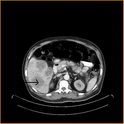
| USG of the abdomen revealed multicystic lesions in the liver. CT scan of the abdomen revealed multifocal, variegated, complex, hypodense space occupying lesions (Figure 1). Fine needle aspiration and biopsy was done from the liver lesions. Purulent material was aspirated from the lesion. Chest rotegenogram was normal. Suspecting bacteria sepsis the patient was started on broad spectrum antibiotics (piperacillin/tazobactam and teicoplanin). He was put on inotropes (Nor adrenalin 4 ml/hour) due to worsening hypotension. Continuous veno-venous haemofiltration was initiated to combat renal failure. |
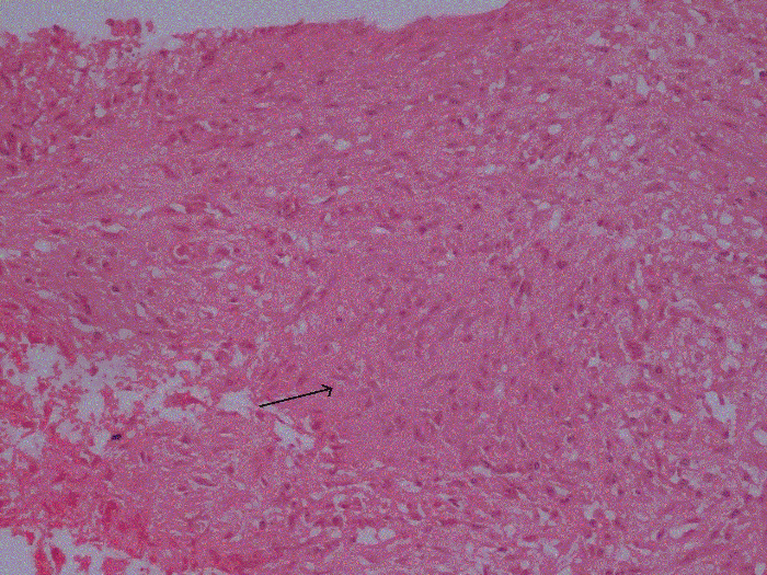
| Histopathological examination of the tissue from the hepatic lesion showed wide spread areas of caseous necrosis and few preserved epitheloid cell clusters consistent with Mycobacterial tuberculosis infection (Figure 2). No organism was seen in the gram and the Ziehl-Nelson (Z-N) stained smear. The pus culture was negative for common aerobic and anaerobic bacterial pathogens. Two sets of blood culture drawn at the time of admission were also sterile. No other source of sepsis was identified. |
| He was promptly started on Anti Tubercular Drugs (ATD) with Rifampicin (600 mg OD), Isoniazid (300 mg OD), Ethambutol (400 mg OD) and Pyrizinamide (750 mg OD) on day 7 of hospitalization. The patient gradually improved with ATD and was successfully taken off pressor agents three days later. He became afebrile and discharged in a haemodynamically stable condition on day 14th of hospitalization. He continued to be on haemodialysis for the chronic renal failure. |
| The pus from the liver lesion grew acid fast bacilli three weeks later following culture in the MGIT system (Becton Dickinson). The acid fast bacillus was identified as Mycobacterium tuberculosis complex sensitive to rifampicin and isoniazid (Geno Type MTBDR plus, Hain Lifescience GmbH). A repeat ultrasound after 4 weeks showed significant reduction in the size of the liver lesions. |
| Discussion |
| The manifestations of Mycobacterium tuberculosis infection are manifold. But the less recognized entity is septic shock. There have been approximately 25 cases of Mycobacterium tuberculosis septic shock sporadically reported in the literature [2]. Mycobacterial septic shock has been mostly reported in patients with severe immunosuppression especially in advanced stages of AIDS [3]. Respiratory distress and shock attributable to tuberculosis has also been reviewed in paediatric patients [4]. |
| In a recent and first of its kind study, Kethireddy et al. has retrospectively analyzed cases of septic shock across 28 centers in Canada, the United States and Saudi Arabia. They have found 1% of all culture positive bacterial septic shock was due to MTB (range 0% in several North American sites to 3.3% in Saudi Arabia) [2]. The incidence of Mycobacterial septic shock is likely to be higher in tuberculosis endemic countries. |
| All forms of immunosuppression predispose to tuberculosis due to primary infection, endogenous reactivation and exogenous infection. In the background of immunosuppression non pulmonary lesions due to unrestricted bacillary dissemination are frequent. Dissemination may present as one or more solitary lesions, as widespread lymphadenopathy or as multiorgan involvement [5]. In this patient with compromised immune status due to chronic kidney disease and diabetes there was extensive involvement of the liver. There was no evidence of any pulmonary or any other similar lesions elsewhere. In case of mycobacterial septic shock, involvement of the respiratory tract is frequently reported and extra pulmonary involvement without evidence of lung involvement as seen in this case appears to be a less common occurrence. |
| Septic shock is generally associated with gram positive and gram negative bacteria. It is generally believed that Mycobacterial septic shock occurs in the setting of miliary tuberculosis. Mycobacterial septic shock can occur even with single organ involvement without widespread dissemination. The Lipoarabinomannan (LAM) in the cell wall of mycobacteria can stimulate macrophages to secrete and release TNF a thereby causing hypotension and shock. |
| Mycobacterial septic shock carries a grave prognosis. Kethireddy et al. reported in-hospital mortality rates of 79.2% as compared to 49.7% for other bacterial shock [2]. An important reason for this seems to be delay in initiating appropriate therapy with anti-mycobacterial drugs due to inability to recognize the etiological agent promptly. It is a well-established fact that administration of appropriate antimicrobial therapy early in the course of septic shock is associated with a favourable outcome. Smear examination by Z-N stain is an insensitive test and may not reveal the bacilli in the lesion. Isolation of Mycobacteria in culture, the gold standard for diagnosing tuberculosis infection requires between 2-6 weeks for the bacilli to grow in culture. Due to low index of suspicion rapid tests such as nucleic acid based tests are not performed routinely. Repeated culture negative for common bacterial and fungal pathogens is often attributed wrongly to the broad spectrum antibiotics that are empirically administered to these septic patients. Many of the cases reported in the literature have been diagnosed post mortem [6,7]. Fortunately in our patient, the histopathological examination pointed towards Mycobacterial etiology and antibiotics were changed to anti tubercular drugs. Similarly, Vadillo et al. reported diagnosing a case of Mycobacterial septic shock on the basis of results of bronchial and bone marrow biopsies on day 2 of hospitalization [8]. The confirmation of the Mycobacterial etiology in this case was obtained when the Mycobacteria grew in culture after 4 weeks. |
| Conclusion |
| Septic shock due to Mycobacterium tuberculosis is probably an iceberg phenomenon in that a large number of cases go undetected. Though mycobacterial septic shock is detected and reported infrequently this entity may not be so rare given the high burden of tuberculosis cases worldwide. Mycobacterial septic shock carries a high mortality due to delay in diagnosis owing to low index of suspicion. Mycobacterial septic shock should be considered in patients with compromised immune system if cultures are negative for bacterial and fungal pathogens. Tests which will rapidly provide a clue to the underlying aetiology such as histopathological/cytopathological examination or nucleic acid based tests should be considered. In particular, clinicians and intensivists should be well aware of this possibility. Furthermore prospectively designed studies are required to gain more insights into this phenomenon. |
References
- World Health Organization Global Tuberculosis Report 2013
- KethireddyS, Light RB, Mirzanejad Y,Maki D, Arabi Y, et al. (2013) Mycobacterium tuberculosis septic shock. Chest 144: 474-482.
- Gachot B, Wolff M, Clair B, Régnier B (1990) Severe tuberculosis in patients with human immunodeficiency virus infection. Intensive Care Med 16: 491-493.
- Mazade MA, Evans EM, Starke JR, Correa AG (2001) Congenital tuberculosis presenting as sepsis syndrome: case report and review of the literature. Pediatr Infect Dis J 20: 439-442.
- Grange JM (1998) Tuberculosis. In: Collier L, Balows A, Sussman M, eds. Topley& Wilson's Microbiology and Microbial Infections. London, England: Arnold; 1998.
- Ahuja SS, Ahuja SK, Phelps KR, Thelmo W, Hill AR (1992) Hemodynamic confirmation of septic shock in disseminated tuberculosis. Crit Care Med 20: 901-903.
- Pene F, Papo T, Brudy-Gulphe L, Cariou A, Piette JC et al. (2001) Septic shock and thrombotic microangiopathy due to Mycobacterium tuberculosis in a nonimmunocompromised patient. Arch Intern Med 161:1347–1348.
- Vadillo M, Corbella X, Carratala J (1994) AIDS presenting as septic shock caused by Mycobacterium tuberculosis. Scand J Infect Dis 26: 105-106.
Figures at a glance
 |
 |
|
| Figure 1 | Figure 2 |
Post your comment
Relevant Topics
- Advanced Therapies
- Chicken Pox
- Ciprofloxacin
- Colon Infection
- Conjunctivitis
- Herpes Virus
- HIV and AIDS Research
- Human Papilloma Virus
- Infection
- Infection in Blood
- Infections Prevention
- Infectious Diseases in Children
- Influenza
- Liver Diseases
- Respiratory Tract Infections
- T Cell Lymphomatic Virus
- Treatment for Infectious Diseases
- Viral Encephalitis
- Yeast Infection
Recommended Journals
Article Tools
Article Usage
- Total views: 14182
- [From(publication date):
December-2014 - Jul 12, 2025] - Breakdown by view type
- HTML page views : 9600
- PDF downloads : 4582
