Schwannoma of Floor of the Mouth: A Case Report
Received: 31-Jan-2020 / Accepted Date: 14-Feb-2020 / Published Date: 21-Feb-2020 DOI: 10.4172/2476-2024.1000162
Abstract
Schwannomas, which are benign tumors that arise from Schwann cells in the myelinated nerves, are common soft tissue tumors in the head and neck region. However, the occurrence of schwannomas in the oral cavity is very rare, accounting for only 1% of all schwannomas in the head and neck region. We report a case of a slowly growing schwannoma exceeding 3 cm in size that occurred on the right side of the floor of the mouth in a 21-year-old woman. The patient underwent a magnetic resonance imaging MRI examination and surgical resection. The patient was definitively diagnosed by histopathological examination and immunohistochemical examination.
Keywords: Schwannoma; Neurinoma; Oral cavity
Introduction
Schwannomas are benign tumors that arise from Schwann cells in the myelinated nerves, are covered by a membrane, and grow slowly. In general, they are solitary tumors. Approximately 25%-45% of schwannomas develop in the head and neck region. Of them, approximately 1%-10% occurs in the oral cavity, and the tongue is the most common site [1].
There are two types of peripheral nerve sheath tumors, namely, neurofibroma and schwannoma. Neurofibroma is composed of neurites, Schwann cells, and fibroblasts that produce collagen and mucoid substances. Schwannomas are derived from Schwann cells [2,3]. These tumors can occur as genetic disorders, in which case they are known as neurofibromatosis type 1 (NF1) and type 2 (NF2) and Schwannomatosis. The tumors arise from the cranial nerves, spinal nerves, and nerves covered with Schwann cells in the autonomic nervous system [1]. In the oral cavity region, it may be difficult to identify the nerve associated with the origin of the tumor. We report a case of a schwannoma that occurred on the floor of the mouth in a 21- year-old woman.
Case Presentation
A 21-year-old woman complained of a painless lump between the floor of the mouth and the underside of the tongue on the right side that had been present for approximately two years. The lump gradually became enlarged but otherwise was asymptomatic. No serious personal or family medical history was reported. An oral cavity examination showed a swelling measuring 4 × 3 cm on the right side of the floor of the mouth. The surface of the lump was smooth and covered by normal mucosa. It was elastic and soft upon palpation.
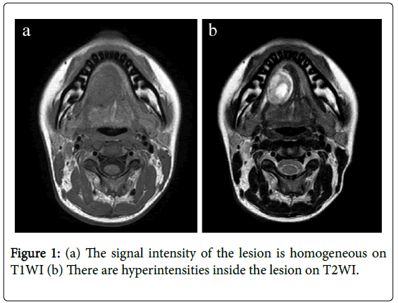
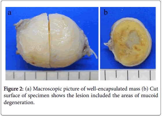
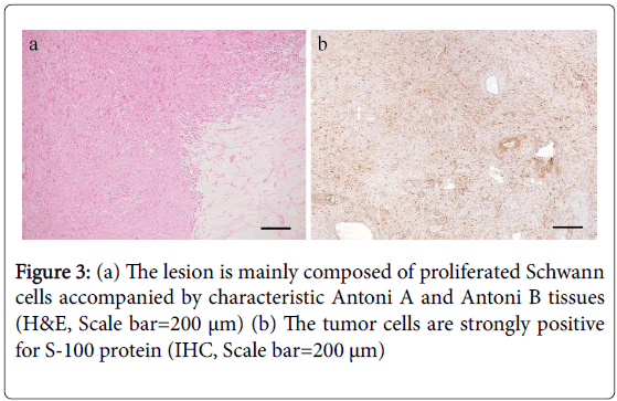
Magnetic Resonance Imaging (MRI) was performed to clarify the features of the lesion and revealed the presence of an egg-shaped node on the right side of the floor of the mouth. On T1-Weighted Imaging (T1WI), the lesion had a clear boundary, and the signal intensity of the inside was homogeneous. On T2-weighted imaging (T2WI), there were hyperintensities inside the lesion (Figure 1). The lesion was judged to be benign on imaging. Based on the clinical findings, the characteristics on MRI, and the anatomical position, we made a provisional diagnosis of a benign tumor occurring under the tongue. The patient underwent surgical removal of the lump under general anesthesia. Gross pathology of the resected specimen showed that the lesion was an encapsulated mass measuring approximately 3.5 cm × 2.5 cm × 2.0 cm, which included the areas of mucoid degeneration and mucus-filled areas (Figure 2). A histopathological examination showed that the lesion had cystic areas and areas of mucoid degeneration, and the lesion was mainly composed of proliferated Schwann cells accompanied by characteristic Antoni A and Antoni B tissues. The tumor was surrounded by a fibrous cap. Immunochemical staining (IHC) revealed that the tumor cells were strongly positive for S-100 protein (Figure 3). Based on these histological findings, the patient was diagnosed with schwannoma. The patient was followed up for six months after surgery with no recurrence or complications.
Discussion
Schwannoma was first described by Verocay in 1910 as a tumor that arises from Schwann cells in the myelinated nerves and named a neurinoma. Subsequently, the term schwannoma has been used for this lesion.
Schwannomas are benign tumors that arise from Schwann cells of the neurilemma. The growth is slow, and the tumors are encapsulated by fibrous tissue. The tumors are seen in the head and neck region in approximately 25%-45% of all cases of schwannomas. The majority of schwannomas that occur in the head and neck region are derived from the vestibular nerve. Approximately only 1-10% of schwannomas in the head and neck region occur in the oral cavity [1]; the incidence is the highest in the tongue, followed by the palate, floor of the mouth, buccal mucosa, and gingiva [4]. A review article in recent years showed no gender difference in the incidence of the tumors and a high incidence in patients aged 20 to 49 years. The mean size of the tumor is 2.4 cm, and the tumors commonly occur in the front of the tongue [1]. In the present case, a tumor appeared on the right side of the floor of the mouth as an asymptomatic lump in a 21-year-old woman.
In general, schwannomas are solitary lesions. However, multiple lesions are seen as part of NF1 in some cases [5]. Most solitary schwannomas are associated with the nerve trunk and grow slowly. They put pressure on the nerves as they grow.
The tumors are generally asymptomatic. However, they can cause pain in some cases [6]. In the previously reported reviews of schwannomas, there have not been sufficient clinical data related to pain with schwannomas and neurological disorders. Although our patient experienced a swelling under the tongue, she did not have pain while swallowing, and she did not have any neurological disorder.
Schwannomas occurring under the tongue need to be discriminated from other benign lesions that commonly occur in this region [5]. Differential diagnosis of swelling on the floor of the mouth includes mucocele, dermoid cyst, epidermoid cyst, salivary gland tumor, and swollen submandibular gland. In the case of a schwannoma in the submandibular gland, discrimination from pleomorphic adenoma in the submandibular gland needs to be carefully performed. The present case histologically had Antoni A and Antoni B patterns, and the tumor tissue was clearly surrounded by fibrous tissue. Antoni A areas are composed of spindle cells with twisted nuclei, and the boundary of the cytoplasm is unclear. The spindle cells are arranged in bundles or interlacing fascicles, and intranuclear vacuoles may be present [2]. In the Antoni A region in this case, the nuclei were aligned and palisading, and Verocay bodies were formed between the alignments of palisading nuclei. In the Antoni B region, there were a few cells, and the alignment was poorly regulated. The stroma was in a state of myxoma and contained a lot of substances including fine collagen fibers. Such tumors can cause hyalinization, calcification, bleeding, sacculation, and denaturation such as dyskaryosis. However, these findings do not suggest malignancy.
S-100 is highly expressed in most cells of schwannomas, whereas the expression varies in neurofibromas. Since the immunostaining of this protein is very consistent, and the intensity is high, it functions as an important diagnostic tool. The expression of S-100 decreases in Antoni B areas. In the present case, most tumor cells were positive for S-100 protein, which indicates that proliferated cells are mainly composed of Schwann cells. Based on the immunostaining results and histological characteristics, we definitively diagnosed schwannoma.
Conclusion
Resection is a common treatment for this tumor. The malignant transformation of the tumor is very rare, and the prognosis is favorable. Although there are a few reports of the clinical significance of neurosensory impairment in schwannomas in the oral cavity, the present patient has shown no signs of recurrence, and the postoperative course is favorable.
Conflict of Interests
The authors declare that there is no conflict regarding the publication of this paper.
Source of Funding
This work was supported by JSPS KAKENHI Grant Numbers JP18K09789, JP18K17224, JP19K29094.
References
- Cohen M, Wang MB (2009) Schwannoma of the tongue: two case reports and review of the literature. Eur Arch Otorhinolaryngol 266: 1823-1829.
- Goldblum JR, Folpe AL, Weiss SW (2014) Enzinger and Weiss’s Soft Tissue Tumors (6th edn), ELSEVIA SAUNDERS, Philadelphia.
- Butler RT, Patel RM, McHugh JB (2016) Head and neck schwannomas: 20-Year experience of a single institution excluding cutaneous and acoustic sites. Head Neck Pathol 10: 286-291.
- Moreno-GarcÃa C, Pons-GarcÃa MA, González-GarcÃa R, Monje-Gil F (2014) Schwannoma of tongue. J Maxillofac Oral Surg 13: 217-221.
- Marx RE, Stern D (2012) Oral and Maxillofacial Pathology: A Rationale for Diagnosis and Treatment (2nd edn) Quintessence Publishing co inc, Batavia.
- Neville BW, Damm DD, Allen CM, Bouquot JE (2009) Soft tissue tumors. Oral and Maxillofacial Pathology (3rd edn), Elsevier, St. Louis.
Citation: Omori H , Nakano K, Nagata Y, Takabatake K, Kawai H, et al. (2020) Schwannoma of Floor of the Mouth: A Case Report. Diagn Pathol Open 5: 162. DOI: 10.4172/2476-2024.1000162
Copyright: © 2020 Omori H, et al. This is an open-access article distributed under the terms of the Creative Commons Attribution License, which permits unrestricted use, distribution, and reproduction in any medium, provided the original author and source are credited.
Share This Article
Open Access Journals
Article Tools
Article Usage
- Total views: 4376
- [From(publication date): 0-2020 - Apr 03, 2025]
- Breakdown by view type
- HTML page views: 3611
- PDF downloads: 765


