Sacroiliac Joint Dysfunction
Received: 06-May-2019 / Accepted Date: 17-May-2019 / Published Date: 24-May-2019 DOI: 10.4172/2165-7025.1000412
Abstract
Sacroiliac joint dysfunction is a painful musculoskeletal condition, responsible for 5%-10% cases of low back pain, also called Sacroiliac Syndrome. It usually occurs due to hypermobility or hypomobility and pathologic conditions. The diagnosis of SIJD made by clinical features, diagnostic test, radiographic, CT scan, MRI and Ultrasound Doppler Imaging. Management of SIJD includes nonsurgical and surgical interventions. Non-surgical management includes medication and physical therapy intervention. Surgical management includes surgical to release pain.
Keywords: Sacroiliac joint dysfunction; MRI; CT; US
Introduction
Sacroiliac joint dysfunction is changing of the structure in the joint (ligaments, cartilage or muscles), which make impairment or limitation on the movement, it can be a result of Low back pain. Sacroiliac joint dysfunction is a condition that is may be misdiagnosed, so it is important to be aware of the specific symptoms associated with SIJ pain; it has a lot of terms such as SIJ Syndrome, SIJ Strain, SIJ Inflammation and Sacroilitis [1].
Epidemiology
The research showed that SI joint dysfunction is more common in women than man with gender ratio female: male (4:1), the general population reported between 15%-38%, the studies indicated SIJ Dysfunction is the main reason of the pain in 5%-10% of patient with LBP [1].
Inclusion criteria
Sacroiliac joint is the joint between sacrum and ilium bone. It connects the spinal vertebral with pelvic, SIJ is a synovial joint (diarthrodial), it has an irregular ridges (elevation and depressions) that gives the joint stability and interlocking of the two bones, The joint cover by Hyaline cartilage (fibrocartilage) [2,3].
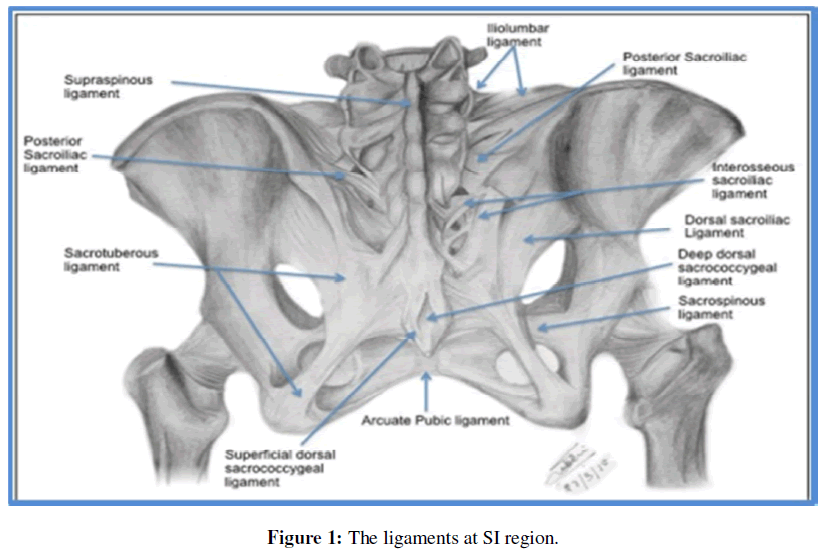
The SI joint attributed to many ligaments such as anterior sacroiliac ligament, Interosseous SI ligament, it is responsible for preventing abduction or distraction of SI joint and helps to bear the weight. Posterior sacroiliac ligaments, Sacrotuberous ligament, Sacrospinous ligament, they give a rotary movement in the joint, and iliolumber ligament, it connects the fifth lumber process to the iliac crest. It is more common damage or torn. Otherwise, during pregnancy these ligaments stretch in long periods in Figure 1 [3,4].
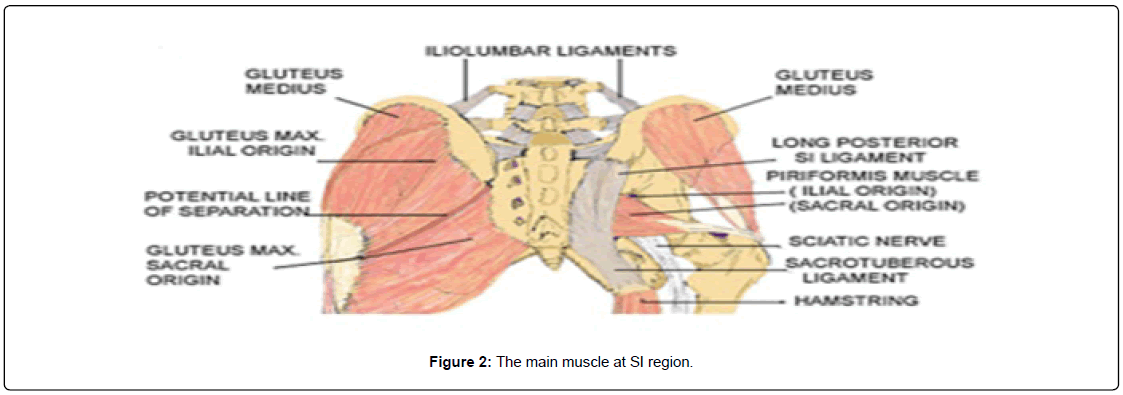
There are several myofascial structures attachment to SI joint (latissimus dorsi, gluteus maximus and Piriformis), they influence movement and stability. This Figure 1 joint innervation by branches of sacral spinal nerve, mainly L4-L5 ventral rami, L5 - S1/S2 dorsal rami in Figure 2 [3,4].
Etiology Of SIJ
Leg length discrepancy
SIJ Dysfunction may occur as result of leg length discrepancy; it is caused by asymmetrical innominate bone which stretch SI ligaments, it also association between LLD and scoliosis, it is affects gait, so it increase ground reaction force and energy consumption, at standing limbs redress these trouble by compensatory mechanism at the short leg by supination and planter flexion in foot and extension in the knee and hip, on another hand, at the long leg pronation in the foot and flexion in the hip and knee, if there is no compensatory the sacroiliac joint lower to the short side, which make change of sacral base [5,6].
Mechanical dysfunction
Hypermobility: SIJ instability is rare in general, instability often occurs as a result of the loss of the functional of any systems of the pelvic region that provide stability, and may be abnormal alignment is a consequence of weakened, injured, or sprained ligaments. While the joint itself is structurally healthy, or there is a different factor to the hypermobility such as change of hormones during pregnancy hypomobility [6,7].
SIJ infection: It’s rare, for example erosion and osteological deformity [1].
Ankylosing spondylitis: It’s bone surface articulation fuse together, its make pain and stiffness at buttock and hip starts around SIJ, its occur over growth of the bone (bony fusion), effect the ligaments and tendons which cause a limitation of the movement of the joint. The main reason of these diseases is genetic [8].
Crystal arthropathy: Its abnormal formation of calcium pyrophosphate in hyaline cartilage, its happen with people have a thyroid condition, kidney failure and disorders that’s effect on calcium, phosphate or iron, signs and symptoms for this disease are pain, stiffness, tenderness, warmth and swelling, Its unusual attack the pelvis, hip, shoulder, and elbow joint [9].
Pregnancy: The change of the weight cause a stretch ligament surround the pelvis region spatial SIJ [10].
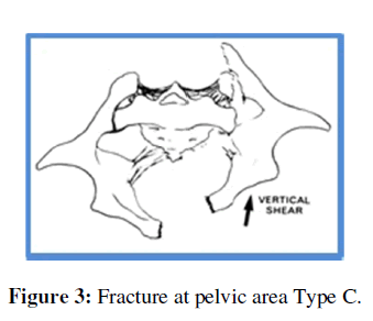
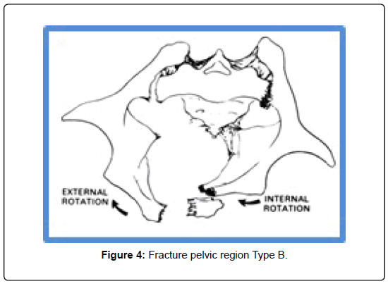
Stress fractures of the sacrum. In general, there are a different kinds of bone fracture in the pelvic such as first type is displaced and stable in the broken area, second is unstable rotatory but vertical is stable, finally is unstable vertical and rotatory. There are three type of fracture of iliac first type 1, it is injury lateral to the sacral foramina, second type 2, it is injury involving the sacral foramina and spinal canal, finally type 3 injury extend to spinal canal two (longitudinal/transverse) in Figures 3 and 4 [11].
Clinical Features
Main symptoms of sacroiliac joint dysfunction are low back pain, pain referred to other region like hip, buttocks, groin and thigh, tenderness aggravation factor such as getting out of bed or chair, climbing stairs, tingling or numbness, activities that put all loads on one side (asymmetric loading), cough and heels [12-15].
Diagnostic Test
There are more than 54 tests for diagnosis sacroiliac joint dysfunction put no research support specific test.
SIJ test categories
Palpation test: Palpation test has two type positional palpations, motion palpation.
a) Positional palpation test: By observation and palpation bony landmark (ASIS, PSIS, greater trochanter), the patient can do it by himself, the position of the test supine, prone, sitting, standing.
b) Motion palpation test: Test is to diagnosis abnormal movement of the sacroiliac joint using land mark its can be active or passive motion, for example, Gillet test [13,15].
Provocation test: Tests that stimulate pain use for diagnosis SIJ dysfunction by stretching and compression surround structure of the SIJ, for example, Clusters test and its include SIJ compression test, SIJ distraction, Posh or thigh thrust, Sacral clearing test, Faber test, Resisted abduction test and Other test, Glanselen test, REAB test and yeoman test [13,15].
Glanselen test: Patient lies in supine position one leg hang of the edge and ask the patient to flex the hip and knee of the other leg toward the chest (effected side) and then apply pressure above knee on the both side, the test be positive when the patient experiences pain on the posterior part of the pelvis (SIJ region) on the hyper extended leg [13,15].
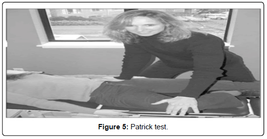
Patrick (Faber) test: It has different name such as Faber and figure four test FABER mean (F: Flexion, AB: Abduction, ER: External Rotation), patient lies in supine position and passively put the effected side in flexion, abduction and external rotation parallel to the knee of the other leg and then fix the ilium of the opposite side and slowly lower the test leg to the treatment table, the test is positive if the patient experiences pain on the posterior pelvis in Figure 5 [13,15].
Investigation
Radiographic
Beam of X-rays is produced by an X-ray generator, it is used to discover bone change such as fracture and may be degeneration at the joint and assessment spinal disease, of course, it is contraindication such as pregnancy, bleeding and pacemaker.
CT Scan (Computed Tomography)
They use ionizing radiation (X-ray) integration with a computer to create images of both soft and hard tissues.
MRI (Magnetic Resonance Imagining)
Use to check out the soft tissue pathology, change, fracture or tumor. Ultrasound Doppler Imaging: Health care professionals use it to view organs, it works with pregnant women because it is safe ray [1,7,11].
Treatment
Non-surgical
Early conservative treatment is very important point, rest, avoid excitation activities, use a local modalities such as ice at acute stage and give the patient a medication such as, Lidoderm Patch, Acetaminophen, NSAIMs (Nonsteroidal Anti-Inflammatory Medications) and muscle relaxants, at chronic stage should be avoid NSAIMs because of side effects on gastric, renal and cardiac systems. Physiotherapists can help patients in different ways such as use electrical stimulation, mobilization technical, postural educationo, exercise, and use a SI belt it can work out with overweight during pregnant and avoid hypermobility [1,7,12].
Manipulation SIJ
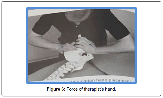
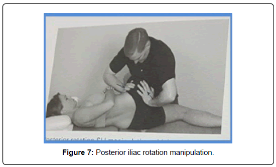
Posterior iliac rotation SIJ manipulation: Gold is manipulated as anti-iliac rotation displacement and restore posterior rotation of ilium, the position of the patient is side lying, the caudal hand’s palm is connect with patient’s ischial tuberosity, the cranial hand’s palm is connect with ASIS, it try to push ASIS posteriorly and pay ischial tuberosity anteriorly for 10-30 seconds in Figures 6 and 7 [16].
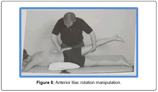
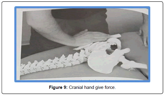
Anterior iliac rotation SI joint manipulation: Aim is to manipulate posterior iliac rotation displacement SI joint dysfunction and restore ant. Rotation of the ilium, the position of the patient is prone and put a pillow under the pelvis, caudal hand on the ant. Thigh proximal to the knee and cranial hand used to connect the PSIS with fingers pointing toward of patient thigh, the procedure is caudal hand try to extend the hip, and cranial hand forces the PSIS towards the table in Figures 8 and 9 [16].
Surgical Intervention
It is not common in SIJD but surgical need when fusion of the sacroiliac joint [1].
Conclusion
There is lack of information in SIJD in many of us, increase in knowledge of SIJ is necessary for optimal treatment. Further, researches and studies are needed focusing anatomic and pathologic change, however, there are an obvious paucity of high quality research, conservation therapies can be used an attempt to avoid surgical intervention, which we have to support.
References
- Frontera WR, Silver JK, Rizzo TD (2008) Essentials of Physical Medicine and Rehabilitation: Musculoskeletal Disorder pain and Rehabilitation. Elsevier, USA
- Forst SL, Wheeler MT, Fortin JD, Vilensky JA (2006) The sacroiliac joint: Anatomy, physiology and clinical significance. Pain Physician 9: 61-67.
- Snell RS (2006) Clinical anatomy by systems. 1st edition, Lippincott Williams & Wilkins, USA.
- Tilvawala K, Kothari K, Patel R (2019) Sacroiliac joint: A review. Indian J Pain 32: 4-15.
- Cummings G, Scholz JP, Barnes K (1993) The effect of imposed leg length difference on pelvic bone symmetry. Spine (Phila Pa 1976) 18: 368-373.
- Gunnar BP, Kozar AJDO, Greg CDO (2003) Sacroiliac Joint Dysfunction in Athletes. Curr Sports Med Rep 2: 47-56 .
- Edavalath M (2010) Ankylosing spondylitis. J Ayurveda Integr Med 1: 211–214.
- HY Lam, Cheung KY, Law SW, Fung KY (2007) Crystal arthropathy of the lumbar spine: a report of 4 cases. J Orthop Surg (Hong Kong) 15: 94-101.
- Maclennan AH, Maclennan SC (1997) The Norwegian Association For Women With Pelvic Girdle Relaxation, Symptom-giving pelvic girdle relaxation of pregnancy, postnatal pelvic joint syndrome and developmental dysplasia of the hip. Acta Obstetricia Et Gynecologica Scandinavica 76: 760-764.
- Slipman CW, Whyte WS, Chow DW, Chou L, Lenrow D, Ellen M (2001) Sacroiliac joint syndrome. Pain Physician 4: 143-152.
- Huijbregts P (2004) Sacroiliac joint dysfunction: evidence-based diagnosis. Feature Article of the Orthopedic Division Canadian Physical Therapy Association.
- Zelle BA, Gruen GS, Brown S, George S (2005) Sacroiliac Joint Dysfunction: evaluation and Management. Clin J Pain 21: 446-455.
- Cohen SP, Chen Y, Neufeld NJ (2013) Sacroiliac joint pain: a comprehensive review of epidemiology, diagnosis and treatment. Expert Rev Neurother 13: 99-116.
Citation: Alarab A, Shameh RA, Radwan A, Al-Daghaghmeh A (2019) Sacroiliac Joint Dysfunction. J Nov Physiother 9: 412. DOI: 10.4172/2165-7025.1000412
Copyright: © 2019 Alarab A, et al. This is an open-access article distributed under the terms of the Creative Commons Attribution License, which permits unrestricted use, distribution, and reproduction in any medium, provided the original author and source are credited.
Share This Article
Recommended Journals
Open Access Journals
Article Tools
Article Usage
- Total views: 4599
- [From(publication date): 0-2019 - Mar 31, 2025]
- Breakdown by view type
- HTML page views: 3728
- PDF downloads: 871

