Case Report Open Access
Root Canal Treatment of a Maxillary First Molar with an Un-instrumented 5th Canal: A Clinical Case Report
Reid V Pullen*Private Practice, Brea, California
- *Corresponding Author:
- Reid V Pullen
DDS, Brea, California 92821
Tel: 714- 529-9209
E-mail: pullendds@gmail.com.
Received Date: May 04, 2017; Accepted Date: June 13, 2017; Published Date: June 17, 2017
Citation: Pullen RV (2017) Root Canal Treatment of a Maxillary First Molar with an Un-instrumented 5th Canal: A Clinical Case Report. J Oral Hyg Health 5: 219. doi: 10.4172/2332-0702.1000219
Copyright: © 2017 Pullen RV. This is an open-access article distributed under the terms of the Creative Commons Attribution License, which permits unrestricted use, distribution, and reproduction in any medium, provided the original author and source are credited.
Visit for more related articles at Journal of Oral Hygiene & Health
Abstract
Objective: Failure to identify and treat all canals within the root canal system is a major reason for endodontic failure. The morphological variations of the maxillary first molar have been the subject of many studies, with most studies focusing on multiple canals within the mesiobuccal root, but maxillary molars may also have fins, isthmi, lateral canals, and in this case, additional palatal canals. Identification and treatment of all canals and anatomical variations is vital for a successful endodontic outcome. Methods: A maxillary first molar with pulpal necrosis and asymptomatic apical periodontitis due to a deep carious lesion was accessed for root canal treatment under a dental operating microscope. Four canals were minimally instrumented to an apical size #20 and treated with the GentleWave® Procedure. The GentleWave Procedure involves Multisonic Ultracleaning™ that creates cavitation with activated distilled water, sodium hypochlorite and ethylenediaminetetraacetic acid (EDTA). After the GentleWave Procedure, a fifth canal, not previously realized, was detected in the palatal root. The fifth canal had been visibly cleaned of debris. Five canals were obturated utilizing gutta-percha and warm vertical condensation with an epoxy resin based sealer. Results: Post-operative radiographs show 5 root canals: 2 palatal canals, 2 mesiobuccal canals, and 1 distobuccal canal. A cone-beam computed tomography (CBCT) scan shows a three-dimensional fill and obturation of complex anatomy. Three-month follow-up evaluation shows radiographic and clinical healing, resolution of periapical lesions, and complete resolution of the clinical symptoms. Conclusion: This case reports describes successful endodontic treatment of a rare maxillary molar with 5 canals, one of which was uninstrumented, utilizing an advanced technology, the GentleWave Procedure. This report highlights the importance of identifying and cleaning complex anatomy to increase successful outcomes.
Keywords
Maxillary first molar; Five root canals; Root canal morphology; Un-instrumented canal; GentleWave procedure; Multisonic ultracleaning
Introduction
Successful root canal treatment depends on the identification, cleaning, shaping, and obturation of the entire root canal system [1]. Failure to identify and treat all root canals lowers the long-term prognosis of the tooth and is a major reason for endodontic failure [2]. Variations in canal configurations make root canal systems a challenge to locate and negotiate. Maxillary molars most commonly have 3 roots, but there is considerable variation and complexity in the root canal systems within those roots [2-6]. The majority of palatal roots in maxillary molars have single canals, but 1.2-3.4% of maxillary molars have two palatal canals [7,8].
A recent advance in endodontic technology, the GentleWave Procedure (Sonendo®, Laguna Hills, CA), offers clinicians the ability to clean areas of the root canal system often untouched or undetected by conventional techniques [9]. The GentleWave Procedure utilizes Multisonic Ultracleaning technology, in which advanced fluid dynamics, acoustics, and tissue dissolution chemistry are applied to remove tissue and debris from the entire root canal system, even in areas that conventional endodontic treatment cannot reach [9]. Recent in vivo clinical studies with 6- and 12-months follow-up demonstrate a high rate of success (97.3-97.4%) after the GentleWave Procedure [10,11]. The GentleWave® System removed 100% of calcium hydroxide from mesial canals instrumented to size #25 in vitro [12]. The GentleWave System was also shown to be effective in removing separated instruments from the apical and middle thirds of molar root canal systems without the need for increased dentin removal for access [13].
This case report describes successful endodontic treatment of a rare maxillary first molar with five canals, including two palatal canals, one of which was un-instrumented, two mesiobuccal canals, and one distobuccal canal, after the GentleWave Procedure and three-months follow-up.
Case Presentation
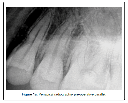
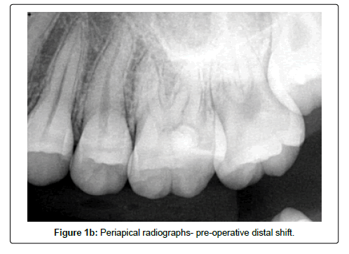
A 21-year-old female was referred to the clinic by a general dentist with a chief complaint of prior severe, constant, dull, throbbing pain causing headaches. After temporary restoration placement and a course of oral antibiotics, the pain had resolved. The patient’s medical history was non-contributory. Extraoral examination was within normal limits. Intraoral examination found no swelling or sinus tracts. Oral hygiene was fair. On clinical examination, the left maxillary first molar (#14) was found to have large occlusolingual caries under a temporary restoration. The tooth did not respond to Endo-Ice® (Coltene®/ Whaledent, Cuyahoga Falls, OH) and tested normally to percussion, palpation, mobility, periodontal probing, and bite testing. Radiographic examination showed tooth #14 with a periapical radiolucency, deep carious lesion closely approximating the dental pulp, a pulp chamber reduced in size, and multiple pulp stones (Figures 1a and 1b). Based on clinical and radiographic findings, the diagnosis of pulpal necrosis and asymptomatic apical periodontitis was made. Prognosis for tooth #14 was fair. Root canal treatment was recommended in an attempt to save the tooth, and the patient consented.
The patient was anesthetized locally with 4% articaine (72 mg) with 1:100,000 epinephrine and 2% lidocaine (36 mg) with 1:100,000 epinephrine via buccal and lingual infiltrations. The tooth was isolated with a dental dam. All treatment was completed under high power magnification with a dental operating microscope and LED illumination. Caries were excavated into the pulp and complete removal was confirmed with caries indicator dye. Coronal tooth structure was restored with bonded micro-hybrid composite. The tooth was conservatively accessed with removal of all pulp horns, ledges and overhangs. Examination of the pulp chamber floor revealed four distinct canal orifices. To maximize preservation of tooth structure, orifice shapers were not utilized during the instrumentation process. Sodium hypochlorite irrigation (0.5%) was employed merely for lubrication and to assist in the prevention of microbial transfer to extra-radicular tissue during shaping [14,15]. Working lengths were estimated with an electronic apex locator (Root ZX II, J. Morita, Irvine, CA). Glide paths were created with K-files up to size #20. Rotary instrumentation, up to an apical size #20, was utilized to minimally shape the canals to create a path for irrigation to reach the apex and to facilitate the placement of root canal filling. The GentleWave Procedure was completed to deliver activated distilled water, sodium hypochlorite and ethylenediaminetetraacetic acid (EDTA) throughout the root canal system. Canals were dried using a combination of 100% ethanol irrigation, micro-suction tips, and paper points.
After the GentleWave Procedure, a fifth canal orifice in the apical third of the palatal root was detected. This canal was not previously realized or instrumented but now appeared free of debris and tissue. The fifth canal was fitted for root canal filling and confirmed by radiograph. All five canals were obturated with gutta-percha by warm vertical condensation and thermo-plasticized gutta-percha backfill. The canal orifices and access cavity were sealed with bonded composite.
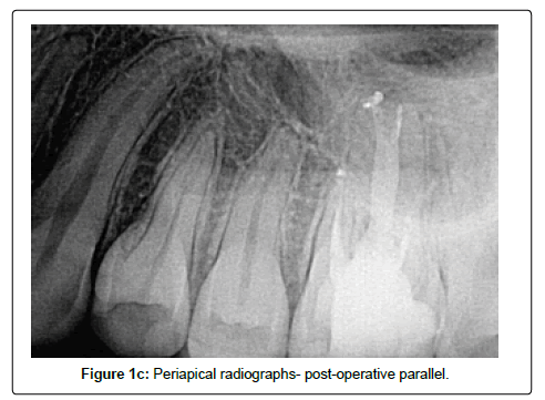
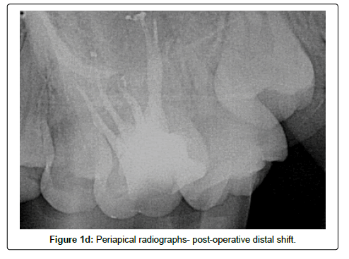
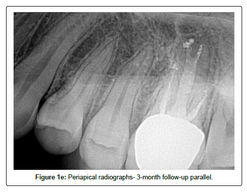
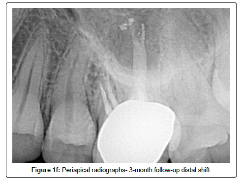
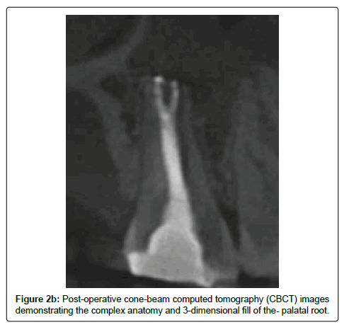
Post-operative radiographs demonstrate root canal filling in five root canal systems, with two canals in the mesiobuccal root, one canal in the distobuccal root, and two canals in the palatal root (Figures 1c- 1f). The palatal root canal system exhibits type V anatomy, in which canals diverge within the root and exit the tooth via two separate foramina [16].
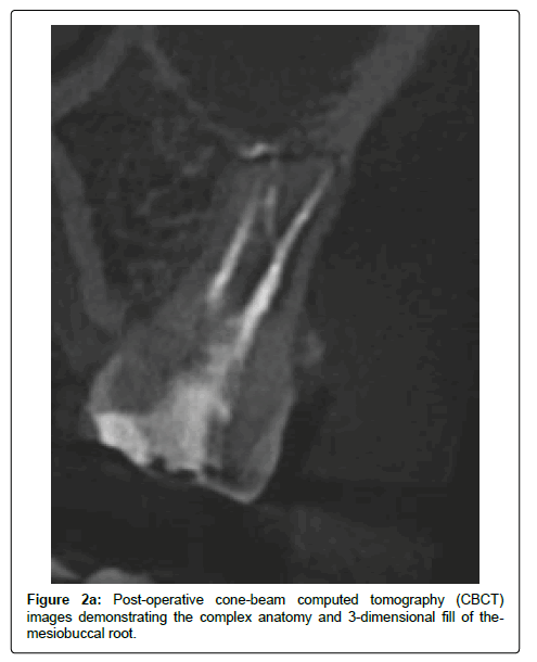
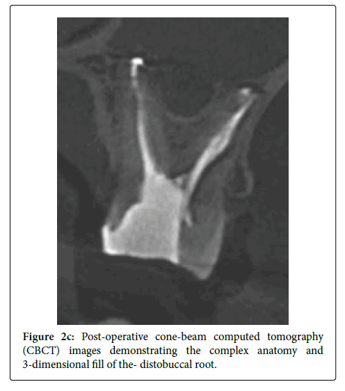
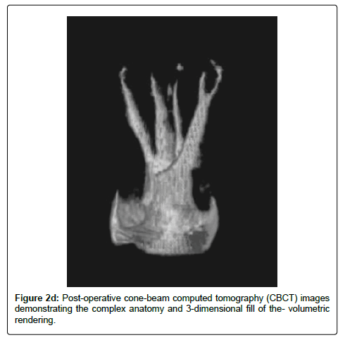
A high-resolution CBCT scan with a focused field of view was acquired post-operatively to better visualize the obturation of all root canal systems and rule out complications (Carestream 8100 3D, Carestream Health, Inc. Rochester, NY). The post-operative CBCT scan further demonstrates the complicated anatomy and five canals (Figures 2a-2d). The mesiobuccal root canal systems were noted to have type XVI anatomy, originating at two separate orifices but diverging into 3 apical exits [17].
The patient was advised to obtain a crown restoration from their general dentist and an endodontic follow-up evaluation was scheduled three-months post-procedure to monitor healing. At the three-month follow-up visit, the patient was asymptomatic and reported no pain or clinical symptoms. Tooth #14 was in function with a crown restoration and intact margins. Three-month follow-up radiographs show a healed tooth with no periapical radiolucency (Figures 1e and 1f). The patient reported a subsequent move out of state and therefore would be unavailable for further follow-up evaluations. The patient was advised to continue comprehensive dental care with a general dentist.
Discussion
The presence of four root canal systems in maxillary molars has been well-documented, usually with two canals in the mesiobuccal root [2,18], but the presence of five root canal systems is quite rare [8]. A review of published case reports shows that the majority of palatal roots contain a single root canal system, but 1.2-3.4% of maxillary molars contain two palatal root canal systems [7,8]. In this case, the second palatal canal was not visualized during instrumentation. Conventional root canal treatment might have resulted in the inability of irrigants to reach within the un-instrumented canal, the inability of sealer or guttapercha to fill the un-instrumented canal, and possibly the persistence of a bacterial reservoir in the uninstrumented canal, which may have compromised the outcome. However, in this case the previously undetected canal was cleaned during the GentleWave Procedure allowing it to be visualized, dried, and filled with gutta-percha and sealer, leading to the complete resolution of the patient’s symptoms and healing by three months of the treatment.
The successful outcome of this case is consistent with previously published clinical studies for the GentleWave System in which molars treated with the GentleWave Procedure healed rapidly with high success rates [10,11]. The presence of sealer puffs at the mesiobuccal and palatal apices suggests the lack of an apical plug of debris and is consistent with a recent study that found significantly less debris in the middle and apical thirds of the root canals after the GentleWave Procedure as compared with conventional root canal treatment [9].
Packed debris in the canals, particularly in the apical third and in complex anatomy, is often seen after conventional instrumentation and inhibits irrigation fluids from reaching the entire root canal system [19,20]. Studies have shown that conventional instrumentation to a minimum size #30 or #35 is required for endodontic irrigating needles to reach the apical third [20]. In this case, instrumentation did not involve coronal flaring or orifice openers and was kept minimal at an apical size #20, consistent with clinical and in vitro studies, to preserve tooth structure, maintain root dentin thickness, and stay natural to the canal anatomy, while still removing apical debris through Multisonic Ultracleaning with the GentleWave System [9-11]. Molina et al. showed that GentleWave System could remove debris from canals and isthmi in the apical third of the root canal system even at smaller file sizes of 15.04 [9]. Identification and cleaning of all canals and complexities of the root canal system utilizing the GentleWave technology may reduce the chance for retreatments and extractions and increase the successful outcome of root canal treatment [10,11].
Conclusion
This case demonstrates the successful endodontic management of a rare case of a maxillary molar with 5 canals, one of which was uninstrumented after root canal treatment utilizing the GentleWave Procedure. Clinicians should be aware of potential encountered anatomical complexities that, if left untreated, could compromise outcomes.
References
- Marquis VL, Dao T, Farzaneh M, Abitbol S, Friedman S (2006) Treatment Outcome in Endodontics: The Toronto Study. Phase III: Initial Treatment. J Endod 32:299-306.
- Weine FS, Healey HJ, Gerstein H, Evanson L (2012) Canal Configuration in the Mesiobuccal Root of the Maxillary First Molar and its Endodontic Significance. J Endod 38:1305-1308.
- Kuttler Y (1955) Microscopic Investigation of Root Apexes. J Am DentAssoc 50: 544-552.
- Pineda F, Kuttler Y (1972) Mesiodistal and BuccolingualRoentgenographic Investigation of 7,275 Root Canals. Oral Surg 33:101-110.
- Acosta SA, TrugedaSA (1978) Anatomy of the Pulp Chamber Floor of the Permanent Maxillary First Molar. J Endod 4(7):214-219.
- Gilles J, Reader A (1990) An SEM Investigation of the Mesiolingual Canal in Human Maxillary First and Second Molars. OralSurg Oral Med Oral Pathol 70:638-643.
- Naseri M, Safi Y, Akbarzadeh BA, Khayat A, Eftekhar L (2016) Survey of Anatomy and Root Canal Morphology of Maxillary First Molars Regarding Age and Gender in an Iranian Population Using Cone-Beam Computed Tomography. Iranian Endod J 11: 298-303.
- Cleghorn BM, Christie WH, Dong CC (2006) Root and Root Canal Morphology of the Human Permanent Maxillary First Molar: A Literature Review. JEndod 32: 813-821.
- Molina B, Glickman G, Vandrangi P, Khakpour M (2015) Evaluation of Root Canal Debridement of Human Molars Using the GentleWave System. J Endod 41:1701-1705.
- Sigurdsson A, Garland RW, Le KT, Woo SM (2016) 12-month Healing Rates after Endodontic Therapy Using the Novel GentleWave System: A Prospective Multicenter Clinical Study. J Endod 42: 1040-1048.
- Sigurdsson A, Le KT, Woo SM, Rassoulian SA, McLachlan K, et al (2016) Six-Month Healing Success Rates After Endodontic Treatment Using the Novel GentleWave™ System: The PURE Prospective Multi-center Clinical Study. J ClinExp Dent 8: e290–e298.
- Ma J, Shen Y, Yang Y, Gao Y, Wan P, et al. (2015) In Vitro Study of Calcium Hydroxide Removal from Mandibular Molar Root Canals. J Endod 41:553-538.
- Wohlgemuth P, Cuocolo D, Vandrangi P, Sigurdsson A (2015) Effectiveness of the GentleWave System in Removing Separated Instruments. J Endod 41(11):1895-1898.
- Molander A, Warfvinge J, Reit C, Kvist T (2007) Clinical and Radiographic Evaluation of One- and Two-visit Endodontic Treatment of Asymptomatic Necrotic Teeth with Apical Periodontitis: A Randomized Clinical Trial. J Endod 33: 1145-1148.
- Salgado RJ, Moura-Netto C, Yamazaki AK, Cardoso LN, de Moura AA, et al. (2009) Comparison of Different Irrigants on Calcium Hydroxide Medication Removal: Microscopic Cleanliness Evaluation. Oral Surg Oral Med Oral Pathol Oral RadiolEndod 107:580-584.
- Vertucci FJ (1984) Root Canal Morphology of the Human Permanent Teeth. Oral Surg58:589-599.
- Sert S, Bayirli GS (2004) Evaluation of the Root Canal Configurations of the Mandibular and Maxillary Permanent Teeth by Gender in the Turkish Population. J Endod 30:391-398.
- Kulild JC, Peters DD (1990) Incidence and Configuration of Canal Systems in the Mesiobuccal Root of Maxillary First and Second Molars. J Endod 16:311-317.
- Gu LS, Kim JR, Ling J, Choi KK, Pashley DH, et al. (2009) Review of Contemporary Irrigant Agitation Techniques and Devices. J Endod 35: 791-804.
- Khademi A, Yazdizadeh M, Feizianfard M (2006) Determination of the Minimum Instrumentation Size for Penetration of Irrigants to the Apical Third of Root Canal Systems. J Endod 32:417-420.
Relevant Topics
- Advanced Bleeding Gums
- Advanced Receeding Gums
- Bleeding Gums
- Children’s Oral Health
- Coronal Fracture
- Dental Anestheia and Sedation
- Dental Plaque
- Dental Radiology
- Dentistry and Diabetes
- Fluoride Treatments
- Gum Cancer
- Gum Infection
- Occlusal Splint
- Oral and Maxillofacial Pathology
- Oral Hygiene
- Oral Hygiene Blogs
- Oral Hygiene Case Reports
- Oral Hygiene Practice
- Oral Leukoplakia
- Oral Microbiome
- Oral Rehydration
- Oral Surgery Special Issue
- Orthodontistry
- Periodontal Disease Management
- Periodontistry
- Root Canal Treatment
- Tele-Dentistry
Recommended Journals
Article Tools
Article Usage
- Total views: 5962
- [From(publication date):
July-2017 - Jul 06, 2025] - Breakdown by view type
- HTML page views : 4944
- PDF downloads : 1018

