Review Article Open Access
Role of Regulatory T-cells in Oral Tolerance and Immunotherapy
| Mark Farrugia and Byron Baron* | |
| Centre for Molecular Medicine and Biobanking, Faculty of Medicine and Surgery, University of Malta, Msida, MSD2080, Malta | |
| *Corresponding Author : | Byron Baron Centre for Molecular Medicine and Biobanking Faculty of Medicine and Surgery, University of Malta Msida, MSD2080, Malta Tel: 00356 21639913 E-mail: byron.baron@um.edu.mt |
| Received: January 28, 2016;Accepted: February 09, 2016; Published: February 16, 2016 | |
| Citation: Farrugia M, Baron B (2016) Role of Regulatory T-cells in Oral Tolerance and Immunotherapy. Biochem Physiol 5:199. doi: 10.4172/2168-9652.1000199 | |
| Copyright: © 2016 Farrugia M, et al. This is an open-access article distributed under the terms of the Creative Commons Attribution License, which permits unrestricted use, distribution, and reproduction in any medium, provided the original author and source are credited. | |
Visit for more related articles at Biochemistry & Physiology: Open Access
Abstract
Food allergies encompass a range of disorders ranging from being an inconvenience to even causing fatalities, mainly due to anaphylaxis. A large number of individuals are affected and this presents great health and economic implications. However as yet, apart from dietary avoidance, effective treatment strategies are practically non-existent. The immune environment related to allergen-tolerance is highly complex and the role of regulatory T-cells in allergenspecific tolerance, their interaction with other cells in inflamed tissues, and their role in antibody regulation have been demonstrated in several studies. Regulatory T-cells are able to control acquired immunity and achieve oral tolerance to food allergens. Immunotherapy for food allergies focuses on desensitisation by increasing the allergen reactivity threshold. So far, the only long-term curative treatment used effectively is allergen-specific immunotherapy which involves the administration of increasing doses of the causative allergen, such that a state of allergen-specific immune tolerance is induced over the course of the treatment. This review covers various forms of allergen-specific immunotherapy, focusing on the role of regulatory T-cells in such therapies, and includes a number of small studies providing ideas for future work in the area.
| Keywords |
| Regulatory T-cells; Immunotherapy; Immune tolerance; Oral tolerance |
| Abbreviations |
| AIT: Allergen-specific Immunotherapy; APCs: Antigen-Presenting Cells; CCR: C-C Chemokine Receptor; CSR: Class Switch Recombination; CD: Cluster of Differentiation; CTLA- 4: Cytotoxic T-Lymphocyte-Associated Protein 4; DCs: Dendritic Cells; EPIT: Epicutaneous Immunotherapy; FPIES: Food-Protein- Induced Enterocolitis Syndrome; SPT: Skin Prick Test; OFC: Oral Food Challenge; ELISAs: Enzyme-Linked Immunosorbent Assays; FOXP3: Forkhead Box P3; GITR: Glucocorticoid-Induced Tumour Necrosis Factor Receptor-Related Protein; HKLM: Heat-Killed Listeria moncytogenes; HR1/2: Histamine Receptor 1/2; Ig: Immunoglobulin; iTregs: Inducible Tregs; IFN-γ: Interferon-Gamma; IL-10: Interleukin-10; nTregs: Natural Tregs; NO: Nitric Oxide; OIT: Oral Immunotherapy; PBMC: Peripheral Blood Mononuclear Cell; PD-1: Programmed Cell Death Protein-1; Bregs: Regulatory B-cells; Tregs: Regulatory T-cells; SCIT: Subcutaneous Immunotherapy; SLIT: Sublingual Immunotherapy; Th cells: T-Helper Cells; TGF-β: Transforming Growth Factor-β; ILC2s: Type 2 Innate Lymphoid Cells |
| Introduction |
| Food allergies are defined as an immunologically-mediated adverse reaction to particular foods, most commonly milk (mostly in children), eggs, nuts (including peanuts, walnuts, almonds and brazil nuts), wheat and other grains with gluten (including barley and oats), fish and shellfish (mostly in adults). Depending on the type of allergy and the severity, a range of disorders may arise including Immunoglobulin E (IgE)-mediated anaphylaxis, food-protein-induced enterocolitis syndrome (FPIES), and other gastrointestinal disorders such as vomiting, reflux, abdominal pains, diarrhea or constipation [1]. Food allergies are quite widespread and affect approximately 5% of adults and 8% of young children in countries with a Western lifestyle [2,3]. Food allergy is one of the most common causes of anaphylaxis that may lead to fatalities. Studies in the United States and United Kingdom showed that the number of hospitalisations for food-induced anaphylaxis has increased more than 3-fold in the past decade [4]. The health and economic effect of food allergies is not only reflected in the cost of healthcare expenditures, but also in the effect it has on the workplace, food industry, food regulatory agencies, and, most importantly, patients and their families, whose lives are affected daily. |
| Food allergies are generally diagnosed by means of a skin prick test (SPT) or blood tests, although the oral food challenge (OFC), or elimination diet may also be used as highly specific diagnostic tests. Both the SPTs and blood tests measure the presence of IgE, the antibody that triggers food allergy symptoms. SPTs are the preferred method of testing because they are inexpensive, produce results in under an hour, and can be performed in the clinic. On the other hand, blood tests consisting of enzyme-linked immunosorbent assays (ELISAs) have a higher cost and do not provide immediate results but are much more accurate and quantitative. |
| The only validated remedy for food allergies is the identification and elimination of the foods responsible [5], and the use of selfinjectable epinephrine to reverse the acute and severe allergic reaction [1]. However, it is not easy to totally avoid contact with food allergens as these tend to be used in most food manufacturing processes. Therefore, developing effective treatment strategies outside of dietary avoidance of antigens has been a high priority for research teams in recent years. |
| Oral Tolerance |
| Food allergies can be induced by both IgE-mediated and non-IgEmediated pathways. In the case of IgE-mediated food allergies, foodspecific IgE antibodies are produced after exposure to particular food allergens that then bind to the Fc receptors on mast cells, basophils, and macrophages [6]. Mediators from the activated cells are released and result in local or systemic symptoms immediately. These mediators may attract other cells, like eosinophils and lymphocytes, to prolong the inflammation [7]. |
| Exposure to a food antigen also generates a regulatory T-cell (Treg) response that can suppress allergic sensitisation to that food allergen. The adoptive transfer of Treg cells was shown to prevent or cure several T-cell–mediated diseases, including allergy, asthmatic lung inflammation, and autoimmune diseases by restoring immune tolerance to allergens, self-antigens, or alloantigens in animal models, and this has demonstrated the pivotal role of Treg cells in inducing and maintaining immune tolerance [8]. Hadis et al. [9] showed that oral tolerance could suppress experimental food allergy through the development of antigen-specific forkhead box P3 positive (FOXP3+) T-cells in mice. In humans, antigen-specific cluster of differentiation (CD) 25+ FOXP3+ Tregs are associated with the onset of clinical tolerance to milk [10]. Tolerance is initiated by dendritic cells (DCs) residing in the gastrointestinal lamina propria. CD103+ DCs capture antigens in the lamina propria, migrate, and initiate oral tolerance in the draining lymph node by activating antigen-specific Tregs that then migrate back to the lamina propria. |
| Subsets of Tregs |
| There are at least five subsets of Tregs identified so far. This makes Tregs one of the most complicated and diverse T-cell groups. More novel subsets of Tregs will surely be discovered. All subsets discovered so far are derived from naïve T-cells and develop under different conditions. For the purpose of this review we shall only focus on those subsets that are directly linked to the food allergy sensitisation scenario. |
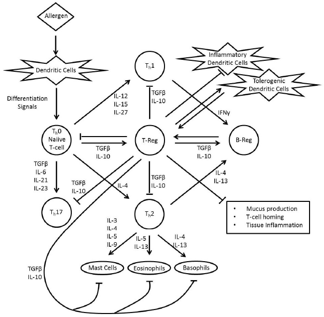
| The largest subset of Tregs is the so-called natural Tregs (nTregs). These are CD4+CD25+FOXP3+ cells which secrete Interleukin-10 (IL-10) and Transforming Growth Factor-β (TGF-β). These Tregs originate from the thymus in response to self-antigens [11]. Two mechanisms have been invoked to describe the function of these Tregs: a contactdependent mechanism in which membrane-bound TGF-β blocks T-cell proliferation and a contact-independent mechanism involving soluble TGF-β and IL-10 [12]. These nTregs have a lot of roles in allergenspecific immune reactions (Figure 1). These include the suppression of dendritic cells (these support the generation of effector T-cells), the inhibition of the production of allergen-specific IgE, the inhibition of both function and migration of effector Th1, Th2, and Th17 cells, the induction of IgG4 secretion and also the suppression of mast cells, basophils, and eosinophils [13]. It is important to keep in mind that IgG4 represents a non-inflammatory Ig isotype that does not activate complement (a system that mediates the specific antibody response) and is thought to block the activation of the more severe IgE [14]. |
| Another type of Tregsis inducible Tregs (iTregs), and these are peripherally-induced Tregs, not produced in the thymus. Naïve CD4+ T-cells in the periphery are induced to express the FOXP3 transcription factor in response to foreign antigens [11] and these cells develop a suppressive function similar to nTregs [15], including the production of IL-10 and TGF-β. |
| There are also CD4+ T-cells that although do not express the FOXP3 gene (which was until recently used as a definitive marker for Tregs) still secrete IL-10 and suppress effector functions of T helper cells (Th cells). These cells therefore still classify as regulatory cells and are known as Tr1 Tregs [16]. Tr1 cells suppress effector T-cell responses by multiple mechanisms that depend on IL-10, TGF-β, programmed cell death protein-1 (PD-1), cytotoxic T-lymphocyte-associated protein 4 (CTLA- 4) [17], and histamine receptor 2 (HR2) [17,18]. |
| The differential regulation of Th 1 and Th 2 cells by histamine is due to the presence of different receptors, as human CD4+ Th1 cells predominantly express histamine receptor 1 (HR1), while CD4+ Th2 cells predominantly express histamine receptor 2 (HR2) [19]. Histamine induces the production of IL-10 by DCs [20] and Th2 cells [18,21] as well as enhances the suppressive activity of TGF-β on T-cells [22], mediated through HR2, suppressing IL-4 and IL-13 production and T-cell proliferation [18,19] This suggest that HR2 acts as a critical receptor in the peripheral tolerance to allergens. |
| It is also reported that in human peripheral blood and lymphoid tissue (but not in the thymus) there also exist CD4+FOXP3+ T-cells that also express the CCR6 gene (which is translated into the C-C chemokine receptor type 6 protein) and are also able to produce IL- 17 upon activation [23]. The CCR6+ IL-17-producing FOXP3+ Tregs strongly inhibit the proliferation of CD4+ effector T-cells. A recent report however shows that IL-17-producing FOXP3+ Tregs are a new crossover immune cell population that could be converted from Tregs to Th17 cells, and thus associated with a decreased suppressive function of T lymphocytes [24]. |
| Recently another subset of Tregs has been discovered that are induced by Nitric Oxide (NO), and fittingly these are called NO-Tregs. NO-Tregs are distinct from other Treg subsets in that apart from lacking the expression of FOXP3, they also express Glucocorticoid-induced tumour necrosis factor receptor-related protein (GITR) and are CD27+, thus having a T-helper phenotype while still maintaining suppressive properties against effector cells, mainly through the production of IL- 10 [25]. IL-10 is the only detectable cytokine produced by NO-Tregs. |
| It has been repeatedly shown that all subsets of Tregs coexist and overlap in many immune tolerance-related situations in humans,including allergies. Tregs share major non-lymphoid tissue trafficking receptors, such as C-C chemokine receptor type 4 (CCR4), CCR5, CCR6, CXCR3, and CXCR6, with Th17 cells [26]. This implies that these T-cells migrate to and within lymphoid tissue. The mechanisms used by Treg cells to suppress a large number of target immune cell types can be broadly divided into two: those that target T-cells (by means of suppressor cytokines, IL-2 consumption, and granzyme/perforininduced cell death pathways) and those that target antigen-presenting cells (APCs) (by inhibiting antigen presentation or down-modulating the expression of CD80 and CD86) [27]. |
| Tregs Action in the Allergy Scenario |
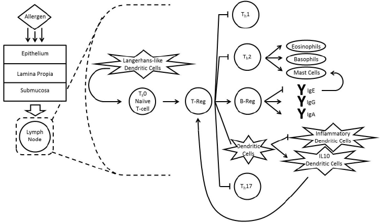
| The suppressive actions of Tregs on other immune cells, including effector T-cells, B-cells, DCs and mast cells, may shed light on the complex nature of how Tregs are able to control acquired immunity and achieve oral tolerance to food allergens. Figure 2 summarizes the effects of Tregs on the other immune cells involved in allergen-immunity. |
| The Tregsuppressive nature seems to focus a lot on affecting the activity of other effector T-cells derived from CD4+ cells, mainly the Th cells. Th cells are thought to differentiate into three main subsets, which are Th1, Th2 and Th17 effector cells. Th1 are Interferon-gamma (IFN-γ) T-cells, Th2 cells produce IL-4 and IL-5, while Th17 secrete IL-17 [28]. These three Th cell types are all responsible in invoking an immune response resulting in an allergic reaction, and are kept in check by Tregs in healthy non-allergic individuals. TGF-β-producing Tregs inhibit Th1 cell differentiation and instead promote Th17 cell differentiation [29]. However, NO-Tregs inhibit Th17 cell production but not Th1 cell differentiation and function [25]. Tregs can also selectively inhibit IFN-γ synthesis [30]. It is important to note however that the nature of Tregs and their effects on other T-cells in vivo is still under debate and several experimental factors might play a role in different results. |
| FOXP3+ T-cells have been known to also affect B-cell function. They have been discovered to exist in the T-B area borders and within germinal centres in secondary lymphoid organs (the areas where B-cells interact with Th cells and undergo Ig production) [31]. It was also shown that Tregs can suppress B-cells without needing to suppress Th cells, and that suppression of B-cells was accompanied by a reduced Ig Class Switch Recombination (CSR). |
| A recently discovered subset of B-cells which express FOXP3 and secrete IL-10 and/or TGF-β has also been discovered, and these cells are referred to as regulatory B-cells (Bregs) [32]. The functional purpose of Breg cells seems to be similar to that of Tregs [33]. Bregs seem to act earlier than Tregs, making it easier for the recruitment of Tregs to occur [34]. |
| Tregs also target DCs, and they are one of the major targets of Treg- mediated suppression. IL-10 producing Tregs seem to induce production of the tolerogenic CD11c+ DCs, and this also leads to the generation of hapten-specific CD8+ Treg cells [35]. CD8+ Tregs are yet another subset of Tregs which protect against contact hypersensitivity. The activation of tolerogenic DCs also induces the production of yet further Tregs. The active role of DCs in the induction of different subsets of Tregs has been supported by several studies [36,37], showing that Tregs are induced via a TGF-β and retinoic acid mechanism. Moreover, DCs from the lamina propria of the small intestine and also from the mesenteric lymph nodes are better than other DCs at inducing the expression of FOXP3 in the presence of exogenous TGF-β in naïve T-cells, suggesting an intrinsic system favouring the production of Tregs in the gut. |
| It was also discovered that FOXP3+ Tregs can suppress the symptomatic phase of mast cell activation and thus control IgEdependent anaphylaxis in mice [38]. Mast cells are the primary effector cells, and are responsible largely (if not completely) for the initiation of allergic pathological damage and clinical symptoms, therefore the degranulation of mast cells is distinctive of allergies [39]. Tregs seem to inhibit mast cell degranulation via CD134 (OX40)/CD252 (OX40- ligand) interactions and also inhibit IL-6 release via TGF-β [40]. |
| One can notice that the immune environment revolving around allergen-tolerance is a very complex one, and more specific subsets of cells are still being discovered. All cell types have their own effect on the overall immune system, but it was observed that the triangle interaction of Treg cells, T-effector cells and DCs is at the heart of such a system [41]. Most types of Treg cells inhibit allergen-specific effector cells in experimental models, and therefore understanding the nature of Treg cells is key to better understanding the possible therapies to immunediseases, including allergies. |
| A key event in the development of a healthy immune response to allergens is the shifting of allergen-specific effector T-cells to a regulatory phenotype, and this appears to have a successful outcome in allergen-specific immunotherapy. Consequently, understanding the immune mechanisms that prevent allergic reactions in healthy, non-allergic individuals and evidence of altered regulation in allergic individuals, offers a better understanding of the mechanisms involved in immune tolerance and how this can be applied for the design of new immune therapies. |
| It has been shown that both healthy and allergic individuals exhibit Th1, Th2, and Tr1 cells, but in different proportions, where in healthy individuals showing detectable IgG antibodies against an allergen, Tr1 cells represent the dominant subset, whereas in allergic individuals, a high frequency of allergen-specific IL-4–secreting T-cells is found. This outlines the importance of the frequency of effector Th2 cells or Tr1 cells in the development of a healthy or allergic immune response [17]. |
| Immunotherapy |
| The development of food allergies tends to be the result of a deregulation of immune tolerance, which can develop against any immune-activating substance, and is known to be mediated by multiple mechanisms. It is generally characterised by an altered allergen-specific memory T- and B-cell response [42-45], consequent to the induction of a type 2 immune response that includes Th2 cells and type 2 innate lymphoid cells (ILC2s), together with the production of allergen-specific IgE antibodies and increased eosinophil numbers in the affected tissues and sometimes in peripheral blood [46]. |
| Generally in healthy individuals, T-cells do not show any proliferative response to allergens in peripheral blood mononuclear cell (PBMC) cultures. This can be due to a low frequency of specific T-cells because of a lifetime lack of exposure. Moreover, if a detectable allergenspecific T-cell response is mounted in a non-allergic individual, active suppression against allergens takes place in cultures by Tr1 cells or CD4+FOXP3+ Treg cells [17,18,47,48]. |
| The prevention of sensitisation to new antigens [49] and prevention of progression to a more severe allergic state, are the major clinical implications of immune tolerance. The main aim of immunotherapy for food allergies is desensitisation by increasing the allergen reactivity threshold in subjects receiving the immunotherapy and retention of this increased reactivity threshold after the therapy has been discontinued. So far, the only long-term curative treatment used effectively is allergenspecific immunotherapy (AIT), which involves the administration of increasing doses of the causative allergen, such that a state of allergenspecific immune tolerance is induced over the course of AIT treatment. |
| The concept of using AIT for food allergy has long been tested through several studies. Current drug development and therapeutic strategies exploit Treg control of allergen-specific immune responses to induce a tolerant state in peripheral T-cells, showing potential for preventive therapies and cures for allergic diseases. The aim is to generate allergen-specific Treg cells and suppress the proliferative and cytokine responses against the major allergen [50]. The basis of AIT, similar to treatment with glucocorticoids or b2-agonists, promotes the numbers and activity of IL-10–secreting Tr1-like cells [24,51,52]. The signaling of Treg cells in AIT is initiated by the production of IL-10 and TGF-β by the antigen-specific Tr1 cells [29,53,54]. However, since these cells are CD4+ and CD25+, it is still unclear whether these are inducible Tr1 cells upregulating CD25 or naturally occurring CD4+ CD25+ Treg cells that produce suppressive cytokines [55]. It has been shown that circulating CD4+ CD25+ Treg cells and IL-10– and TGF-β–secreting Tr1 cells represent overlapping populations in adults. Moreover, it has been shown that CD4+CD25+ Treg cells from atopic donors are less effective at suppressing the proliferation of CD4+ CD25- T-cells after allergen AIT [29,32]. As a consequence, it has been suggested that upregulation of CD4+ CD25+ Treg cells plays a role in allergen AIT. |
| In the 1980s, subcutaneous immunotherapy (SCIT) was tested for peanut allergy. SCIT is a mode of therapy that has been proven effective and safe for the treatment of allergies to environmental factors and insect stings [56,57]. These trials had shown the efficacy of SCIT against peanut allergy, however there was an unacceptably high rate of severe allergic reactions [58]. Other routes of immunotherapy have therefore been investigated, although no such methods are yet ready for routine clinical practice [59]. |
| Oral Immunotherapy (OIT) is the most studied immunotherapy treatment so far and is the method which shows the greatest efficacy for the treatment of food allergy. This method invokes an immune response to antigens that are delivered orally, and the subject gradually gains immune tolerance mediated by regulatory T-cells. The presumed mechanism of action for OIT is that of the activation of gut mucosal dendritic cells, which in turn affect the allergic response through immunomodulation of tissue and circulating effector cells [60]. This immunomodulation is done via the interaction of Tregs with effector T-cells, as previously explained. Other mechanisms which are presumed to be important include the modulation of IgE responses, polyclonal increases in specific IgG4 levels [61], as well as suppression of basophils through an IgE receptor pathway [62]. |
| Although at the moment OIT is the most researched of any immunotherapy treatments for food allergy, yet to date there is still limited evidence of long-term follow-up of subjects after OIT, with mixed results on the achievement of long-term tolerance. In 2013, it was demonstrated for the first time that sustained unresponsiveness developed in half of the subjects with peanut allergy able to complete treatment after years of OIT, and it was still not entirely proved that this was due to OIT [63]. Another report issued in 2013 assessing the long-term follow-up of cow’s milk immunotherapy concludes that long-term outcomes after cow’s milk immunotherapy are mixed, with some patients losing desensitisation over time and not more than a third of the subjects in these studies tolerating a full servings of cow’s milk without symptoms [64]. The conclusion of these reports and similar investigations agree that only a small fraction of those starting treatment achieve long-term tolerance, and therefore the need to develop better therapies for treatment of food allergies is evident. |
| Even though OIT seems to be the most promising regarding its efficacy, safety is a major concern with the oral route since it is also the route that normally leads to food-induced allergic reactions. Sublingual immunotherapy (SLIT) and epicutaneous immunotherapy (EPIT) were thus proposed as alternative routes that could have a significantly better safety profile yet still retain the ability to induce tolerance. |
| The APCs present in the sublingual environment induce Tregs similar to those of the intestinal tract [65], whilst the limited antigen dose applied through this route improves the safety of these trials [66]. This improvement in safety comes at the price of being less effective than OIT, although some groups report promising efficacy with SLIT for treatment of peanut allergy [67,68]. The suggested mechanism of how SLIT affects the Treg cells and subsequently modulates the Th2/Th1 balance is that of allergen interaction with protolerogenic Langerhans cells in the oral mucosa, and these in turn lead to downregulation of the allergic response [69] (Figure 2). |
| The findings observed in SLIT seem to be similar to those seen with injection AIT. Increases in serum allergen-specific IgG4 levels [70,71], decreases in allergen-stimulated T-cell proliferation [72], induction of IL-10 in T-cells [72-74], suppression of Th2 cells [73], and decreased eosinophilia and eosinophil migration in response to allergen challenge [75,76] have been reported. However, a significant number of studies have not detected any immunologic changes [70,77]. In a recent study after 4 weeks of SLIT, higher frequencies of circulating CD4+CD25+ T-cells were detected together with increased FOXP3 and IL-10 and reduced IL-4 and IFN-γ mRNA expression compared with expression seen before SLIT [74]. Proliferation to all 3 antigens was markedly reduced but increased significantly after depletion of CD25+ cells or addition of anti-IL–10 antibodies. Neither TGF-b levels nor cell-cell contact–mediated suppression of CD4+CD25+ cells changed during the course of SLIT. |
| EPIT compares to the other methods described in that its delivery of allergen is done to the skin surface through the application of an allergen-containing patch. This activates the skin Langerhans cells, and these then migrate to lymph nodes and eventually downregulate the effector cell responses by activating iTregs [78,79] . These induced Tregs are thought to be able to disperse to a wider range of target areas in subjects when compared to OIT and SLIT. Pre-clinical studies in mice have shown that EPIT leads to the suppression of allergic inflammation in the lung and also in the gastrointestinal tract, with reduction of IgE, enhancement of IgG, and suppression of Th2 effector responses [80]. In mice, it was observed that the application of the antigen to nondamaged skin led to cutaneous dendritic cells to acquire the antigen and promote the development of Tregs [81]. |
| Future Work |
| So far, most immunotherapy trials which have been done, focus on the use of allergen-mediated immunotherapy mechanisms. Although they are promising, their safety profile makes it difficult for more rigorous and widespread testing. Using recombinant technology, food allergens can not only be produced in large quantities with standard quality, but the IgE-binding epitopes of such recombinant proteins can be further modified. Food antigens have already been modified by adding sugar structures that allowed binding to the receptor SIGNR1 on gastrointestinal DCs, and this enhanced tolerance through induction of IL-10-producing Tregs [82], presenting a potential future approach for immunotherapy. Other modifications may include for example sitedirected mutagenesis to reduce the allergenic power of these proteins. |
| In addition to humeral immunity, allergen-specific T-cells, especially Tregs themselves, also play an important role in allergy and are another therapeutic target. Synthetic peptide-based vaccines have been developed and clinically evaluated [83-86]. Mixtures of short peptides demonstrated downregulation of systemic Th1 and Th2 cell responses to allergen [83], together with concomitant induction of IL-10 production [86]. Studies of immunotherapy using synthetic peptides containing immuno-dominant T-cell epitopes from an allergen have shown that this can induce T-cell non-responsiveness [87]. In these studies it was shown that upon the exposure of the epitope, IL-4 and secretion of IgE and IgG1 was reduced, while CD4+CD25+ Treg cells increased, coupled with an increase in IL-10 [88]. Further studies should investigate whether Tregs can be used therapeutically once sensitisation has already been achieved. |
| There are also ideas of using DNA vaccines and other forms of gene therapy for allergies, but at the moment they are still in their infancy and none seem to be targeting Tregs specifically yet [89]. However, there have been other novel approaches which are intended to modulate the Tregs in immunotherapy, including the introduction of adjuvants such as heatkilled Listeria moncytogenes (HKLM), CpG motifs, and mannoside used with modified allergens during immunotherapy, which were showed to enhance the type I helper T-cells and/or regulatory T-cell responses [90]. There was also a recent study on airway allergic inflammation that involved the use of recombinant DNA immunotherapy to introduce bacterial HSP in the subject, and this led to an increase in Treg activity [91]. Such studies, although not directly tested in the food allergy scenario, might shed some light on new approaches of how to stimulate further the Treg activity in a non-allergen-related manner. |
References
|
Figures at a glance
 |
 |
| Figure 1 | Figure 2 |
Relevant Topics
- Analytical Biochemistry
- Applied Biochemistry
- Carbohydrate Biochemistry
- Cellular Biochemistry
- Clinical_Biochemistry
- Comparative Biochemistry
- Environmental Biochemistry
- Forensic Biochemistry
- Lipid Biochemistry
- Medical_Biochemistry
- Metabolomics
- Nutritional Biochemistry
- Pesticide Biochemistry
- Process Biochemistry
- Protein_Biochemistry
- Single-Cell Biochemistry
- Soil_Biochemistry
Recommended Journals
- Biosensor Journals
- Cellular Biology Journal
- Journal of Biochemistry and Microbial Toxicology
- Journal of Biochemistry and Cell Biology
- Journal of Biological and Medical Sciences
- Journal of Cell Biology & Immunology
- Journal of Cellular and Molecular Pharmacology
- Journal of Chemical Biology & Therapeutics
- Journal of Phytochemicistry And Biochemistry
Article Tools
Article Usage
- Total views: 10368
- [From(publication date):
March-2016 - Nov 21, 2024] - Breakdown by view type
- HTML page views : 9658
- PDF downloads : 710
