Make the best use of Scientific Research and information from our 700+ peer reviewed, Open Access Journals that operates with the help of 50,000+ Editorial Board Members and esteemed reviewers and 1000+ Scientific associations in Medical, Clinical, Pharmaceutical, Engineering, Technology and Management Fields.
Meet Inspiring Speakers and Experts at our 3000+ Global Conferenceseries Events with over 600+ Conferences, 1200+ Symposiums and 1200+ Workshops on Medical, Pharma, Engineering, Science, Technology and Business
Case Report Open Access
Rhino-Cerebral Mucormycosis: Improving the Prognosis of a Life-Threatening Disease
| Victoria Vilalta1, Josep Munuera2, Gema Fernandez-Rivas3 and Josep M Llibre1* | ||
| 1 Internal Medicine Department, University Hospital Germans Trias i Pujol, Badalona, Spain | ||
| 2 MRI Unit Badalona, Institut de Diagnòstic per la Imatge, Badalona, Spain | ||
| 3 Microbiology Department, University Hospital Germans Trias i Pujol, Badalona, Spain | ||
| Corresponding Author : | Josep M Llibre Hospital Universitari Germans Trias i Pujol Ctra de Canyet, Badalona, Spain Tel: +34934978887 E-mail: jmllibre.fls.germanstrias@gencat.cat |
|
| Received March 25, 2014; Accepted May 27, 2014; Published June 03, 2014 | ||
| Citation: Vilalta V, Munuera J, Fernandez-Rivas G, Llibre JM (2014) Rhino-Cerebral Mucormycosis: Improving the Prognosis of a Life- Threatening Disease . J Infect Dis Ther 2:144. doi:10.4172/2332-0877.1000144 | ||
| Copyright: © 2014 Llibre JM, et al. This is an open-access article distributed under the terms of the Creative Commons Attribution License, which permits unrestricted use, distribution, and reproduction in any medium, provided the original author and source are credited. | ||
Related article at Pubmed Pubmed  Scholar Google Scholar Google |
||
Visit for more related articles at Journal of Infectious Diseases & Therapy
| Introduction |
| Rhinocerebral mucormycosis is a rare life-threatening fungal infection particularly jeopardized by delayed diagnosis and initiation of treatment. The incidence could be increasing due to longer survival of immune-suppressed patients. With the advent of newer antifungals, improvements in surgical techniques, and advances in diagnostic technology (both in the microbiology lab and in radiology), we have now the potential to ameliorate the ominous prognosis of this disease. Therefore, early clinical suspicion, diagnosis and empirical initiation of antifungal treatment are of paramount interest, now more than ever. We present a case of Rhinocerebral mucormycosis and review the clinical clues in magnetic resonance imaging and clinical suspicion tips for earlier diagnosis. |
| Case Report |
| Mucormycosis is a rare, emerging and life-threatening infection caused by inhaled spores of fungi of the order of Mucorales [1]. The incidence is 1.7 cases/million people/year with prevalence in autopsy series of 1-5 cases per 10,000 [2]. Usually, diagnosis and therapy are performed too late because of low index of suspicion, with up to half of the cases only diagnosed postmortem. The advent of magnetic resonance imaging (MRI), liposomal amphotericin B, and technical advances in surgical debridement, have the potential to improve the prognosis even in severe cases with central nervous system (CNS) involvement. |
| A 53-year-old woman with poorly controlled diabetes mellitus and treated with azathioprine and metil-prednisone for autoimmune cholangitis, came to our emergency department for having two episodes of tonico-clonic generalized seizures. She had a three-week history of left facial pain and headache, low-grade fever, and occasional epistaxis through the left nose. The physical examination revealed no fever, no meningeal signs, left maxillary sinus pain and three painless, papulo-erythematous scabbing lesions on the scalp of <10 mm diameter. |
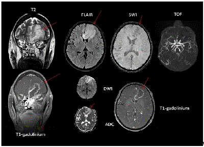
| Laboratory results showed a 732 mg/dl glycaemia with negative ketonuria, normal pH and HCO3, 7000 leucocytes/µL (95% neutrophils), and low hemoglobin (10.1 g/dl). Blood and urine cultures were negative. A MRI (Figure 1) showed an infiltrative lesion T2 hyperintense, with multiple microhemorraghic foci and peripheral enhancement, with radial strands inside the lesion. Coronal (right) views on T2 and contrast-enhanced T1 showed association with left sinusitis and orbital celulitis (fat of extraconal compartment). No arterial or venous extension was seen. |
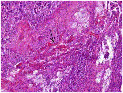
| The patient received high-dose insulin and anticonvulsant treatment with IV levetiracetam. With the suspicion of mucormycosis (high risk inmunosuppressed and diabetic patient with sinusitis, altered mentation, seizures and consistent MRI findings), high-dose IV liposomal amphotericin B (6 mg/kg/day) was immediately started, oral metilprednisolone was withdrawn, and azathioprine dose halved. An endonasal biopsy (figure 2) showed extensive ischemic necrosis with abundant hyphae and focal acute inflammatory reaction. Inoculated onto Sabouraud dextrose agar plates at 25ºC, after 24 hours a massive growth of a filamentous fungus, non-septated and with wide mycelium, was identified as Rhizopus spp. |
| A debridement of inflammatory tissue in left ethmoid, maxillary antrostomy and drainage of the single brain collection through frontal craniotomy was performed. A 6-week course of full dose IV liposomal Amphotericin B was completed and the patient was successfully switched to oral posaconazole with clinical resolution and complete solving of MRI signs at 10 weeks. |
| Many sites of infection have been described in mucormycosis including pulmonary, cutaneous, gastrointestinal, disseminated and rhinocerebral, which is the most common in diabetics [3]. The pathogenic mechanism is vascular invasion resulting in tissue infarction, necrosis and thrombosis. Hyperglycemia and acidosis are known to impair the ability of phagocytes, which are by means of oxidative and non-oxidative mechanisms the principal host defence against mucor [3]. |
| The mycological culture of a biopsy is only diagnostic if the fungus is cultured from sterile tissues, or repeatedly or massively isolated from non-sterile sites in large numbers of colonies, because mucosal colonization can exist in patients without infection and it is a relatively common contaminant in laboratories [4]. The definitive diagnosis is therefore better supported by a biopsy showing the presence of extensive ischemic necrosis with abundant hyphae and focal acute inflammation. Hyphae are characteristically wide, non-septate with right angles [5]. |
| We could not carry out the biopsy on the erytematous papular lesions because they were solved with earlier treatment. Cutaneous lesions in disseminated rhino-cerebral mucormycosis must be deliberately searched, because they can be a helpful clue in the diagnosis, as it happens with many fungal infections. |
| The sinus contents have a variety of MRI signal characteristics, including T2 hyperintensity or marked hypointensity (“black turbinate”) on all sequences, possibly due to the presence of iron and manganese in fungal elements [6]. For rhino-orbito-cerebral mucormycosis, thickening and lateral displacement of the medial rectus muscle are characteristics of orbital invasion from the ethmoid sinuses. A lack of enhancement of the superior ophthalmic vein or ophthalmic artery and internal carotid artery may be seen, related to vasculitis and thrombosis [7]. The lesions have variable enhancement patterns with gadolinium, ranging from homogeneous to heterogenous or non-enhancing at all. Contrast-enhanced T1-weighted images are helpful in delineating the parechymatous, meningeal or cavernous spread. |
| The CNS may be invaded directly through the cribiform plate, superior orbital fissure or basal foramina, but also indirectly via the invasion of vessels. In these cases infarcts may be added to the cerebritis [8]. Thus, it is also recommended to include in the study the main intracraneal and cervical vessels (Figure 1). |
| Proper debridement of infected sinus and orbital space leads to high cure rates (>85%) [9]. In a case series of 49 patients with rhinocerebral mucormycosis, mortality with antifungal agents alone was 70% compared to 17% when surgery was associated. However, these data could be biased, indeed favouring the performance of surgery in individuals with better prognosis and surviving the first days of treatment [10]. |
| Early initiation (within 5 days after strong suspicion or diagnosis) of polyene therapy is of paramount importance, usually with high dose liposomal amphotericin B (5 to 8 mg/kg).3 Doses and duration of antifungal therapy are not well established due to the lack of comparative studies, but should be continued until clinical resolution of signs, symptoms of infection and radiographic signs on serial imaging [1]. The combination of a polyene and an echinocandin has not proven to increase outcomes yet, even though Rhizopus expresses the target enzyme for echinocandins [1]. |
| Therefore, in diabetic or immunosuppressed patients with epistaxis, acute/subacute history of headaches, maxillary pain or sinusitis, neurological deficits or seizures, rhino-orbito-cerebral mucormycosis must be included in the differential diagnosis. Early diagnosis, reversal of underlying predisposing factors, correct antifungal therapy, and early surgical debridement are the four steps required for healing this deadly fungal disease, with an ominous prognosis that could improve significantly with the recent advances in antifungal treatment and diagnostic techniques. |
| Funding |
| The present work received no funding. |
| Declaration of interest |
| The authors declare that they have no conflicts of interest. |
References
- Spellberg B, Walsh TJ, Kontoyiannis DP, Edwards J Jr, Ibrahim AS (2009) Recent advances in the management of mucormycosis: from bench to bedside. Clin Infect Dis 48: 1743-1751.
- Tietz HJ, Brehmer D, Jänisch W, Martin H (1998) [Incidence of endomycoses in the autopsy material of the Berlin Charité Hospital]. Mycoses 41 Suppl 2: 81-85.
- Spellberg B, Edwards J Jr, Ibrahim A (2005) Novel perspectives on mucormycosis: pathophysiology, presentation, and management. ClinMicrobiol Rev 18: 556-569.
- Greenberg RN, Scott LJ, Vaughn HH, Ribes JA (2004) Zygomycosis (mucormycosis): emerging clinical importance and new treatments. CurrOpin Infect Dis 17: 517-525.
- Bhansali A, Bhadada S, Sharma A, Suresh V, Gupta A, et al. (2004) Presentation and outcome of rhino-orbital-cerebral mucormycosis in patients with diabetes. Postgrad Med J 80: 670-674.
- Chan LL, Singh S, Jones D, Diaz EM Jr, Ginsberg LE (2000) Imaging of mucormycosis skull base osteomyelitis. AJNR Am J Neuroradiol 21: 828-831.
- Bae MS, Kim EJ, Lee KM, Choi WS. Rapidly Progressive Rhino-orbito-cerebral Mucormycosis Complicated with Unilateral Internal Carotid Artery Occlusion: A Case Report. Neurointervention 2012; 1:45-9.
- Dussaule C, Nifle C, Therby A, Eloy O, Cordoliani Y, et al. (2012) Teaching neuroimages: brain MRI aspects of isolated cerebral mucormycosis. Neurology 78: e93.
- Nithyanandam S, Jacob MS, Battu RR, Thomas RK, Correa MA, et al. (2003) Rhino-orbito-cerebral mucormycosis. A retrospective analysis of clinical features and treatment outcomes. Indian J Ophthalmol 51: 231-236.
- Peterson KL, Wang M, Canalis RF, Abemayor E (1997) Rhinocerebralmucormycosis: evolution of the disease and treatment options. Laryngoscope 107: 855-862.
Figures at a glance
 |
 |
|
| Figure 1 | Figure 2 |
Post your comment
Relevant Topics
- Advanced Therapies
- Chicken Pox
- Ciprofloxacin
- Colon Infection
- Conjunctivitis
- Herpes Virus
- HIV and AIDS Research
- Human Papilloma Virus
- Infection
- Infection in Blood
- Infections Prevention
- Infectious Diseases in Children
- Influenza
- Liver Diseases
- Respiratory Tract Infections
- T Cell Lymphomatic Virus
- Treatment for Infectious Diseases
- Viral Encephalitis
- Yeast Infection
Recommended Journals
Article Tools
Article Usage
- Total views: 15515
- [From(publication date):
August-2014 - Jul 12, 2025] - Breakdown by view type
- HTML page views : 10856
- PDF downloads : 4659
