Make the best use of Scientific Research and information from our 700+ peer reviewed, Open Access Journals that operates with the help of 50,000+ Editorial Board Members and esteemed reviewers and 1000+ Scientific associations in Medical, Clinical, Pharmaceutical, Engineering, Technology and Management Fields.
Meet Inspiring Speakers and Experts at our 3000+ Global Conferenceseries Events with over 600+ Conferences, 1200+ Symposiums and 1200+ Workshops on Medical, Pharma, Engineering, Science, Technology and Business
Case Report Open Access
Retropharyngeal Abscess: Its Evolution and Imaging Assessment
| Yongdong Wang*, Nina Sigh and John Mernagh | |
| Department of Radiology, McMaster University Medical Center, Hamilton, ON, Canada | |
| Corresponding Author : | Yongdong Wang Department of Radiology McMaster University Medical Center Hamilton, ON, Canada E-mail: wangyon@HHSC.CA |
| Received April 10, 2013; Accepted May 14, 2013; Published May 20, 2013 | |
| Citation: Wang Y, Sigh N, Mernagh J (2013) Retropharyngeal Abscess: Its Evolution and Imaging Assessment. OMICS J Radiology 2:127. doi: 10.4172/2167-7964.1000127 | |
| Copyright: © 2013 Wang Y, et al. This is an open-access article distributed under the terms of the Creative Commons Attribution License, which permits unrestricted use, distribution, and reproduction in any medium, provided the original author and source are credited. | |
Visit for more related articles at Journal of Radiology
| This is a case of a 12 year old female who presented initially with neck pain which progressed to right neck swelling, difficulty swallowing and right sided Horner’s syndrome for 3 days. There was also development of a muffled voice, paralysis of the right XII nerve. There was no cough, difficulty breathing or strider. |
| The initial X-ray of the neck showed thickening of the retropharyngeal soft tissue. There was significant leukocytosis (28.1) with increased neutrophils (25.8). |
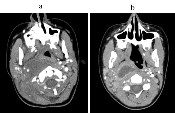
| An emergency Contrast Enhancement CT (CECT) scan demonstrated right retropharyngeal subtle hypodensity with mass effect and faint intermittent peripheral rim enhancement suggestive of early organizing retropharyngeal abscess. The CT value of the central hypodensity measured 34 House field units (Figure 1a). In addition there was edematous change of the retropharyngeal space (RPS), Right Parapharyngeal Space (PPS) and Right Carotid Space (CS). Multiple reactive enhancing lymph nodes were seen along the right side of the neck. The right jugular vein was attenuated. Non-occlusive thrombus was seen in the right transverse and sigmoid sinus extending into the right upper jugular vein. |
| Doppler Ultrasound subsequently demonstrated a patent, but narrowed right mid jugular vein. |
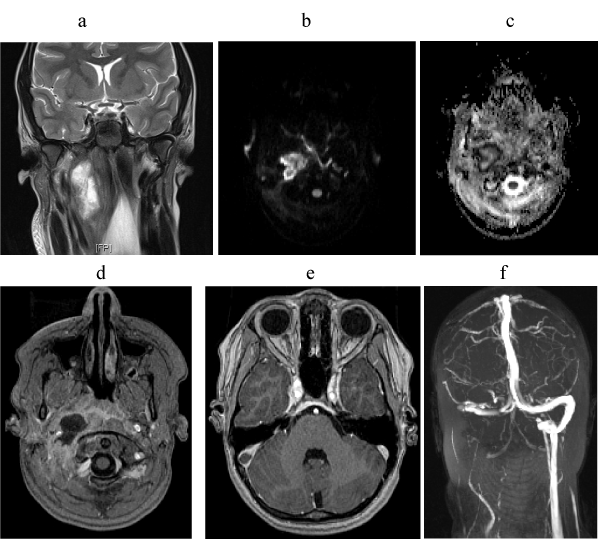
| This was followed by multiplanar multisequence MRI with and without Gadolinium performed at day 5 which confirmed the central necrosis of retropharyngeal adenopathy with peripheral restricted diffusion (Figures 2a-2d). Significant surrounding soft tissue enhancement was seen indicative of phlegmon. In addition, MRI confirmed the presence of non-occlusive thrombosis in the right transverse and sigmoid sinuses and extension into the upper jugular vein (Figures 2e and 2f). |
| Transcervical incision/drainage and trans-oral aspiration were performed, all failed to drain pus or fluid from the right retropharyngeal region by the Surgical team. The patient was treated continuously with Ceftriaxone and Metronidazole, as well as subcutaneous Enoxparin for observation. |
| By the day 10 there was development of trismus, and increasing pain and fullness of the neck. Repeat CT exam showed presence of mature abscess surrounding by complete ring enhancing wall (Figure 1b). The central abscess content measured 15 HU. |
| Transoral aspiration and incision at site of fluctuance was performed. 15 ml of brownish dishwasher like fluid was aspirated. With further antibiotic and anti-coagulant treatment, the patient symptoms improved. Subsequent follow-up MR exams showed gradual resolution of the phlegmon, abscess and associated venous sinus thrombus. |
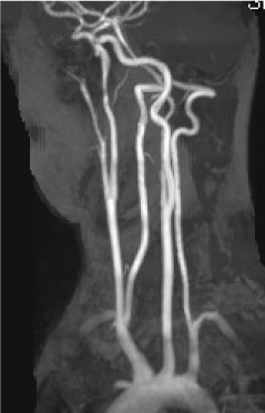
| At the one month follow up there was luminal narrowing of the upper jugular vein and mild stenosis of the right internal carotid artery (Figure 3). The patient complained of headache that worsened with lying down. A lumbar puncture was performed proving mild increased intracranial pressure (55 mmHg) which was treated well with Diamonx. This was thought to be insufficient venous return due to luminal narrowing of the jugular vein. |
| Discussion |
| The retropharyngeal space is a potential space outlined between the middle and deep layers of deep cervical fascia [1]. It is located anterior to the Alar space (also known as the “danger space”, since infection can spread directly into thorax) and prevertebral space, laterally adjacent to carotid space, anteriorly adjacent to the pharynx and esophagus, superiorly to the skull base and inferiorly to the upper mediastinum to T3 level [2]. |
| There are multiple lymph nodes in the retropharyngeal space, also named Nodes of Rouviere. There is high incidence of retropharyngeal infections involving these Rouviere nodes especially in patients that are under 5 years of age, boys more than females [3]. Some lymphadenitis may progress to suppuration and some may need surgical treatment [4-6]. The etiology is usually polymricrobial containing both anaerobic and aerobic flora [7]. Early parental antibiotic treatment is important to control the infection [5,8]. |
| Clinically the patients usually present with fever, neck pain, swelling, difficulty swallowing and leukocytosis. Severe cases will present with upper airway compromise and ominous drooling [3]. |
| The retropharyngeal abscess can be diagnosed and treated with surgical drain/aspiration. The presurgical imaging exam may show a high false positive result up to 25% [4]. Daya et al. [5] reported that it is more appropriate to call it retropharyngeal infection to guide appropriate treatment. |
| There is a high complication rate of the retropharyngeal abscess, up to 10-20% [7] and mortality of 1% [9]. Appropriate imaging is important to detect the source of infection and the extent, as well as monitoring its progression. |
| CT and MRI can define the specific anatomic landmarks of the deep neck potential spaces [1,2]. The epicenter of the retropharygeal abscess is located in the paramedial location of the upper cervical region with mass effect. It may displace the parapharyngeal space anterolaterally, carotid space laterally and the esophagus and pharynx anteriorly. In addition there is surrounding phlegmon involving the adjacent structures. According to Davis et al. [10], there is predominant nodal involvement in the suprahyoid RPS disease and non-nodal involvement in the infrahyoid RPS disease. |
| Initially there is presentation of lymphadenitis with RPS edema or phlegmon. With disease progression, there might be formation of retropharyngeal abscess. Early abscess may show subtle central hypodensity on CT (hyperintense on T2W images on MRI) and an enhancement pattern with intermittent incomplete rim enhancement which is nonspecific [9]. At later stage, there is evolution of central fluid density surrounded by complete ring enhancing wall representing typical liquification of the necrotic component or maturation of abscess. |
| Previous studies reported hypodensity with surrounding rim enhancement can be the diagnostic criteria for retropharyngeal abscesses [5,6,9,11]. Daya reported CT sensitivity of 81% and specificity of 57% [5]. Rim enhancement alone has a high sensitivity for abscess of 89% but is not specific. Kirse and Roberson reported irregularity and scalloping enhancing wall gave a higher PPV of 94% for abscess [11]. Christenson et al. classified the retraphryngeal abscess into 3 types of CT enhancement pattern and reported an overall accuracy of 88% [6]. |
| In our case we found that even though there was clear evidence of necrosis in the retropharyngeal space during the early stage both interpreted by CT and MRI, there was no successful aspiration and incision drainage. Upon further liquification of the central necrosis (CT value drop to 15 HU from 34HU) and with formation of complete ring enhancing wall, palpable fluctuance became evident. It was only at this point in time that there was successful drainage of the abscess contents. |
| The CT value measurement did contribute in proper assessment of maturation of the abscess. We think that in appropriate patients who don’t respond well with antibiotic treatment or clinical worsening, follow-up CT is helpful to assess the maturation of the abscess and help to determine the timing and successfulness of surgical intervention. |
| MRI may be more sensitive to demonstrate the evolution features of necrosis with non-enhancement pattern, restricted diffusion of the necrotizing component, and the latter stage of ring enhancing wall of the organizing abscess. However, MRI is a time consuming modality often requiring GA in these pediatric patients and may restrict its widespread clinical application for non-complicated cases, depending on availability. |
| The extension of the retropharyngeal abscess may spread to adjacent spaces and down to the thorax via the mediastinum [12,13]. It is critical to include the posterior cranial fossa and upper mediastinum in the CT scan. In this case there was Lemierre’s syndrome [7], a complication of thrombophlebitis involving the internal jugular vein via adjacent bacteria infection and subsequent upper sigmoid/transverse sinus thrombosis. |
| CT and MRI both successfully depict the extent of the thrombosis, including both intrinsic thrombosis and extrinsic jugular compression by surrounding abscess/phlegmon and luminal narrowing. Doppler Ultrasound is more convenient and accurate in assessing the patency/ severity of luminal narrowing and blood flow dynamics in the mid to distal jugular vein. |
| In addition, appropriate contrast enhanced pulmonary CT can be arranged in suspected cases with positive thrombophlebitis, given the potential risk of septic embolism involving the lungs. In our case there was also mild internal carotid arterial stenosis detected by MRA, a rare sequela of focal infection [14]. |
| In conclusion, CT and MRI play an important role in defining the specific location and extent of the retropharyngeal infection and monitoring the evolution of abscess formation. Combining the numerical evaluation of the central fluid hypodensity, with the CT Houndsfield value, and the complete ring enhancing wall allows for more appropriate interpretation of the maturation of the abscess contents. This will help guide successful surgical drainage. |
| A combination of CECT, MRI and MRV with Gadolinium and Doppler Ultrasound are all valuable modalities to evaluate Lemierre’s syndrome. |
References |
|
Figures at a glance
 |
 |
 |
| Figure 1 | Figure 2 | Figure 3 |
Post your comment
Relevant Topics
- Abdominal Radiology
- AI in Radiology
- Breast Imaging
- Cardiovascular Radiology
- Chest Radiology
- Clinical Radiology
- CT Imaging
- Diagnostic Radiology
- Emergency Radiology
- Fluoroscopy Radiology
- General Radiology
- Genitourinary Radiology
- Interventional Radiology Techniques
- Mammography
- Minimal Invasive surgery
- Musculoskeletal Radiology
- Neuroradiology
- Neuroradiology Advances
- Oral and Maxillofacial Radiology
- Radiography
- Radiology Imaging
- Surgical Radiology
- Tele Radiology
- Therapeutic Radiology
Recommended Journals
Article Tools
Article Usage
- Total views: 16836
- [From(publication date):
July-2013 - Mar 29, 2025] - Breakdown by view type
- HTML page views : 12174
- PDF downloads : 4662
