Case Report Open Access
Retroperitoneal Schwannoma: When EUS-Guided FNA can Avoid Surgery
Claudio Zulli1*, Nadia Alberghina1, Giuseppe Grande1, Mauro Manno1, Luca Reggiani Bonetti2, Flavia Pigò1, Vincenzo Giorgio Mirante1, Santi Mangiafico1, Rita Conigliaro1 and Carmelo Barbera1
1Gastroenterology and Digestive Endoscopy Unit, NOCSAE Hospital, Baggiovara di Modena, Italy
2Department of Diagnostic Medicine and Public Health, University of Modena and Reggio Emilia-Section of Pathology, Modena, Italy
- Corresponding Author:
- Claudio Zulli
Gastroenterology and Digestive Endoscopy Unit
NOCSAE Hospital, via Pietro Giardini 1355
Baggiovara di Modena, ZIP Code 41126, Italy
Tel: +39 0593961260
Fax: +39 0593961216
E-mail: mailto:zulli.claudio@gmail.com
Received date: March 25, 2016; Accepted date: June 23, 2016; Published date: June 26, 2016
Citation: Zulli C, Alberghina N, Grande G, Manno M, Bonetti LR, et al. (2016) Retroperitoneal Schwannoma: When EUS-Guided FNA can Avoid Surgery. J Gastrointest Dig Syst 6:443. doi:10.4172/2161-069X.1000443
Copyright: © 2016 Zulli C, et al. This is an open-access article distributed under the terms of the Creative Commons Attribution License; which permits unrestricted use; distribution; and reproduction in any medium; provided the original author and source are credited.
Visit for more related articles at Journal of Gastrointestinal & Digestive System
Abstract
Schwannomas are rare benign tumor that arises from peripheral or cranial nerve. Commonly, they occur into the head or neck and rarely into the retroperitoneum or pancreas. Usually they are asymptomatic tumor, discovered incidentally. Final diagnosis is generally confirmed after surgical intervention. The possibility to reach the lesion by EUS and to perform FNA can avoid invasive procedures. Here we discuss a rare case of retroperitoneal schwannoma diagnosed by Endoscopic ultrasound (EUS) guided Fine needle aspiration (FNA).
Keywords
Schwannoma; Retroperitoneal tumor; Neurilemmoma; EUS-FNA
Case Report
Schwannomas are rare benign tumor that arises from peripheral or cranial nerve. Commonly, they occur into the head or neck and rarely into the retroperitoneum or pancreas [1]. Usually they are asymptomatic tumor, discovered incidentally [2]. Final diagnosis is generally confirmed after surgical intervention. The possibility to reach the lesion by EUS and to perform FNA can avoid surgical diagnostic procedures [3]. Actually, a surgical approach can be considered only with a therapeutic purpose, in case of presence of large symptomatic masses.
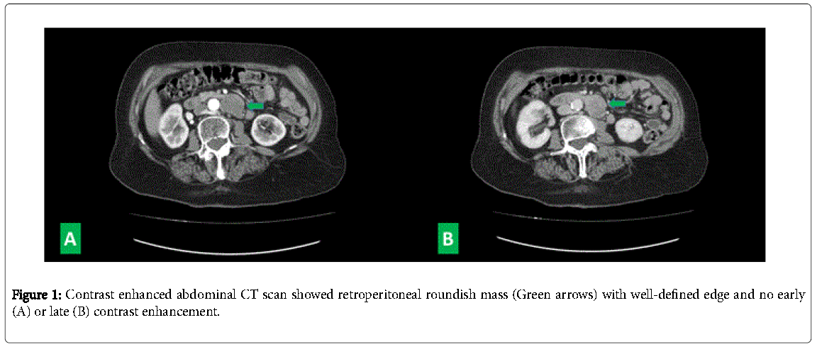
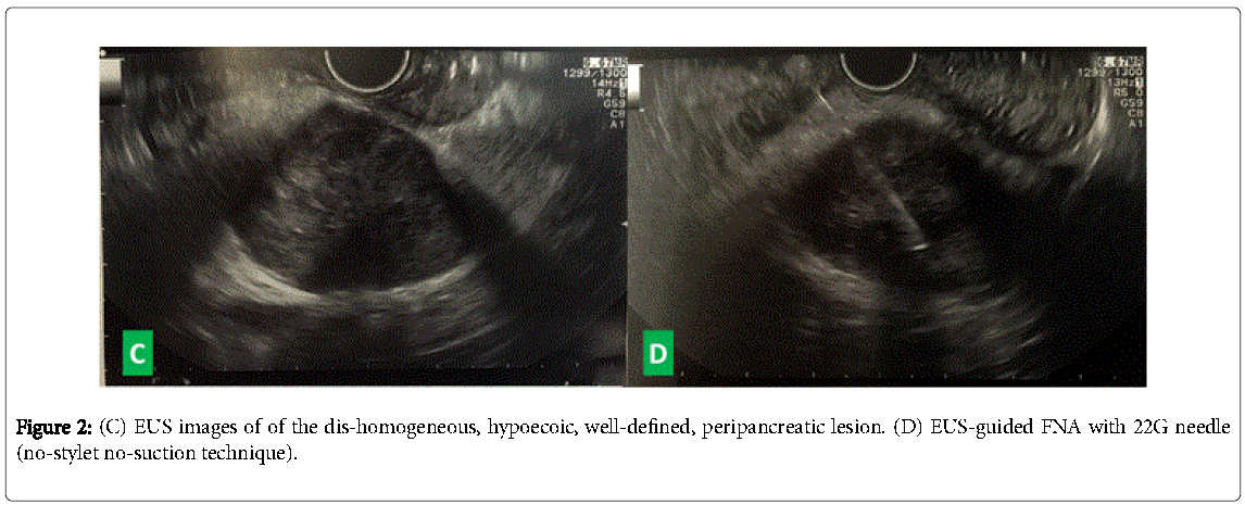
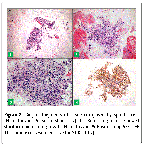
A 80 years-old woman referred to our unit to perform EUS-guided FNA of a retroperitoneal mass, discovered during follow-up abdominal US for HCV-related hepatitis. CT scan confirmed the presence of a 35 × 28 mm solid, ovoid, dis-homogeneous lesion located dorsally between the left side of abdominal aorta, left renal vein and the ascending part of duodenum (Figure 1). The patient was asymptomatic, and neoplastic markers were negative. She underwent a EUS (GF-UCT 180; Olympus Co., Japan) under deep sedation, revealing a dis-homogeneous, hypo-echoic, well-bordered, 40 × 28 mm diameter mass, located between the dorsal part of duodenum and the head of the pancreas. A EUS-FNA was performed using a 22 Gauge needle, through the duodenal wall, with the no-stylet no-suction technique (Figure 2). Histological findings included fragments of tissue composed by spindle cells showing a specific immunoreactivity to S-100; DOG1 and CD117 stains were both negative (Figure 3). Final diagnosis was a schwannoma and patient was referred to a radiological follow-up.
Schwannomas, also named neurilemmoma, are rare, benign tumors originating from Schwann cells. Retroperitoneal location is uncommon (1-3% of all Schwannomas and almost 1% of all retroperitoneal tumors). The diagnosis can be delayed due its location, so it could appear as a giant mass [4]. Radiological findings are not pathognomonic and tissue sampling is necessary for a final diagnosis [3]. However, definitive diagnostic result is generally obtained by surgery. Our experience suggests that EUS-guided FNA represents a reliable alternative for a noninvasive diagnosis of retroperitoneal mass [5]. Surgery is mandatory only in case of symptomatic masses causing abdominal discomfort or pain.
References
- Barresi L, Tarantino I, Granata A, Traina M (2013) Endoscopic ultrasound-guided fine-needle aspiration diagnosis of pancreatic schwannoma. Digestive and liver disease 45: 523.
- Dede M, Yagci G, Yenen MC, Gorgulu S, Deveci MS, et al. (2003) Retroperitoneal benign Schwannoma: report on three cases and analysis of clinicoradiologic findings. Tohoku J Exp Med 200:93-97.
- Stelow EB, Lai R, Bardales RH (2004) Endoscopic ultrasound-guided fine-needle aspiration cytology of peripheral nerve-sheath tumors. DiagnCytopathol 30:172-177
- Fu H, Lu B (2015) Giant retroperitoneal schwannoma: a case report. Int J ClinExp Med 8:11598-11601
- Kudo T, Kawakami H, Kuwatani M, Ehira N, Yamato H, et al. (2011) Three cases of retroperitoneal schwannoma diagnosed by EUS-FNA. World J Gastroenterol17:3459-3464.
Relevant Topics
- Constipation
- Digestive Enzymes
- Endoscopy
- Epigastric Pain
- Gall Bladder
- Gastric Cancer
- Gastrointestinal Bleeding
- Gastrointestinal Hormones
- Gastrointestinal Infections
- Gastrointestinal Inflammation
- Gastrointestinal Pathology
- Gastrointestinal Pharmacology
- Gastrointestinal Radiology
- Gastrointestinal Surgery
- Gastrointestinal Tuberculosis
- GIST Sarcoma
- Intestinal Blockage
- Pancreas
- Salivary Glands
- Stomach Bloating
- Stomach Cramps
- Stomach Disorders
- Stomach Ulcer
Recommended Journals
Article Tools
Article Usage
- Total views: 14713
- [From(publication date):
June-2016 - Jul 11, 2025] - Breakdown by view type
- HTML page views : 13828
- PDF downloads : 885


