Rectal Carcinoma with Osteosclerotic Metastases-A Rare Occurrence
Received: 26-Apr-2016 / Accepted Date: 03-May-2016 / Published Date: 12-Nov-2016 DOI: 10.4172/2476-2253.1000104
Abstract
Colorectal carcinoma is one of the most deadly cancers worldwide. Osseous metastases are unusual until late in the disease and usually follow hepatic & pulmonary metastases. Osseous metastases in rectal carcinoma are usually osteolytic or mixed with a minor proportion presenting solely as osteosclerotic metastases. In this article, we present a rare case of rectal carcinoma showing pure, multifocal, osteosclerotic metastases at first diagnosis without any pulmonary/hepatic metastases emphasizing the role of osseous evaluation in all patients whether early or late in the disease.
Keywords: Rectal; Carcinoma; Osteosclerotic; Metastases
5544Introduction
Colorectal carcinoma ranks third among the cancer-related deaths which are primarily related to recurrence and metastases [1]. Osseous metastases from colorectal carcinoma are rare and usually occur in the late stage of the disease with vast majority following pulmonary or hepatic metastases [2,3]. Very few cases of rectal carcinoma with osseous metastases escaping pulmonary/hepatic circulation have been reported in the literature [4,5]. Osteolytic metastases is commonest followed by mixed (osteolytic & osteosclerotic) and osteosclerotic metastases in the decreasing order of frequency [6].
Case Report
A forty-year old male presented with history of off & on, bright-red, partially-mixed bleeding per rectum in stools since last 3-4 months associated with weakness, anorexia and weight loss. Clinical examination revealed signs of anemia and fixed annular mass on digital per rectal examination causing luminal stenoses that bled on touch. Routine laboratory tests revealed raised ESR (40 mm) and reduced total RBC count with microcytosis and hypochromia signifying iron deficiency anemia. Serum CEA levels were within normal limits.
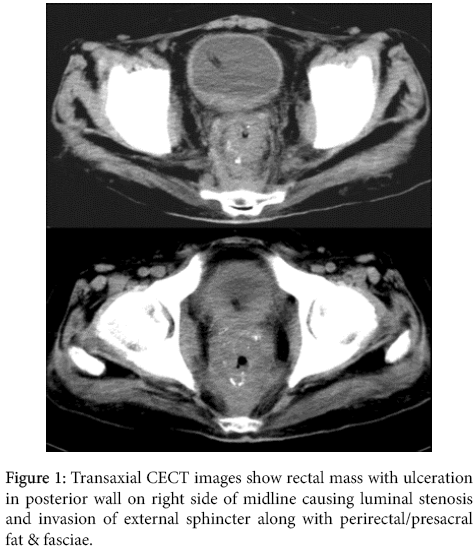
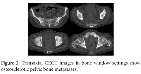
Posteroanterior radiograph of the chest was unremarkable while ultrasonography revealed rectal mass without obvious retroperitoneal adenopathy & hepatic parenchymal lesions. Contrast-enhanced computed tomography of the patient was then performed which revealed significant circumferential mural thickening in rectum showing focal ulceration and multifocal, punctate calcifications causing luminal stenoses associated with involvement of external sphincter; few small perirectal nodes and invasion of perirectal and presacral fat / fasciae without obvious pelvic / retroperitoneal adenopathy (Figure 1). Note was also made of multifocal, ill-defined, sclerotic lesions in the iliac bone on both sides of midline without obvious hepatic or pulmonary parenchymal metastases (Figure 2). Based on the radiological findings, high suspicious of rectal mucinous adenocarcinoma with osteosclerotic pelvic metastases was suggested.
Histopathological examination of the rectal and pelvic bone biopsy specimen revealed moderately differentiated mucinous adenocarcinoma with bone metastases confirming the radiological diagnosis. The patient was then referred to a dedicated cancer hospital for further management.
Discussion
Osseous metastases from colorectal carcinoma are rare with higher rarity in absence of pulmonary or hepatic metastases [4,7]. As the postulated route of spread is direct invasion of paravertebral venous plexus of Batson, the common sites of metastases are vertebrae followed by pelvic bones, sacrum, skull and long bones in the decreasing order of frequency.
Nozue et al. reported very low overall incidence of skeletal metastases in colorectal cancers i.e. 1.3% with majority of them being coexistent with hepatic/ pulmonary metastases and 0.32% incidence of isolated skeletal metastases with majority associated with rectosigmoid carcinoma [8,9]. Roth et al. also concluded in their study that bone metastases in colorectal cancers in the absence of visceral metastases are extremely rare [3]. Prognosis of patients with skeletal metastases is poor but better than when associated with visceral metastases [3,8,10]. Solitary bone metastasis is however associated with better prognosis than multifocal metastases [11].
Osseous metastases from colorectal carcinoma are predominantly osteolytic with osteoblastic or mixed type being very rare [6] making our index case rare because it was predominantly osteosclerotic in the absence of visceral metastatic lesions. Very few cases have been reported in the medical literature as ours [4,5,12]. Osteolytic lesions may demonstrate reactive bone formation in pre or postchemotherapy states, at times mimicking primary bone tumor known as pseudo-sarcomatous change [7,13].
Diagnosis can be suspected by radiography in disseminated disease and by computed tomography or magnetic resonance imaging in radiographically occult disease. Though nonspecific yet bone scintigraphy using Tc-99 is the most sensitive method of detected osseous metastases in the entire body. Final confirmation requires tissue biopsy.
Median survival of patients with skeletal metastases has been described to be around 10 months [2,8] but can be increased to up to 32 months with multimodality treatment including surgical excision, chemotherapy & radiotherapy [14].
Palliative treatment with radiation therapy or biphosphonates is the mainstay in all patients of rectal carcinoma presenting with osseous metastases (whether osteolytic or osteosclerotic) as latter is an ominous sign indicating impending death especially when accompanied with visceral metastases. Biphosphonates are more effective with osteolytic than osteosclerotic metastases as they predominantly inhibit osteoclast activity. With increasing age of survival and medical insurance coverage, more and more cases may be detected & treated with this rare form of disease. Hence recognizing the rare presentation of rectal carcinoma is not only essential in reducing the patient as well as the social financial burden but also in understanding behaviour of the tumor that help in inventing new drugs/forms of treatment [15].
Conclusion
Though skeletal metastases especially osteosclerotic type are rare in rectal carcinoma yet they do occur. Hence, in addition of prostate & breast carcinoma, colorectal carcinoma should also be kept in the differential diagnosis of osteosclerotic metastases. Better prognosis of osseous metastases alone (both solitary as well as multifocal) in colorectal carcinoma makes their early detection imperative in improving the survival of these patients.
References
- Aich RK, Suman G, Phalguni G, Jibak B, Diptimoy D (2014) Bone metastasis. J Cancer Res Ther 10: 413-415.
- O'Connell JB, Maggard MA, Ko CY (2004) Colon cancer survival rates with the new American Joint Committee on Cancer sixth edition staging. J Natl Cancer Inst 96: 1420-1425.
- Roth ES, Fetzer DT, Barron BJ, Joseph UA, Gayed IW, et al. (2009) Does colon cancer ever metastasize to bone first? a temporal analysis of colorectal cancer progression. BMC Cancer 9: 274.
- Chalkidou AS, Boutis AL, Padelis P (2009) Management of a Solitary Bone Metastasis to the Tibia from Colorectal Cancer. Case Rep Gastroenterol 3: 354-359.
- Ellington JK, Kneisl JS (2009) Acrometastasis to the foot: three case reports with primary colon cancer. Foot Ankle Spec 2: 140-145.
- Santini D, Tampellini M, Vincenzi B, Ibrahim T, Ortega C, et al. (2012) Natural history of bone metastasis in colorectal cancer: final results of a large Italian bone metastases study. Ann Oncol 23: 2072-2077.
- Udare A, Sable N, Kumar R, Thakur M, Juvekar S (2015) Solitary Osseous Metastasis of Rectal Carcinoma Masquerading as Osteogenic Sarcoma on Post-Chemotherapy Imaging: A Case Report. Korean J Radiol 16: 175-179.
- Nozue M, Oshiro Y, Kurata M, Seino K, Koike N, et al. (2002) Treatment and prognosis in colorectal cancer patients with bone metastasis. Oncol Rep 9: 109-12.
- Sundermeyer ML, Meropol NJ, Rogatko A, Wang H, Cohen SJ (2005) Changing patterns of bone and brain metastases in patients with colorectal cancer. Clin Colorectal Cancer 5: 108-113.
- Kanthan R, Loewy J, Kanthan SC (1999) Skeletal metastases in colorectal carcinomas: a Saskatchewan profile. Dis Colon Rectum 42: 1592-1597.
- Jemal A, Bray F, Center MM, Ferlay J, Ward E, et al. (2011) Global cancer statistics. CA Cancer J Clin 61: 69-90.
- Rodrigues J, Ramani A, Mitta N, Joshi P (2012) A rare case of colon carcinoma with metastases to the bone with review of the literature. The Internet Journal of Oncology 8: 2.
- Oh YK, Park HC, Kim YS (2001) Atypical bone metastasis and radiation changes in a colon cancer: a case report and a review of the literature. Jpn J Clin Oncol 31: 168-171.
- Choi SJ, Kim JH, Lee MR, Lee CH, Kuh JH, et al. (2011) Long-term disease-free survival after surgical resection for multiple bone metastases from rectal cancer. World J Clin Oncol 2: 326-328.
- Assi R, Mukherji D, HaydarA, Saroufim M, Temraz S , et al. (2016) Metastatic colorectal cancer presenting with bone marrow metastasis: a case series and review of literature. J Gastrointest Oncol 7: 284-297.
Citation: Rastogi R, Meena GL, Wani AM, Gupta Y, Joon P, et al. (2016) Rectal Carcinoma with Osteosclerotic Metastases-A Rare Occurrence. J Cancer Diagn 1:104. DOI: 10.4172/2476-2253.1000104
Copyright: © 2016 Rastogi R, et al. This is an open-access article distributed under the terms of the Creative Commons Attribution License, which permits unrestricted use, distribution, and reproduction in any medium, provided the original author and source are credited.
Share This Article
Recommended Journals
Open Access Journals
Article Tools
Article Usage
- Total views: 13723
- [From(publication date): 6-2016 - Apr 05, 2025]
- Breakdown by view type
- HTML page views: 12809
- PDF downloads: 914


