Case Report Open Access
Re-activation of IPF and Appearance of Cancer on the Native Lung after Single Lung Transplantation
| Di Scioscio V1, Cecchelli C1, Greco L1, Guerrieri A2, Morelli A2, Anagnostopoulou G2, Maleddu A3, Garzillo G1* and Zompatori M1 | |
| 1Department of Radiology, University of Bologna, Italy | |
| 2Department of Pneumology, University of Bologna, Italy | |
| 3Department of Oncology, University of Bologna, Italy | |
| Corresponding Author : | Garzillo G Department of Radiology University of Bologna, Italy E-mail: giorgio.garzillo@gmail.com |
| Received May 04, 2013; Accepted May 13, 2013; Published May 17, 2013 | |
| Citation: Di Scioscio V, Cecchelli C, Greco L, Guerrieri A, Morelli A, et al. (2013) Re-activation of IPF and Appearance of Cancer on the Native Lung after Single Lung Transplantation. OMICS J Radiology 2:125. doi: 10.4172/2167-7964.1000125 | |
| Copyright: © 2013 Di Scioscio V, et al. This is an open-access article distributed under the terms of the Creative Commons Attribution License, which permits unrestricted use, distribution, and reproduction in any medium, provided the original author and source are credited. | |
Visit for more related articles at Journal of Radiology
Abstract
IPF is an interstitial pneumonia of unknown origin with a very bad prognosis. A universally accepted therapeutic approach has still not been elaborated. Lung transplantation represents in many cases the only possible therapy for these patients. This report describes a case of IPF treated by lung transplantation. Radiological follow up of the patient after immunosuppressive therapy showed a re-activation of the primitive pathology on the native lung, a possible relapse of IPF on the graft and the appearance of two neoplastic lesions, one in the native lung and one in the left maxillary sinus.
| Keywords |
| Idiopathic pulmonary fibrosis; Lung transplantation; Immunosuppressive therapy |
| Abbreviation |
| IPF: Idiopathic Pulmonary Fibrosis; ATS-ERS: American Thoracic Society-European Respiratory Society; UIP: Interstitial Usual Pneumonia; HRCT: High Resolution Computed Tomography |
| Introduction |
| IPF (Idiopathic pulmonary fibrosis) is the most common form of interstitial lung disease of unknown origin (7-10 cases/100.000/pro year) [1]. Previous guidelines suggested lung biopsy for diagnosis of IPF, whereas the new 2011 evidence-based IPF-guidelines have defined for the first time a radiologic criterion for a UIP pattern, allowing IPF diagnosis without surgical lung biopsy. Furthermore, an IPF diagnosis can be made only if other causes of interstitial lung disease are excluded [2]. The clinical presentation is often characterized by exertional dyspnea and a restrictive pattern at the pulmonary function tests. The histological pattern of IPF is known as UIP (Interstitial Usual Pneumonia), which is characterized by a heterogeneous combination of normal lung areas, interstitial inflammation, fibroblastic proliferation and interstitial fibrosis. UIP is not specific for IPF, being recognizable in other lung pathologies such as asbestosis. The radiological suspicion of IPF is based on the following HRCT signs [3]: |
| • Honey combing lung with a mostly basal/sub-pleural and sometimes asymmetrical distribution. |
| • Areas of ground glass attenuation associated with traction bronchiectasis. |
| • Irregular reticular thickening |
| The median survival of these patients is very low (2-4 years) because of the dramatic progression of the pathology into respiratory failure often complicated by infection. There is still no evidence of an effective therapeutic approach to IPF [4]. The combination of azathioprine and prednisone was suggested as standard therapy by the ATS and ERS, despite very low evidence. Lung transplantation for IPF seems to have a worse outcome in comparison with other indications [5]. Combination of cyclosporine A, corticosteroids and azathioprine is the most used immunosuppressive therapy after lung transplantation. Recent studies have demonstrated that this approach does not seem to prevent the fibrotic involution of the native lung in single lung transplantation [6]. In addition the triple long term immunosuppressive regimen increases the overall risk of cancer and in particular of native lung cancer [7]. |
| Case Report |
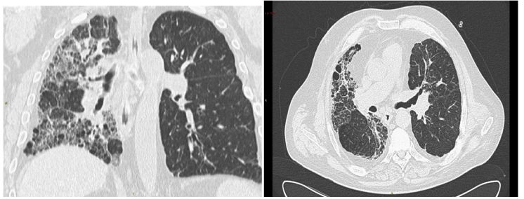
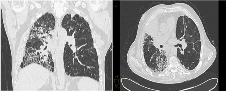
| G.L. (male, 62 years old, ex smoker, life-long welder) knows to have IPF since 1993, when the pathology was suspected through a workmedicine visit. At the time the patient did not present any particular symptomatology and was not treated until 1999. At the end of this year poliglobulia was diagnosed and he began drug therapy based on corticosteroids and undefined immunosuppressive agents. In 2003, due to the worsening of the exertional dyspnea, the quantities of corticosteroids were increased and he started domiciliary oxygentherapy. In 2004 G.L. was inserted in the waiting list for single lung transplantation and underwent a surgical lung biopsy which confirmed the diagnosis of IPF. The left lung transplantation was executed in June 2005 at the S. Orsola-Malpighi hospital of Bologna. A second biopsy of the extirpated lung showed a histological pattern compatible with UIP. The patient started the immunosuppressive therapy based on cyclosporine A, corticosteroids and azathioprine, which was then substituted with mycophenolatemofetil owing to the appearance of ancholestatichepatopaty. In May 2006 the graft developed a stenosis of the bronchial anastomosis that required application of an endobronchial stent, which caused several bacterial infections. Due to the immunosuppressive therapy the patient developed also recurrent re-activation of Cytomegalovirus infection, which required antiviral therapy with Valganciclovir. In January 2008 the endobronchial stent was removed and was not substituted with a new one. Between 2006 and 2008 the respiratory functionality of G.L. decreased progressively, on account of a condition of chronic rejection that caused a bronchiolitis obliteranssindrome, documented at a CT scan of April 2008 (Figures 1-4). |
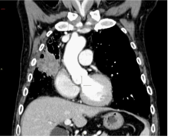
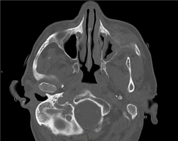
| In the meanwhile, cyclosporine A was substituted by tacrolimus and the oxygen-therapy was increased. In March 2010 another CT scan showed an interstitial fibrotic thickening of the transplanted lung that should be referred to the fibrotic evolution of the bronchiolitis obliteranssindrome. Compared to the previous CT scan of April 2008, the native lung showed an extension of the honeycombing areas, due to a worsening of the IPF. In addition the CT examination documented a consolidative area of 4.5 cm, involving the median and upper lobe of the native lung, associated with peri-tracheal adenopaties. The histological examination classified the lesion as a barely differentiated lung carcinoma. The CT study of the head demonstrated finally an erosive neoplasm of the left maxillary sinus, subsequently classified as a squamous cell carcinoma. Thoracic-Surgical and ORL evaluation excluded any possibility of a therapeutic resection of the two cancers. |
| Discussion |
| Grgic et al. [6] have demonstrated that combined use of azathioprine, cyclosporine A and corticosteroids after single lung transplantation is not able to stop the progression of IPF on the native lung [6]. Their retrospective study included 13 patients with IPF who underwent a single lung transplantation and were followed up through TC scans in the following years. According to these results we revealed an important evolution of the primary disease in our patient’s native lung, which was not avoided by the immunosuppressive therapy. |
| Bronchiolitis obliterans is a form of rejection to lung transplantation [8]. The fibrotic involution of our patient’s transplanted lung should be probably related to the end stage of this complication. The recurrence of IPF in the transplanted lung was excluded. In medical literature there’s only one described case of recurrence of IPF (subgroup “desquamative interstitial pneumonia”) in a single lung transplant recipient [9]. |
| According to different studies the rate of cancer in the native lung after single lung transplantation varies between 0.46 and 6.9% [10]. The new immunosuppressive therapy schemes can undoubtedly lead to carcinomas of the native lung. Furthermore, there is a 10% rate of lung cancer in IPF patients, especially adenocarcinoma [11], which is independent from the risk related to the immunosuppressive therapy. As documented by our patient’s clinical history this association seems not to be eliminated by single lung transplantation. |
| At the time, only a biopsy of the transplanted lung could definitely exclude the hypothesis of a recurrence of IPF. Besides, we still have very few clinical and functional data that support this rare complication. Anyway, the primary disease is undoubtedly in progression on the native lung, showing the incapacity of the immunosuppressive therapy in preventing it. In addition, the above-mentioned therapy should be considered partly responsible of the appearance of the two cancers. |
| In conclusion, we believe that the singularity of this case demonstrates how the immunosuppressive schemes should be reconsidered, as they can lead to carcinoma of the native lung, and that therefore, single lung transplantation should be abolished for IPF. |
References |
|
Figures at a glance
 |
 |
 |
 |
| Figure 1 | Figure 2 | Figure 3 | Figure 4 |
Relevant Topics
- Abdominal Radiology
- AI in Radiology
- Breast Imaging
- Cardiovascular Radiology
- Chest Radiology
- Clinical Radiology
- CT Imaging
- Diagnostic Radiology
- Emergency Radiology
- Fluoroscopy Radiology
- General Radiology
- Genitourinary Radiology
- Interventional Radiology Techniques
- Mammography
- Minimal Invasive surgery
- Musculoskeletal Radiology
- Neuroradiology
- Neuroradiology Advances
- Oral and Maxillofacial Radiology
- Radiography
- Radiology Imaging
- Surgical Radiology
- Tele Radiology
- Therapeutic Radiology
Recommended Journals
Article Tools
Article Usage
- Total views: 14089
- [From(publication date):
July-2013 - Jul 16, 2025] - Breakdown by view type
- HTML page views : 9484
- PDF downloads : 4605
