Rare Presentation of Renal Squamous Cell Carcinoma as Pyo-Nephrosis without Nephrolithiasis- A Case Report
Received: 01-Mar-2023 / Manuscript No. cns-23-87652 / Editor assigned: 04-Mar-2023 / PreQC No. cns-23-87652 / Reviewed: 18-Mar-2023 / QC No. cns-23-87652 / Revised: 24-Mar-2023 / Manuscript No. cns-23-87652 / Published Date: 30-Mar-2023
Abstract
Squamous cell carcinoma (SCC) of the kidney is an extremely rare tumor, which is usually associated with nephrolithiasis and also rarely associated with pyonephrosis,1 representing only 0.5 - 15% of all urological malignancies. The diagnosis of renal SCC is usually unsuspected due to the rarity and inconclusive clinical and radiological features; hence the prognosis is very poor, we are reporting a case of squamous cell carcinoma of the kidney in a 35-year-old female who presented with the right sided abdomen pain for 1 year and intermittent fever and lump right lumbar region for 6 months. The patient was treated with simple nephrectomy for Pyonephrosis, histopathological evaluation reported unsuspected well differentiated squamous cell renal carcinoma.
Keywords
Squamous cell carcinoma; Pyonephrosis; Radical nephrectomy
Introduction
Primary squamous cell carcinoma (SCC) of the kidney is an extremely rare entity, which is usually associated with nephrolithiasis. It is rarely associated with pyo-nephrosis, [1] representing only 0.5 - 15% of all urological malignancies. The diagnosis of renal SCC is usually unsuspected due to the rarity and inconclusive clinical and radiological features. Most of the patients are diagnosed at an advanced stage and with poor outcome. Radical nephrectomy is the mainstay of the treatment2. Review of literature shows that not many cases of primary SCC of kidney have been reported to date [2]. Etiological factors responsible for renal SCC are renal calculi, infection, exogenous chemicals, hormonal imbalance, and vitamin A deficiency, it can occur without any etiological factors1. We report a case of primary SCC of renal parenchyma in a 35-year-old female who presented with chronic right sided dull aching abdominal pain for 1 year and intermittent fever for 6 months. The post-operative histopathological report revealed an unsuspected well differentiated squamous cell carcinoma of kidney with a concomitant pyo-nephrosis, however no renal calculi was present. The patient was treated with right simple nephrectomy. The rarity of this renal SCC tumor associated with absence of renal calculi and its association with pyo-nephrosis is reported.
Case Report
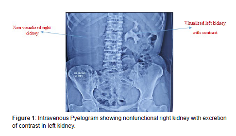
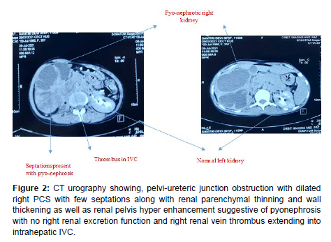
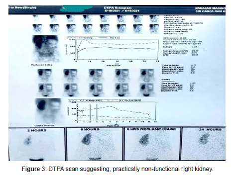
A 35-year lady presented, with complaints of dull-aching pain right lumbar region which was insidious in onset, non-progressive, for 1 year, with complaints of high-grade intermittent fever that started abruptly 6 months back, relieved with paracetamol, fever associated with chills and post febrile sweating, usually at night. She noticed a lump in her right flank region, 5 months back. History of significant weight loss present, No history of pyuria/hematuria. There was no history of tobacco or alcohol use. No other significant past or family history was noted. On per abdominal examination, a retroperitoneal, tender, non-ballotable and bimanually palpable mass of around 15*18 cm in right lumbar region was present, the rest of systemic examination was normal. On investigations the USG Abdomen suggested right sided pyo-nephrosis, gross chronic ascites and echogenic thrombus in IVC. The Intravenous Pyelogram (IVP) showed non-functional right kidney with excretion of contrast in left kidney. The CT urography revealed, right pelvic- ureteric junction obstruction with grossly dilated pelvicalyceal system (PCS) with renal parenchymal thinning and hyper-enhancement suggestive of pyo-nephrosis with right renal vein thrombus. Percutaneous drainage of pus was done and subsequent pus sent for culture and microbiology, revealed field full of pus cells, degenerated neutrophils and no atypical cells, ZN staining for AFB was negative. DTPA scan was done suggested, practically non-functional right kidney. With CT urography suggestive of PUJ obstruction, ESR raised-66 mm/hour and ADA levels 112 U/L, on clinical suspicion a provisional diagnosis of Tubercular pyo-nephrosis was made. The patient was started on anti-tubercular treatment. Subsequently she was planned for simple nephrectomy in view of right sided symptomatic nonfunctional kidney. The right kidney was removed in piecemeals since dense adhesions were formed with surrounding tissues and the residual renal tissue were fragile. Patient was apparently well 10 days in post-operative period; she developed high grade continuous fever subsequently, with hypercalcemia and leukemoid reaction and bilateral pleural effusion and sepsis. She was resuscitated in ICU with iv fluids, inotropes and broad-spectrum iv antibiotics, later antibiotics as per blood culture sensitivity reports and iv zolendronate to control hypercalcemia were started, the patient developed septicemic shock and multi-organ dysfunction and she passed away 16 days later (Figures 1-3).
Figure 2: CT urography showing, pelvi-ureteric junction obstruction with dilated right PCS with few septations along with renal parenchymal thinning and wall thickening as well as renal pelvis hyper enhancement suggestive of pyonephrosis with no right renal excretion function and right renal vein thrombus extending into intrahepatic IVC.
Discussion
The renal cell carcinomas (RCCs) are primarily adenocarcinoma are responsible for 80-85% of all primary renal malignancy, transitional cell carcinomas and other renal epithelial tumors, such as oncocytomas, collecting duct tumors, angio-myolipomas, and renal sarcomas comprise < 8% [3].
Renal SCC is a rare malignancy, comprises <1% of all renal tumors. Chronic irritation, infection and inflammation and proliferation from staghorn calculi causes squamous cell metaplasia, leads to formation of squamous cell carcinoma of the renal PCS. This version of renal malignancy has very poor prognosis with a 5-year survival rate <10% [4].
N Talwar1 et al in 2006, reported the case of a 69-year-old gentleman who had presented with features of pyo-nephrosis and underwent nephrectomy, the postoperative histological report suggested an unsuspected squamous cell carcinoma of renal pelvis with a concomitant pyo-nephrosis.
Yavuz Güler [5] et al reported a SCC of an ectopic nonfunctional pelvic kidney in 73-year-old Caucasian women with no predisposing factor.
Amirreza Fotovat [6] et al reported primary squamous cell carcinoma of renal parenchyma with nephrolithiasis in a 49-year-old woman presenting with complaints of pyelonephritis, radiological findings favored of xantho-granulomatous pyelonephritis. Histological findings revealed the involvement of kidney by malignancy.
Our reporting of this case highlights the importance of awareness of a very rare and aggressive carcinoma in a patient with the rarity of this tumor in the absence of renal calculi and its association with clinical presentation as pyo-nephrosis.
Conclusion
Renal squamous cell carcinoma is a very rare cancer and generally associated with nephrolithiasis. Rarely renal SCC associated with pyonephrosis. This case of 35-year-old lady is probably unique as it showed renal SCC may be present in pyo-nephrotic kidney without Nephrolithiasis.
Declaration
Funding
No funding sources
Conflict of interests
None declared
Ethical approval
Not acquired
References
- Talwar N, Dargan P, Arora MP, Sharma A, Sen AK (2006) Primary squamous cell carcinoma of the renal pelvis masquerading as pyonephrosis : a case report. Indian J Pathol Microbiol 49:418-420.
- Sahoo TK, Das SK, Mishra C, Dhal I, Nayak R, et al. (2015) Squamous cell carcinoma of kidney and its prognosis: a case report and review of the literature. Case Rep Urol 469327.
- Garfield K, LaGrange CA (2021) Renal cell cancer. In: StatPearls [Internet]. Treasure Island (FL): StatPearls Publishing; 2021 [cited 2021 Sep 10].
- Jongyotha K, Sriphrapradang C (2015) Squamous cell carcinoma of the renal pelvis as a result of long-standing staghorn calculi. Case Rep Oncol 8:399-404.
- Guler Y, Ucpınar B, Erbin A (2019) Renal pyelocalyceal squamous cell carcinoma in a patient with an ectopic kidney presenting with chronic pyelonephritis: a case report. J Med Case Reports 13:154.
- Fotovat A, Gheitasvand M, Amini E, Ayati M, Nowroozi MR, et al. (2021) Primary squamous cell carcinoma of renal parenchyma: Case report and review of literature. Urology Case Reports 37:101-627.
Indexed at, Google Scholar, Crossref
Indexed at, Google Scholar, Crossref
Citation: Branco M (2023) Rare Presentation of Renal Squamous Cell Carcinoma as Pyo-Nephrosis without Nephrolithiasis- A Case Report. Cancer Surg, 8: 049.
Copyright: © 2023 Branco M. This is an open-access article distributed under the terms of the Creative Commons Attribution License, which permits unrestricted use, distribution, and reproduction in any medium, provided the original author and source are credited.
Select your language of interest to view the total content in your interested language
Share This Article
Recommended Journals
Open Access Journals
Article Usage
- Total views: 2249
- [From(publication date): 0-2023 - Nov 26, 2025]
- Breakdown by view type
- HTML page views: 1897
- PDF downloads: 352


