Research Article Open Access
Rapid Methods for Isolation and Screening of Methane DegradingBacteria
| Jhala Yogeshvari K*, Vyas Rajababu V, Panpatte Deepak G and Shelat Harsha N | |
| Department of Agricultural Microbiology, Anand Agricultural University, Anand, Gujarat, India | |
| Corresponding Author : | Jhala Yogeshvari K Department of Agricultural Microbiology Anand Agricultural University Anand, Gujarat, India E-mail: yogeshvari.jhala@gmail.com |
| Received: November 06, 2015; Accepted: December 01, 2015; Published: December 12, 2015 | |
| Citation: Yogeshvari JK, Rajababu VV, Deepak PG, Harsha SN (2016) Rapid Methods for Isolation and Screening of Methane Degrading Bacteria. J Bioremed Biodeg 7:322. doi:10.4172/2155-6199.1000322 | |
| Copyright: © 2016 Yogeshvari JK, et al. This is an open-a ccess article distributed under the terms of the Creative Commons Attribution License, which permits unrestricted use, distribution, and reproduction in any medium, provided the original author and source are credited. | |
| Related article at Pubmed, Scholar Google | |
Visit for more related articles at Journal of Bioremediation & Biodegradation
Abstract
There are only few microbial strains available world over having capacity to degrade methane due to lack of rapid methodology for their isolation and efficiency studies. In present study we have isolated five methane degrading bacterial strains by enrichment of soil samples in the flasks containing methane as sole source of carbon in its head space and water as basal medium. Moreover their survival in the evacuated tubes containing methane and presence of soluble methane monooxygenase enzyme by colorimetric plate assay was studied which represents a simplest method of screening of methane utilizing microbial strains as compared to conventional gas liquid chromatographic technique as well as enzymatic assay and molecular detection of the genes encoding methane monooxygenase and methanol dehydrogenase genes. In addition to this presence of a key methane metabolism enzyme methane monooxygenase and ethanol dehydrogenase enzyme was confirmed by detection of genes encoding the enzymes as well as qualitative detection of the enzyme activities in the isolates to provide strong support to the methane degrading capacity of the isolates and to validate the method used in this study to isolate methanotrophs.
| Keywords |
| Methane monooxygenase; Methanol dehydrogenase |
| Introduction |
| Among all greenhouse gases methane account for about 15-20% of total global warming effect (IPCC, 2007). The sources of methane gas emission contributing about 50% of total methane emission includes wetlands, rice fields, enteric fermentation in animals, termites and landfills etc. [1]. The only known biological sink for atmospheric methane is its oxidation in aerobic soils by “methanotrophic (=methylotrophic) bacteria”, this may contribute up to 10-20% to the total methane destruction [2]. So isolating efficient methane degrading bacteria (methanotrophs) from natural habitats may present a near long term advantage in solving global warming problem. Unfortunately till date we have only few numbers of microbial strains which can degrade methane due to lack of methodologies for isolation and screening of such strains. We have tried to find out an easy and rapid method for isolation and screening of methane degrading bacteria from environmental samples. |
| In this study we present a more direct isolation method. A strong initial selection in favour of methane utilising bacteria was obtained by directly injecting methane gas in to enrichment flasks. Moreover methane utilizing capacity of isolates were further evaluated by incubating the isolates in evacuated tubes containing methane as the sole source of carbon and water as basal medium. Methane utilizing nature of the isolates was further evaluated by detecting genes encoding methane monooxygenase and methanol dehydrogenase enzymes as well as corresponding enzyme activities which are a characteristic of methanotrophic bacteria. |
| Materials and Methods |
| Soil samples |
| For isolation of methanotrophic bacteria from wet land paddy, total 15 soil samples were collected at flowering stage representing major traditionally rice growing areas of Middle and South Gujarat, India. Out of fifteen soil samples, 7 samples were collected from Main Rice Research Station, Nawagam, 4 samples from Agricultural Research Station for Irrigated Crops, Thasra, 2 each from farmer’s field at Tarapur and Vyara Taluka, respectively. The soil samples from rhizosphere of rice roots were collected in sterile HDPE bags and brought to the laboratory and processed immediately after collection. |
| Isolation of methane utilizing bacteria |
| 1 gm of soil (wet weight) was inoculated in the flask containing sterile RO water containing desired minerals as basal medium the flasks were than sealed with rubber cock tightly supplemented with 1% methane gas and enriched for 15 days. After enrichment bacterial growth was purified on NMS agar medium incubated in anaerobic jar containing methane gas. |
| In vitro conformation of methane utilizing activity |
| Suspected isolates were tested for utilization of methane gas at the rate of 1% as sole source of carbon in evacuated tubes containing water (basal medium)+methane (head space of the tube) to confirm methylotrophic metabolism. Growth and survival of isolates were measured by recording colony counts from inoculated tubes at 10 days after inoculation on NMS agar and recorded as colony forming units (CFU)/ml after 10 days of inoculation. |
| Detection of Bacterial methane metabolism genes |
| The presence of mmoX, pmoA and mxaF gene in the isolates, encoding sMMO, pMMO and methanol dehydrogenase which are key enzymes in bacterial methane metabolism pathway, was used for authentication of the isolates. The presence of genes in the isolates was detected by partial amplification of the genes using specific primers [3]. Following primer pairs used for mmoX (534f - ccgctgtggaagggcatgaa & 1393r-cactcgtagcgctccggctc); pmoA (A189f – ggngactgggacttctgg & A682r-gaasgcngagaagaasgc) and mxaF (1003f–gcggcaccaactggggctggt & 1561r-gggcagcatgaagggctccc), respectively. The 40 μl PCR reaction mixture contains DNA template 50 ng, 1X Taq buffer, 0.2 mM of each of dNTPs mixture, 1 μM of each primers, 1.5 mM MgCl2, and 2 U of Taq DNA polymerase (Bangalore Genei, India). PCR amplification was performed in a thermal cycler (Eppendorf Master cycler, Germany) using the following conditions: initial denaturation at 95°C for 1 min, 30 cycles consisting of 95°C for 1 min (denaturation), annealing 62- 52°C touchdown - 1 min for mmoX and pmoA, |
| while 55°C–1 min for mxaF gene, 72°C for 1 min (primer extension), and final extension 72°C for 5 min. PCR products were separated by electrophoresis on 1.5% agarose gels stained with ethidium bromide and documented in AlphaImager TM1200 documentation and analysis system. |
| Quantitative estimation of methane mono oxygenase and methanol dehydrogenase activities |
| Cell free extract was prepared by centrifugation of cell mass for 30 min at 10,000 g washed once by suspending in 0.05 M potassium phosphate buffer (pH 7.0), the washed cells were suspended in the same buffer, homogenized and disrupted by ultrasonic treatment of 50 Hz (10 sec/ml). The suspension was than centrifuged at 10,000 g for 30 min and the resulting supernatant was used as crude extract. Proteins were determined by Lowry’s method using bovine serum albumin as standard and methanol dehydrogenase activity was assayed by the method suggested by Anthony (1971). |
| Soluble methane monooxygenase activity: For quantitative determination of soluble methane monooxygenase a slightly modified version of the naphthalene oxidation assay of was followed. Each culture was transferred in 1 ml aliquots to 10 ml screw-cap tubes and 1 ml of pre-filtered saturated naphthalene solution was added to each tube. The samples were prepared in triplicate keeping sterile medium control as blank. The reaction mixtures were incubated at 200 rpm on incubator shaker at 25°C for 1 to 3 hrs. After incubation, 100 μl of freshly prepared 4.21 mM tetrazotized-o-dianisidine solution was added to each tube and the intensity of coloured diazo-dye complex was immediately monitored by recording the A525 by spectrophotometry. The intensity of diazo-dye formation is proportional to the naphthol concentration (1-naphthol and 2-naphthol). The specific activity of sMMO was expressed as nanomoles of naphthol formed per milligram of cell protein per minute. |
| Methanol dehydrogenase activity: Qualitative detection of methanol dehydrogenase was carried out following method of Eggeling and Sahm (1980) with some modifications. The enzyme assays were performed in triplicate and data obtained were subjected to statistical analysis by using Completely Randomized Design (CRD) (Panse and Sukhatme, 1978). |
| Identification of potential methylotrophic isolates through 16S rDNA sequencing |
| Genomic DNA extraction: Five methylotrophic isolates were grown in Luria broth for 24 hrs, and genomic DNA was extracted by C-TAB method. The integrity and concentration of purified DNA was determined by agarose gel electrophoresis. The total genomic DNA extracted was dissolved in sterile distilled water and stored at 4°C. 16S rDNA amplification was performed in a thermal cycler (Eppendorf Master cycler, Germany) with a 25 μl reaction mixture containing 50 ng of genomic DNA, 0.2 mM of each dNTPs, 1 μM of each primer, 2.5 mM of MgCl2, and 1 U of Taq DNA polymerase (Bangalore Genei, India) and the buffer supplied with the enzyme. |
| DNA Sequencing Analysis: DNA fragment from five isolates were chosen to be sequenced based on the dominant isolates that isolated from soil samples. PCR product was purified using DNA Gel Extraction Kit (Banglore Genei, India). Purified products were sequenced using big dye terminator with ABI PRISMTM model 3130 Genetic Analyzer. The sequence data of bacterial isolates were compared to sequence from GenBank in National Center for Biotechnology Information (NCBI, www.ncbi.nlm.nih.gov) using BLASTN program for identification and phylogenetic analysis of the isolates. |
| Results and Discussion |
| Isolation of methane utilizing bacteria from wetland paddy |
| As conventional method of isolation of methane utilizing bacteria generally Nitrate mineral salt media/ammonium mineral salt medium supplemented with 1% methanol were used which do not support growth and isolation of methanotrophs. As a novel media reverse osmosis water was utilized as basal medium which contains all the minerals required for microbial growth and as a selective agent the flasks were supplemented with methane gas as a sole source of carbon. These two together will not allow any undesired microorganisms to grow in the media and organisms capable of utilizing methane as sole carbon source will multiply. In all total 74 isolates were obtained from rhizospheric soil of rice and during screening of isolates’ for their ability to survive by utilizing methane as sole carbon source isolate M 8 (from rhizospheric soil of organically grown rice cv. IR-64 at main rice research station, Nawagam, Gujarat, India), M 10 (from rhizospheric soil of rice grown at farmers’ field of Vyara, Surat, Gujarat, India), M 14 (from rhizospheric soil of rice grown at farmers’ field of Vyara, Surat, Gujarat, India), M 17 (from rhizospheric soil of rice grown at farmers’ field of Tarapur, Gujarat, India) and M 29 (from rhizospheric soil of organically grown rice cv. GR-11 at main rice research station, Nawagam, Gujarat, India) were found to multiply in 1% methane+water. So inspired from the results, these five potent isolates viz. M 8, M 10, M14, M 17 and M 29 were chosen for further study. As the reference standard Methylobacterium extrorquens strain (Cat. No. MTCC 298) obtained from Microbial Type Culture Collection Center, IMTECH, Chandigarh was used as these strain is reported to possess soluble methane monooxygenase and methanol dehydrogenase activities which are involved in bacterial methane metabolism. As a source of isolation rhizospheric soils from wetland field at mid-season were selected as the chances of getting efficient methane degrading bacterial strains are maximum at this stage. Because of water logged conditions the methanogenic bacteria are producing methane at maximum rates during this time and due to abundance of favourable carbon source (methane) the methanotrophic bacteria are flourishing. Till date most of the efforts pertaining to isolate methylotrophic bacteria are concentrated on phyllospheric population but in present investigation the isolation was attempted from rhizospheric samples of wetland paddy fields by using enrichment culture technique isolated Methylobacterium populi sp. nov. a novel aerobic, pink-pigmented, facultatively methylotrophic, methane-utilizing bacterium from poplar trees by enrichment of tissue culture explants in Nitrate mineral salt media supplemented with 1% methanol as carbon source. As a source of isolation rhizospheric soils from wetland field at mid-season were selected as the chances of getting efficient methane degrading bacterial strains are maximum at this stage. Because of water logged conditions the methanogenic bacteria are producing methane at maximum rates during this time and due to abundance of favourable carbon source (methane) the methanotrophic bacteria are flourishing. Till date most of the efforts pertaining to isolate methylotrophic bacteria are concentrated on phyllospheric population but in present investigation the isolation was attempted from rhizospheric samples of wetland paddy fields by using enrichment culture technique isolated a novel, pink-pigmented aerobic, facultatively methylotrophic bacterial strain from the phyllosphere of Funaria hygrometrica using Ammonium Mineral Salt (AMS) media supplemented with 0.5% methanol as sole carbon source. Aken et al. isolated Methylobacterium populi sp. nov. a novel aerobic, pink-pigmented, facultatively methylotrophic, methaneutilizing bacterium from poplar trees by enrichment of tissue culture explants in Nitrate mineral salt media supplemented with 1% methanol as carbon source. |
| In vitro conformation of methane utilizing activity |
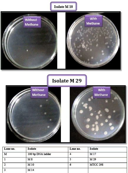
| Results showed that five selected isolates were found capable of utilizing methane as sole source of carbon for their growth and survival. After 10 days of inoculation, there was fourfold increase in cell numbers confirming methanotrophic nature of isolates. (Plate 1; Figure 1). |
| Detection of methane metabolism genes |
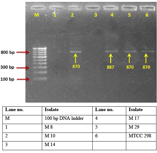
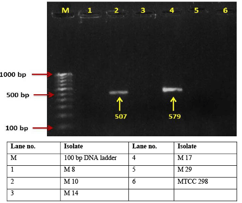
| Functional genes unique to the physiology and metabolism of organisms of interest have been explored in molecular studies. Functional genes for enzymes, such as methane monooxygenase (pMMO and sMMO; genes pmoA and mmoX, respectively) and methanol dehydrogenase (MDH; gene mxaF), involved in the methane oxidation pathway, have been used for detection of methanotrophs. Gene probes that target functional genes have been developed for the pmoA gene [4] coding for the α-subunit of the pMMO and the mxaF gene [5,6] coding for the α-subunit of the MDH present in all methylotrophs [7,8]. In present investigation all the isolates showed either presence of mmoX gene encoding the α subunit (mmoX) of the hydroxylase component of the sMMO cluster or pmoA gene encoding α-subunit of particulate methane monooxygenase cluster which are the two forms of methane mono oxygenase enzyme responsible for methane degradation. Among all the tested isolates M 8, M 10, M 14 and M 17 showed ~ 518 bp, 536 bp, 518 bp and 527 bp respectively, indicating these isolates may possess pMMO enzyme responsible for capacity of methylotrophs to utilize methane (Plate 3). Among the five tested isolates, M 10, M 17, M 29 and M. extrorquens strain gave single band of ~ 870 bp indicating these isolates may also have capacity to utilize methane (Plate 2) using soluble methane monooxygenase enzyme. Overall, from the above results it was clear that isolate M 10 and M 17 possess both sMMO and pMMO genes indicating they are better cultures for methane degradation. The soluble MMO is not universal to all methanotrophs and is found predominantly in the genera Methylosinus and Methylococcus [9]. pmoA gene encoding one of the subunits of the particulate MMO, has also been examined recently [10]. This gene has been found in all methanotrophs studied and reported so far. The use of a second functional gene probe, mxaF, which is found in all methanotrophs including gram negative strains was recommended to confirm methylotrophic nature of isolates. The advantage of using the mxaF gene is that it can indicate larger group of organisms which assimilate C1 compounds for their role in environment. In present study isolate M 10 and M 17 showed single band of ~507 and ~579 bp size indicating presence of mxaF gene (Plate 4). Similarly [3] detected methanotrophic diversity on roots of submerged paddy using primer sets A189/A682 encoding pMMO, 534f/1393r encoding sMMO and f1003/ r1561 encoding methanol dehydrogenase yielding PCR products of 550 bp, 863 bp and 525 bp size respectively. |
| Quantitative estimation of methane mono oxygenase and methanol dehydrogenase activities |
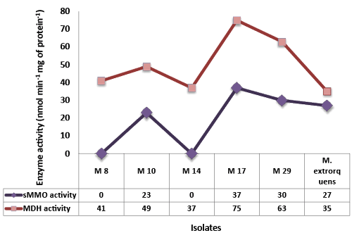
| To confirm methanotrophic nature of isolates detection of enzymatic activity of highly conserved key enzymes viz. particulate methane monooxygenase (pMMO), soluble methane monooxygenase (sMMO) and methanol dehydrogenase (MDH), may offer the possibility of detecting known methanotrophs (Hanson and Hanson, 1996). The ability of methanotrophs to oxidize methane is due to the possession of the enzyme called methane monooxygenase (MMO). This enzyme oxidizes methane to methanol. The MMO enzyme is the subject of extensive biochemical and molecular research. There are two distinct forms of this enzyme, a membrane-bound particulate methane monooxygenase (pMMO) and cytoplasmic soluble methane monooxygenase (sMMO) (Hanson and Hanson, 1996). The soluble methane monooxygenase activity of all the isolates ranged from 22.5 to 37.0 nmol min-1 mg of protein-1 (Figure 2). Among all the isolates, P. illinoisensis showed higher soluble methane monooxygenase activity (37.0 nmol min-1 mg of protein-1) in cell free extract followed by B. megaterium (30.0 nmol min-1 mg of protein-1). All the isolates were found positive for methanol dehydrogenase activity. Methanol dehydrogenase activity of all the isolates ranged from 35-75 nmol min-1 mg of protein-1 (Figure 2). Among all the isolates, P. illinoisensis showed the highest methanol dehydrogenase activity (75 nmol min-1 mg of protein-1) in cell free extract followed by B. megaterium (63 nmol min-1 mg of protein-1). These results confirm the methane degrading ability of the isolates obtained from newer method and thereby verify the efficiency of the method [11]. |
| Identification of isolates through 16S rDNA sequencing |
| From 16S rRNA partial gene sequence isolate M 8 was identified as Bacillus aerius with 100% similarity and 100% query coverage to B. aerius strain 24 K. Isolate M 10 was identified as Rhizobium sp. showing 96% identity with R. selenitireducens strain B1with 100% query coverage which confirms the isolate M 10 belongs to Rhizobium genus but the species was not confirmed. Isolate M 14 was also identified as B. subtilis with 99% similarity and 100% query coverage to B. subtilis strain DSM 10. The phylogenetic tree constructed showed one major clusters showing close similarity with B. subtilis. Isolate M 17 showed about 99% similarity and 100% query coverage with P. illinoisensis strain JCM 9907 indicating M 17 may be the native species of P. illinoisensis. Isolate M 29 showed about 99% similarity and 100% query coverage with B. megaterium QM B1551 [12]. The sequence analysis of partial 16S rRNA gene of isolate M 8, M 10, M 14, M 17 and M 29 have been deposited in NCBI, GeneBank under accession numbers KC787582, KC787583, KC855269, KC787584 and KC787585 respectively. The analysis named isolate M 8 as B. aerius AAU M 8, Isolate M 10 as Rhizobium sp. AAU M 10, isolate M14 as B. subtilis AAU M 14, isolate M 17 as P. illinoisensis AAU M 17 and M 29 as B. megaterium AAU M 29. |
| Conclusion |
| The method standardize in present investigation provides a less expensive and more rapid method for isolation of methane utilizing bacteria from environmental samples as compared to conventional method available for isolation of methane utilizing bacteria which are time consuming, less selective and expensive utilizing gas liquid chromatographic detection of methane utilization capacity of isolate. Among the isolates obtained in this study Rhizobium sp. and P. illinoisensis proved to be more efficient methane degraders followed by B. megaterium, B. aerius and B. subtilis. These novel indigenous methylotrophic PGPR based indicated wide scope and prospects as agriculturally beneficial bioinput to reduce methane emission from rice ecosystem. |
References
|
Figures at a glance
 |
 |
| Figure 1 | Figure 2 |
 |
 |
 |
 |
| Plate 1 | Plate 2 | Plate 3 | Plate 4 |
Relevant Topics
- Anaerobic Biodegradation
- Biodegradable Balloons
- Biodegradable Confetti
- Biodegradable Diapers
- Biodegradable Plastics
- Biodegradable Sunscreen
- Biodegradation
- Bioremediation Bacteria
- Bioremediation Oil Spills
- Bioremediation Plants
- Bioremediation Products
- Ex Situ Bioremediation
- Heavy Metal Bioremediation
- In Situ Bioremediation
- Mycoremediation
- Non Biodegradable
- Phytoremediation
- Sewage Water Treatment
- Soil Bioremediation
- Types of Upwelling
- Waste Degredation
- Xenobiotics
Recommended Journals
Article Tools
Article Usage
- Total views: 17627
- [From(publication date):
January-2016 - Apr 07, 2025] - Breakdown by view type
- HTML page views : 16432
- PDF downloads : 1195
