Radiological Imaging of Choroid Plexus Tumors Systematic Approach towards Diagnosing a Choroid Plexus Tumor
Received: 22-Jun-2021 / Accepted Date: 06-Jul-2021 / Published Date: 13-Jul-2021 DOI: 10.4172/2167-7964.1000334
Abstract
Choroid plexus tumors are rare intraventricular tumors. The common locations are the lateral ventricle in children and the fourth ventricle in adults. In adults, they are commonly located in and generally present before the age of 2 years. The main aim is to give a step by step approach with ruling out the differential diagnosis and reaching a diagnosis of a choroid plexus tumor. With the help of ultrasonography, computed tomography and magnetic resonance imaging sequences and ruling out the differentials in every step and reaching to a diagnosis of a choroid plexus tumor. Points to reliably differentiate a choroid plexus papilloma from a choroid plexus carcinoma on imaging are also highlighted through a systematic approach of a choroid plexus tumor.
Keywords: Choroid plexus papilloma; Intraventricular tumors; Systematic approach
Introduction
Choroid plexus tumors originate from the neuroectoderm of the choroid plexus cells that secrete cerebrospinal fluid. They account for approximately 1% of all brain tumors. The common locations are the lateral ventricle in children and the fourth ventricle in adults. In adults, they are commonly located in the infratentorial region while in children they are supratentorial tumors.
Objective
Our main aim is to give a step by step approach with ruling out the differential diagnosis and reaching a diagnosis of a choroid plexus tumor.
Case report
A 10 month’s old child reported to the pediatrics department with complains of fever and abnormal eye movements. No abnormal body movement was there. The patient was conscious but irritable. The child gave a history of head trauma 17 days back. There was no history of loss of consciousness. Fundus examination of the child revealed bilateral papilledema. Other investigations of the child were unremarkable. The child was advised ultrasound of the cranium. Proper informed consent was taken from the child’s father before every investigation.
Ultrasound of the cranium (Figure 1) revealed a hyper echoic well defined lesion adjacent the trigon and occipital horn of the right lateral ventricle. There was no mass effect. No vascularity could be appreciated on color Doppler evaluation. Differential diagnosis of a periventricular hyper echoic lesion on cranium ultrasonography are hematoma in the germinal matrix (perinatal period only), mass lesion, periventricular leukomalacia. Since, the patient gave a history of fall, the differentials given were a Hematoma and an intraventricular mass lesion the child was advised further investigations. Figure 1
Figure 1: Ultra sound images (a) A hyper echonice lesion (arrow) noted adjacent the occipital horn of right lateral ventricle glossy dilated lateral ventricle (b). Ratio of the diameter of lateral ventricle with the diameter of the brain <60% suggestive of hydrocephalus (c) no vascularity noted on colour Doppler few echogenic foci noted in the lesion
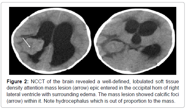
Non contrast computed tomography of the brain was done (NCCT) (Figure 2). Computed tomography of the brain revealed a well-defined lobulated mass lesion epic entered at the choroid plexus of the right lateral ventricle which was hyper dense relative to the brain parenchyma. The diagnosis of an intraventricular mass was confirmed. In view of the specific location and hyper dense mass, a differential diagnosis of intraventricular meningioma and a choroid plexus tumor was given. Figure 2
Figure 2: NCCT of the brain revealed a well-defined, lobulated soft tissue density attention mass lesion (arrow) epic entered in the occipital horn of right lateral ventricle with surrounding edema. The mass lesion showed calcific foci (arrow) within it. Note hydrocephalus which is out of proportion to the mass.
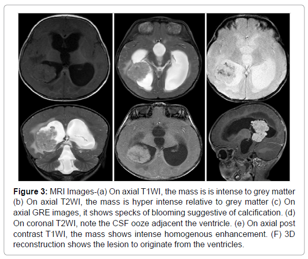
MRI showed a lobulated solid mass lesion originating from the choroid plexus of occipital horn of right lateral ventricle which was is intense to grey matter on T 1WI and hyper intense on T 2/FLAIR images. It’s showed specks of blooming on GRE sequences and intense post contrast enhancement the margins of the mass were well defined and showed frond like appearance. Marked hydrocephalus was seen in bilateral lateral ventricle, third ventricle and fourth ventricle with adjacent periventricular ooze which was more around the trigon of right lateral ventricle. The diagnosis given was a choroid plexus papilloma with a choroid plexus carcinoma as a differential. Figure 3
Figure 3: MRI Images-(a) On axial T1WI, the mass is is intense to grey matter (b) On axial T2WI, the mass is hyper intense relative to grey matter (c) On axial GRE images, it shows specks of blooming suggestive of calcification. (d) On coronal T2WI, note the CSF ooze adjacent the ventricle. (e) On axial post contrast T1WI, the mass shows intense homogenous enhancement. (F) 3D reconstruction shows the lesion to originate from the ventricles.
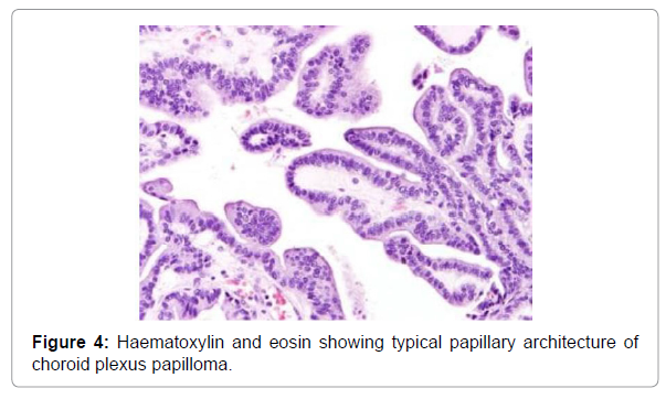
MRI features which suggest that this is a choroid plexus papilloma are a solid tumor with no necrotic area within, multiple flow voids are seen, margins are well defined and show a frond like morphology and contrast enhanced MRI showed homogenous enhancement. The child underwent surgery and histopath revealed a typical papillary architecture on hematoxylin and eosin Figure 4.
Discussion
Differential diagnosis of an intraventricular lesion is central neurocytoma, intraventricular meningioma, choroid plexus carcinoma [1]. Central neurocytoma originate from the septum pellucidum, shows mild to moderate heterogeneous contrast enhancement with numerous bubbly cystic areas and has larger flow voids [2]. Intraventricular meningioma is well circumscribed and show homogenous contrast enhancement and are common in adults [3]. Choroid plexus carcinoma has irregular margins with intense post contrast enhancement. They show parenchymal invasion and moderate vasogenic edema. According to the latest WHO classification system, CPTs can be classified into 3 subtypes: choroid plexus papilloma (CPP grade I), atypical choroid plexus papilloma (aCPP grade II) and choroid plexus carcinoma [4]. CPTs show calcification in about 30% of cases and show intense post contrast enhancement. Most of the choroid plexus tumors are benign (choroid plexus papilloma) and have an indolent progression. However, in children less than 2 years of age, choroid plexus carcinoma is also common [5]. Larger tumor volume, irregular internal morphology, presence of necrosis, marked peritumoral edema, heterogeneous contrast enhancement; invasion into surrounding brain parenchyma could be signs of a choroid plexus carcinoma. However, these cannot be differentiated on imaging alone. [6].
Conclusion
Since, choroid plexus tumors are rare tumors, a systematic approach is needed to reach to a diagnosis of a choroid plexus tumor. Clinically correlating the history with characteristic findings of hydrocephalus out of proportion to the size of lesion, calcifications and intense post contrast enhancement in a mass lesion epic entered in the ventricles in a child can lead us to the diagnosis of a choroid plexus neoplasm. Differentiation between a choroid plexus papilloma and carcinoma can be made based on the enhancement pattern, volume of tumor, involvement of brain parenchyma and presence of necrosis reliably on imaging alone in many cases.
References
- Safaee M, Oh MC, Bloch O, Sun MZ, Kaur G, et al. (2013) Choroid plexus papilloma’s advances in molecular biology and understanding of tumor genesis. Neuro-Oncology 15:255-267.
- Louis DN, Perry A, Reifenberger G, von Deimling A, Figarella-Branger D, et al. (2016)The World Health Organization classification of tumors of the central nervous system: a summary. Acta Neuropathol, 131:803–820
- Dhillon RS, Wang YY, McKelvie PA, O’Brien B. (2013) Progression of choroid plexus papilloma. J Clin Neuro sci 20:1775–1778.
- Osborn AG, Salzman KL, Thurnher MM, Rees JH, Castillo M. (2012) The New World health organization classification of central nervous system tumors: what can the Neuroradiologist really say? AJNR Am J Neuroradiol 33:795–802
- Koeller KK, Sandberg GD. (2002) from the archives of the AFIP cerebral intraventricular neoplasms: radiologic-pathologic correlation. Radiographic 22:1473–1505.
- Sethi D, Arora R, Garg K, Tanwar P. (2017)Â Choroid plexus papilloma. Asian J Neurosurg 12 : 139-141.
Citation: Batra C (2021) Radiological Imaging of Choroid Plexus Tumors Systematic Approach towards Diagnosing a Choroid Plexus Tumor. OMICS J Radiol 10: 334. DOI: 10.4172/2167-7964.1000334
Copyright: © 2021 Batra C. This is an open-access article distributed under the terms of the Creative Commons Attribution License, which permits unrestricted use, distribution, and reproduction in any medium, provided the original author and source are credited.
Share This Article
Open Access Journals
Article Tools
Article Usage
- Total views: 2569
- [From(publication date): 0-2021 - Mar 29, 2025]
- Breakdown by view type
- HTML page views: 1882
- PDF downloads: 687


