Research Article Open Access
Radiological Features of the Brain and Spinal Cord Gliomas on Computed Tomography and Magnetic Resonance Imaging
| Sultan Alshoabi1,2, Moawia Gameraddin1* and JumaaTamboul1 | |
| 1Department of Diagnostic Radiologic Technology, Faculty of Applied Medical Sciences, Taibah University, Almadinah Almunawarah, Saudi Arabia | |
| 2Al-Thawra Modern General Hospital (TMGH), Republic of Yemen | |
| Corresponding Author : | Moawia Gameraddin Department of Diagnostic Radiologic Technology Faculty of Applied Medical Sciences Taibah University, Almadinah Almunawarah Saudi Arabia Tel: + 00966534821130 E-mail: m.bushra@yahoo.com |
| Received: November 06, 2015 Accepted: December 23, 2015 Published: December 28, 2015 | |
| Citation: Alshoabi S, Gameraddin M, Tamboul J (2015) Radiological Features of the Brain and Spinal Cord Gliomas on Computed Tomography and Magnetic Resonance Imaging. OMICS J Radiol 4:211. doi:10.4172/2167-7964.1000211 | |
| Copyright: © 2015 Alshoabi S, et al. This is an open-access article distributed under the terms of the Creative Commons Attribution License, which permits unrestricted use, distribution, and reproduction in any medium, provided the original author and source are credited. | |
Visit for more related articles at Journal of Radiology
Abstract
Aim: The aim of this study is to characterize the site, types and radiological features of gliomas, such as edema, mass effect, and type of enhancement on CT-scan and MRI imaging.
Patients and methods: Thirty-three patients were studied retrospectively from the PACS system of multi-slice CT-scanner and MRI using the protocol of imaging of the head and spine.
Results: Gliomas involved male patients more than females (60.1% vs. 39.9%). About 96.9% of the gliomas were in the brain and only 3.1% was in the cervical spine. Nearly 51.5% of the gliomas showed radiographic features of low-grade gliomas and 48.5% showed radiographic features of high-grade gliomas. Approximately 30% of gliomas were located at the parietal lobe of the brain. Nearly 45.5% of the gliomas showed heterogenous enhancement, 36.4% marginal enhancement and 5.1% had no enhancement after contrast administration.
Conclusion: Gliomas were more common in male than female and high grade in elderly while low-grade gliomas in children. Gliomas were mostly involving the parietal lobes. The majority of gliomas showed heterogeneous enhancement after contrast administration. Magnetic resonance imaging is the most effective imaging method to characterize gliomas.
|
Abstract
Aim: The aim of this study is to characterize the site, types and radiological features of gliomas, such as edema, mass effect, and type of enhancement on CT-scan and MRI imaging.
Patients and methods: Thirty-three patients were studied retrospectively from the PACS system of multi-slice CT-scanner and MRI using the protocol of imaging of the head and spine. Results: Gliomas involved male patients more than females (60.1% vs. 39.9%). About 96.9% of the gliomas were in the brain and only 3.1% was in the cervical spine. Nearly 51.5% of the gliomas showed radiographic features of low-grade gliomas and 48.5% showed radiographic features of high-grade gliomas. Approximately 30% of gliomas were located at the parietal lobe of the brain. Nearly 45.5% of the gliomas showed heterogenous enhancement, 36.4% marginal enhancement and 5.1% had no enhancement after contrast administration. Conclusion: Gliomas were more common in male than female and high grade in elderly while low-grade gliomas in children. Gliomas were mostly involving the parietal lobes. The majority of gliomas showed heterogeneous enhancement after contrast administration. Magnetic resonance imaging is the most effective imaging method to characterize gliomas. Keywords
Radiological features; Gliomas; Brain; Spine; Computed tomography
Introduction
Glioma is the most common primary brain neoplasm that arises from the glial cells that surround the nerve cells of the brain and spinal cord to keep them in place and support their function. Gliomas show a wide diversity with respe ct to their site and radiological features. The glioma can be classified according to the type of the glial cells they arise from into different types such as astrocytoma that arises from astrocytes, ependymoma that arises from the ependymal cells that lines the ventricular system, oligodendroglioma that arises from oligodendrocytes and mixed glioma that arises from a mixture of these cell types [1].
Glioma also categorized by World Health Organization (WHO) grading system into: Low-grade gliomas [WHO grade I-II] are well-differentiated neoplasms and tend to exhibit benign tendencies and have a better prognosis however, they have a significant rate of recurrence and tendency to increase in grade overtime while High-grade gliomas [WHO grade III-IV] are undifferentiated malignant neoplasms with the worst prognosis [2]. Glioblastoma [WHO grade-IV] is the most frequent type of glioma that equals approximately 45% of all types of glioma, and the most malignant glioma that has a five years relative survival of only 5%. Glioblastoma may develop rapidly after a short clinical history and without evidence of a less malignant precursor lesion (primary glioblastoma) or slowly through progression from realy present low-grade, diffuse or anaplastic astrocytoma (secondary glioblastoma) [2,3]. Diffuse astrocytomas (WHO grade II) are slowly growing, well-differentiated tumors, but they diffusely infiltrate into the surrounding brain tissues and have an intrinsic tendency to progress to anaplastic astrocytoma (WHO grade III) and eventually to secondary glioblastoma (WHO grade IV). A small portion of these tumors are caused by Mendelian disorders, including neurofibromatosis and tuberous sclerosis [2,3]. Juvenile Pilocytic Astrocytoma is the most common pediatric central nervous system glial neoplasm and the most common pediatric cerebellar neoplasm. This neoplasm has a benign biologic behaviour that translates into an extremely high survival rate of 10 years in approximately 94% of cases that is really the best survival rate of any glial cells brain neoplasm. The patients of pilocytic astrocytoma usually present in the first 2 decades of life with manifestations directly related to the specific site of the neoplasm. The most common sites of pliocytic astrocytoma include the cerebellum, the optic nerve and the hypothalamic region and the cerebral hemispheres but this neoplasm can also arise in the ventricles and in the cervical spinal cord [4]. Diffuse pontine gliomas are a group of malignant neoplasms with a very poor prognosis that is poorer than the focal brain stem gliomas and they account for approximately 15% of pediatric brain neoplasms and approximately 20-30% of all of the posterior fossa neoplasms. Diffuse pontine gliomas are differentiated by WHO as grade-II (Fibrillary astrocytomas) or grade-III (Anaplastic astrocytoma) [5]. Rare types of glioma such as optic nerve glioma and spinal cord glioma mainly affect children and the spinal cord gliomas usually arise in the cervical part of the spinal cord. The exact presentation of the gliomas will vary according to the site and size of the neoplasm but general manifestations as signs if increased intracranial pressure, convulsions and hydrocephalus may occur in any neoplasm. A combination of ataxia, cranial nerve palsies and long tract signs usually occurs in the posterior fossa gliomas with different duration of symptoms that is short in diffuse glioma [6]. Patients and Methods
Study population
This is a retrospective study conducted during the period of 2008 and 2012 in Al-Thawrah Modern General Hospital (TMGH), Sana'a. In this study, there were 33 cases of gliomas of the brain and spine had been selected to satisfy this study of glioma.
Imaging procedures: Patients were imaged with either one/two imaging modalities, CT scan and MRI, 18 patients of this sample were scanned using a 64 multi-slice CT scan using the protocol of the brain imaging in which the patient was scanned in supine position with the head of the patient is cantered and fixed in the gantry of CT-scan machine. Brain scan was done under selected exposure factors with slice thickness of 8 mm and intravenous low-osmolar water soluble contrast media (LOCM) was injected to the patients. Interpretation of the brain scan was performed by consultant Radiologists to confirm the diagnosis. The remaining 15 patients of this study were imaged with 1.5 tesla magnetic resonance imaging (MRI) by using the protocol of imaging of the head and spine by MRI in which the following parameters of imaging were selected; 10 mm slice thickness of the brain, T1,T1 with contrast, T2 and FLAIR weighted images were done and the contrast media that used is intravenous Gadolinium (0.1 ml/Kg) according to the weight of the patient. Statistical analysis
The collected data in the study was analyzed by using the SPSS software program. The study contains qualitative and quantitative variables. Chi-square test was used to find association of age groups and existence of edema with the types of gliomas. The quantitative data were described using numbers, percentages and tables.
Results
This study included 33 patients that 20 patients (60.1%) were males while 13 patients (39.9%) were females and their ages ranged between 5 to 72 years old. They had been referred for CT-scan or MRI examination of the brain or spine. Gliomas were higher in males (20 patients) than in females (13 patients) (Figures 1 and 2).
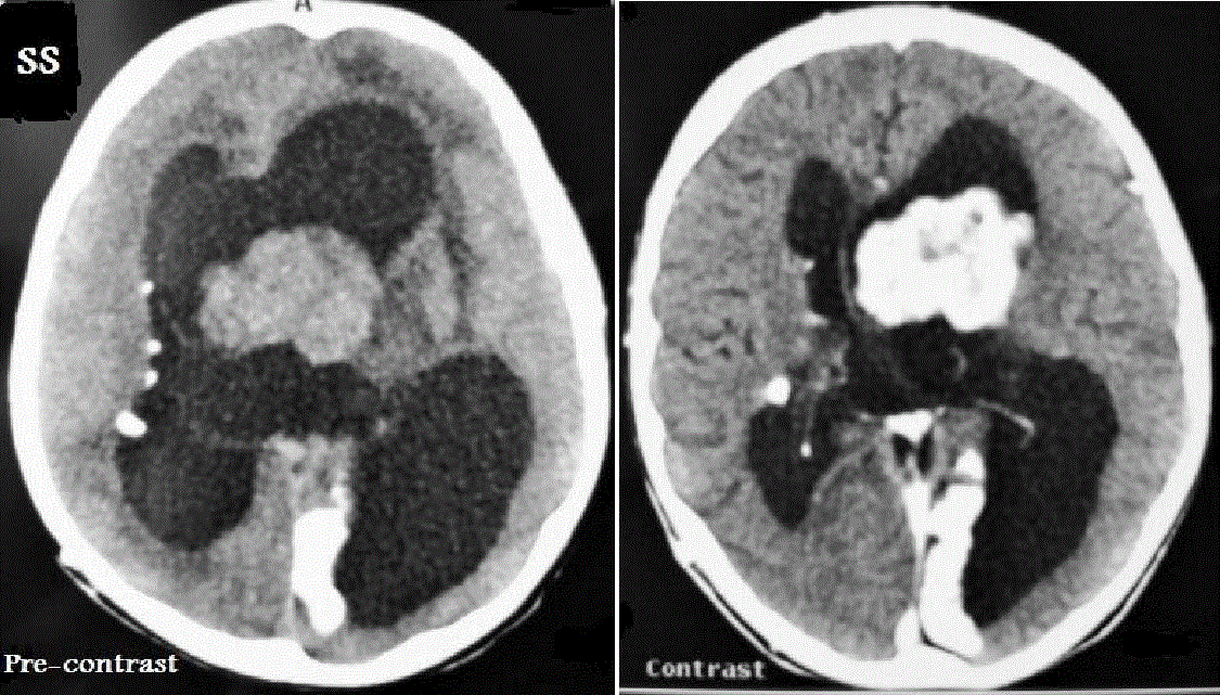
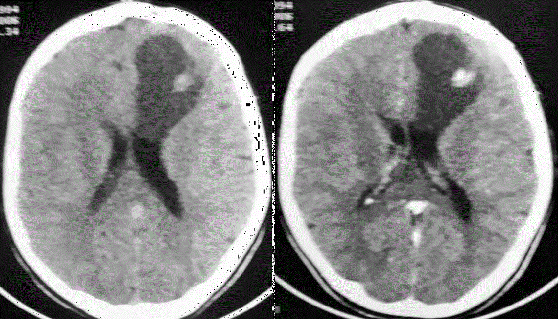
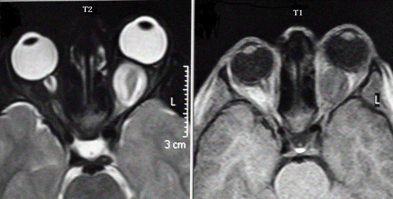
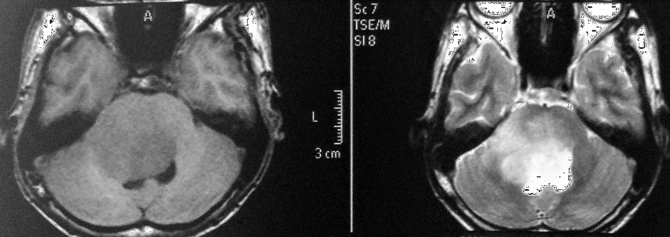
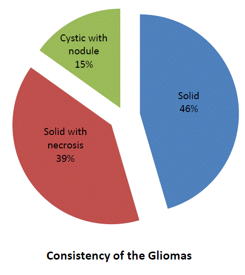
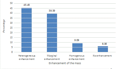
Most cases of the gliomas were in the brain (32 cases) and only one was in the cervical spine. About 20 cases (60%) of the gliomas in this study were large (more than 4 × 4 cm) and 13 cases (40%) were small in size (Figures 3 and 4). About 31 cases of the gliomas (93%) in this study were focal gliomas whereas only 1 case (3%) was multifocal glioma and 1 case (3%) was diffuse glioma. About 17 cases (53%) of the gliomas showed radiographic features of low-grade glioma and 16 cases (47%) showed radiographic features of high-grade glioma (Table 1). Exactly 7 cases of high grade glioma were in old age in the reverse of those showed features of low grade glioma 9 cases were in children. Nine cases of gliomas in this study were found in the parietal lobe (27%), Four cases in the frontal lobe (12%), Four cases in the occipital lobe (12%), Three cases in the temporoparietal lobe (9%), Three cases in the pons (9%), Three cases in the thalamus (9%), Three cases in the cerebellum (9%), Two cases in the optic nerve (6%), One case in the lateral ventricle (3%) and only one case in the cervical spine(3%) (Table 3). Calcification was found only in 2 cases of the gliomas (6%). Nearly 15 cases (45%) of gliomas were solid masses, 13 cases (39%) were solid masses with necrotic areas inside them and 5 cases (15%) were cystic lesions with solid parts (Figure 5). Exactly 15 cases (45%) of the gliomas in this study showed heterogeneous enhancement and 13 cases (39%) showed marginal enhancement after contrast administration whereas 3 cases (9%) showed homogenous enhancement and only 2 cases (6%) showed no enhancement after contrast administration (Figure 6). Approximately 19 cases (59%) of the gliomas showed edema around them that was severe edema in 5 cases (15%), moderate in 7 cases (22%) and mild in 7 cases (22%) while 14 cases (43%) showed no edema around them. All cases of the gliomas in this study showed mass effect on the surrounding soft tissues as the following; 15 case (45%) showed grade-III mass effect that means effacement of the cortical sulci with compression of the ventricles and shifting of the cerebral midline, 14 case (32%) showed grade-II mass effect that means effacement of the cortical sulci and compression of the lateral ventricles and only 4 cases (12%) showed grade-I mass effect that means effacement of the sulci. Discussion
In this retrospective study, the gliomas were more common in males than in females (20 versus 13). These findings are similar to the findings of Laigle-Donadey F et al., who reported that the incidence of brain neoplasms was higher in men than in women (7.6 versus 5.3 per 100,000 person-years) and the lifetime risk of developing a brain tumor is 0.65%in men and 0.5% in women [6].
The anatomic site of a glioma in the brain influences its prognosis and treatment options. Most of the cases of the glioma in this study were involving the parietal lobe, temporoparietal, front parietal or in the occipitoparietal. These findings were differ than the findings of Suvi Larjavaara, et al., who reported that the gliomas were located in the frontal lobe in 40% of the cases, temporal in 29%, parietal in 14%, and occipital lobe in 3%, with 14% in the deeper structures [7]. In this study, there were three cases of pilocytic astrocytoma in 2, 9 and 16 years children and all of these pilocytic astrocytomas were cystic with mural nodules that showed strong enhancement of the mural nodules after contrast administration, these findings are similar with David Sutton, et al., who reported that pilocytic astrocytoma usually present in childhood between 5-15 years old and they present as a cystic lesion in 70% of cases with enhancing mural nodule after contrast administration [8]. In this study, there was only one case of Sub ependymal giant-cell astrocytoma located in the left ventricle compressing the foramen of Monro causing obstructive hydrocephalus with multiple sub ependymal calcified nodules in a male child of 12 years old with tuberous sclerosis(TS), these findings are consistent with David Sutton, et al., who reported that 90% of cases of Giant cell astrocytoma’s are located in the foramen of Monro and usually protrude into the lateral ventricle and usually associated with tuberous sclerosis [9]. In this study, approximately 45% of all cases of the study (14 cases) were Glioblastoma multiform those were distributed between 34 and 72 years old age patients and were consisted of solid lesions with necrotic areas and no calcification and showed heterogenous after contrast administration, all of these cases were focal lesions except one that was multicentric glioblastoma multiforme, these findings are similar with David Sutton, et al., who reported that Glioblastoma usually occurs after 40 years old age as a focal solid lesions with necrotic areas and shows heterogenous enhancement after contrast administration and only 5% of glioblastoma occurs as multifocal lesion and rarely shows calcification [9]. Low-grade gliomas usually showed no edema while high-grade gliomas predominantly showed mild or moderate edema. This result is similar with Marc RJ Carlson, et al., who reported that the presence and grade of edema increase with increase the grade of glioma. The result of his study showed that in grade-III gliomas edema was grade 0 in 79%, grade 1 in 16% and grade 2 in only 5%whereasin the Glioblastoma edema (Grade-IV) was grade 0 in 23% of cases, grade 1 in 23% of cases and were grade 2 in 54% of cases [10]. Contrast enhancement occurred in both low-grade gliomas and high-grade gliomas that was heterogenous enhancement in 15 cases, marginal enhancement in 12 cases, homogenous enhancement in 4 cases and no enhancement in 2 cases. These findings were approximately agreed with Lote K et al., who reported that sensitivity and specificity of contrast enhancement is used as a test for high-grade glioma was 0.87 and 0.79, respectively. Enhancement was a strong negative prognostic factor comparable to high-grade histology in the total patient population as Parentheses reported that tumor contrast enhancement was present in 96% of glioblastoma, 87% in high-grade gliomas, 56.5% in anaplastic gliomas, and 21% in low-grade gliomas [10]. Conclusion
Gliomas are more common in males than females and high grade gliomas are more common in elderly while low grade gliomas are more common in children. Gliomas were mostly involving the parietal lobes. Focal glioma is the most common type of gliomas. Gliomas predominantly show grade-I or grade-II brain edema and grade-III mass effect is the most common. The majority of gliomas showed heterogeneous enhancement or marginal enhancement after contrast administration and rarely show homogenous or no enhancement. Magnetic resonance imaging is the most effective imaging method to characterize gliomas.
|
References
- Ostrom QT, Bauchet L, Davis FG, Deltour I, Fisher JL, et al. (2014) The epidemiology of glioma in adults: a "state of the science review". Neuro-Oncology 16: 896-913.
- https://en.wikipedia.org/wiki/Glioma
- Watanabe T, Nobusawa S, Kleihues P, Ohgaki H (2009) IDH1 mutations are early events in the development of astrocytomas and oligodendrogliomas. Am J Pathol174: 1149-1153.
- Koeller KK, Rushing EJ (2004) From the archives of the AFIP: pilocytic astrocytoma: radiologic-pathologic correlation. Radiographics 24: 1693-708.
- Helton KJ, Phillips NS, Khan RB, Boop FA, Sanford RA, et al. (2006) Diffusion tensor imaging of tract involvement in children with pontinetumors. American journal of neuroradiology 27: 786-793.
- Laigle-Donadey F, Doz F, Delattre JY (2008) Brainstem gliomas in children and adults. CurrOpinOncol 20: 662-667.
- Larjavaara S, Mäntylä R, Salminen T, Haapasalo H, Raitanen J, et al. (2007) Incidence of gliomas by anatomic location. Neuro-oncology 9: 319-325.
- Sutton D, Robinson PJA, Jenkins JPR, Whitehouse RW, Allan PL, Wilde P, et al. (2003) Textbook of Radiology and Imaging. (7th edition) Elsevier Science Ltd, UK, pp. 1739-1744.
- Carlson MR, Pope WB, Horvath S, Braunstein JG, Nghiemphu P, et al. (2007) Relationship between Survival and Edema in Malignant Gliomas: Role of Vascular Endothelial Growth Factor and Neuronal Pentraxin 2. Clin Cancer Res 13: 2592-2598.
- Lote K, Egeland T, Hager B, Stenwig B, Skullerud K, et al. (1997) Survival, prognostic factors, and therapeutic efficacy in low-grade glioma: a retrospective study in 379 patients. J ClinOncol 15: 3129-3140.
Tables and Figures at a glance
| Table 1 | Table 2 | Table 3 |
Figures at a glance
 |
 |
 |
| Figure 1 | Figure 2 | Figure 3 |
 |
 |
 |
| Figure 4 | Figure 5 | Figure 6 |
Relevant Topics
- Abdominal Radiology
- AI in Radiology
- Breast Imaging
- Cardiovascular Radiology
- Chest Radiology
- Clinical Radiology
- CT Imaging
- Diagnostic Radiology
- Emergency Radiology
- Fluoroscopy Radiology
- General Radiology
- Genitourinary Radiology
- Interventional Radiology Techniques
- Mammography
- Minimal Invasive surgery
- Musculoskeletal Radiology
- Neuroradiology
- Neuroradiology Advances
- Oral and Maxillofacial Radiology
- Radiography
- Radiology Imaging
- Surgical Radiology
- Tele Radiology
- Therapeutic Radiology
Recommended Journals
Article Tools
Article Usage
- Total views: 11755
- [From(publication date):
December-2015 - Mar 29, 2025] - Breakdown by view type
- HTML page views : 10769
- PDF downloads : 986
