Research Article Open Access
Quantitative and Morphological Evaluation of Cancellous and Cortical Bone of the Mandible by CT
| Mohammad A Momin1, Tohru Kurabayashi2, Takashi Yosue1 | |
| 1The Nippon Dental University, School of Life Dentistry, Department of Oral and Maxillofacial Radiology, Tokyo, Japan | |
| 2Tokyo Medical and Dental University, School of dentistry, Department of Oral and Maxillofacial Radiology, Tokyo, Japan | |
| Corresponding Author : | Mohammad Abdul Momin Oral and Maxillofacial Radiology The Nippon Dental University 1-9-20, Fujimi, Chiyoda-ku, Tokyo 102-8159, Japan Tel: + 81-3-3261-6521 Fax: +81-3-3261-6516 E-mail: drmomin.orad@hotmail.com |
| Received November 19, 2013; Accepted December 09, 2013; Published December 19, 2013 | |
| Citation: Momin MA, Kurabayashi T, Yosue T (2013) Quantitative and Morphological Evaluation of Cancellous and Cortical Bone of the Mandible by CT. OMICS J Radiology 3:155. doi: 10.4172/2167-7964.1000155 | |
| Copyright: © 2013 Momin MA, et al. This is an open-access article distributed under the terms of the Creative Commons Attribution License, which permits unrestricted use, distribution, and reproduction in any medium, provided the original author and source are credited. | |
Visit for more related articles at Journal of Radiology
Abstract
The morphology and quantity of bone are essential for preoperative planning of implant placement in the mandible. The thickness of the cortical bone has a larger influence on the initial stabilization than the length of the implant fixture. Earlier studies reported that the cancellous (CAB) and cortical (COB) thickness is the key to successful implantation. Implant success rate might be affected by the CAB and COB thickness. The quantity of bone is an important factor for long-term maintenance of the stability of the bone-implant interface. In the present study, we quantitatively and precisely evaluated the CAB and COB thicknesses in five sections from 6 mm anterior to 18 mm posterior to the mental foramen. To our knowledge, this is the first computed tomography study to report on quantitative measurement of the CAB and COB thicknesses and morphology of the mandible in a wider area from anterior to posterior of the mental foramen. For implant installation, careful evaluation and full knowledge of the thicknesses of CAB and COB thickness, as well as the shape of a mandible, are necessary, providing anatomic guidelines to clinicians, which will be helpful for implant surgery.
| Keywords |
| CAB and COB thicknesses; CT Diagnosis; Mandibular Canal |
| Introduction |
| Determinations of morphology and quantity of bone are essential for preoperative planning of implant placement in the mandible [1]. Generally, the cortical bone thickness is important for achieving implant stability. To achieve a predictable clinical outcome, implant stability is necessary for osseointegration [2]. In addition, the thickness of the cortical bone has a larger influence on the initial stabilization than the length of the implant fixture. Earlier studies reported that the cancellous and cortical bone thickness is the key to successful implantation [3,4]. Implant success rate might be affected by the CAB and COB thickness. The quantity of bone is an important factor for long-term maintenance of the stability of the bone-implant interface [5-7]. |
| Bone quantity may settle on the implant insertion angle. Recently, advances have been made in bone grafting for sinus lift and flapless implant surgery. In any case, successful implant operations require precise information about bone size and morphology [8,9]. Implants placed in cortical bone were found to require greater removal torque and remained constant over time for cortically placed implants. The removal torque increased over time for an implant placed in cancellous bone. Adequate cortical engagement is necessary for nplacing dental implants [10]. |
| Various studies have measured precise mandible bone sizes on radiographs. Lindh et al. concluded that CT provides significantly more accurate values than panoramic radiography [11], and the correlation coefficients were 0.36 to 0.91 between the two modalities [12]. Panoramic radiography is used frequently for preoperative diagnosis, but this method exhibits image distortion and magnification, and it consequently provides inexact information [13]. Moreover, it is buccolingually impossible to measure the thickness of CAB and COB by panorama radiography or 2D radiography, but knowledge of CAB and COB thicknesses is essential for successful implant placement. |
| Conventional tomography is more accurate for measuring bone size, but the difficulty of adjusting tomographic objective planes may cause the measured values to be inaccurate [14]. Researchers have concluded that CT can measure the precise size and shape of bone with a multi-thin voluntary sectional image [15,16]. Quirynen et al. [17] and Tepper et al. [18] measured the size of the mandible as limited to the interforaminal region using CT. Because implant treatments are applied in the posterior regions more frequently, in our previous study, we investigated the standard bone size and morphology over a wider region using CT [19]. In the present study, we quantitatively and precisely evaluated the CAB and COB thicknesses in five sections from 6 mm anterior to 18 mm posterior to the mental foramen. A previous paper reported that buccal cortical bone thickness in the mandible was greatest at a site distal to the first molar [20], corresponding to section 4 in this study. We found the thickest cortical bone in section 1, which is 6 mm anterior to the mental foramen. It was speculated that the difference between the present data and those of Zhao et al. might be due to differences in measurement site [20]. |
| To our knowledge, this is the first computed tomography study to report on quantitative measurement of the CAB and COB thicknesses and morphology of the mandible in a wider area from anterior to posterior of the mental foramen. |
| Materials and Methods |
| Patients |
| CT data of the mandible of 79 Japanese patients (52 males and 27 females; age range, 19-77 years; mean age, 49 years) were used in this study. The patients underwent CT examination at Tokyo Medical and Dental University’s Dental Hospital from June 2005 through February 2006. The side of the mandible containing tumors or cysts was excluded from this examination. A total of 136 sides of mandibles were examined (Table 1). |
| Imaging |
| The CT examinations were performed with a Somatom plus S scanner (Siemens Medical Systems, Erlangen, Germany), which operated at 120 kV and 85-110 mA with a 1-mm slice thickness and a table speed of 2 mm/sec. After the examination, contiguous 1-mmthick axial CT images parallel to the inferior border of the mandibles were reconstructed. These images were examined using Dental CT® reformatting imaging software; with 2 mm intervals, cross-sectional CT images were obtained. These images were printed on film with a Fuji Dry imager (Fuji Film Medical Co. Ltd., Tokyo, Japan). |
| Procedure of measurement |
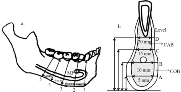
| The CAB and COB thicknesses of the mandibles were measured at the five sections as follows: the cross-sectional image in which the mental foramen was recognized was defined as section 2, and the image 6 mm anterior to section 2 was defined as section 1. These measurement sections are shown in Figure 1a. |
| In each section, the distances between the inner and outer borders of the buccal and lingual sides for cortical bone and from the inner border of the buccal side to the inner border of the lingual side for cancellous bone were measured. The measurement of the cancellous and cortical bone thicknesses from the alveolar crest was avoided because of atrophic changes due to periodontal disease [21] or tooth extraction, [22] and it is believed this exclusion enhances the reproducibility of the results [19]. |
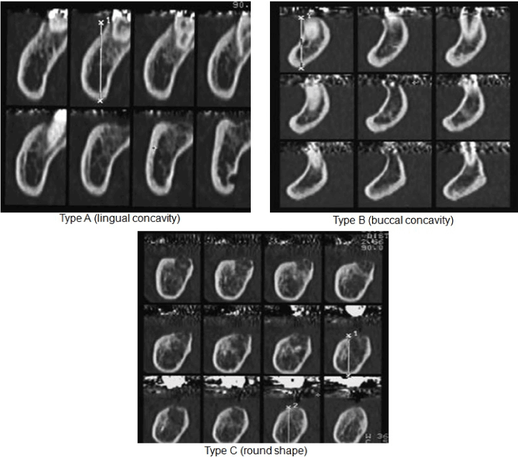
| The CAB and COB thicknesses A, B, C, and D were measured at 5, 10, 15, and 20 mm, respectively, above the inferior border of the mandible toward the alveolar crest, as shown in Figure 1b. There were three types of shape of the mandibles: type A (lingual concavity), type B (buccal concavity), and type C (round shape) [19]. To identify the location of the mandibular canal, the distance from the superior border of the canal to the alveolar crest (SAC) was also measured, and the distance from the inferior border of the canal to the bottom of the mandible (IBM) and the diameter of the canal (DOC) at 5 sections were measured. The measurements were performed by two radiologists and each point was measured three times using a sliding caliper (Nakamura Mfg., Tokyo, Japan). |
| Statistics |
| All data are presented as means and standard deviations (SD). The statistical differences were tested using Student’s t-test or chi-square test. A P value less than 0.05 indicated significance. |
| Results |
| Table 1 shows the distribution of patients according to age, sex, number of measurement sides, and CAB and COB thicknesses of the mandible. A total of 136 sides of mandibles, 680 heights, and 2720 CAB and 2720 COB thicknesses were measured. |
| CAB and COB thicknesses of the mandible |
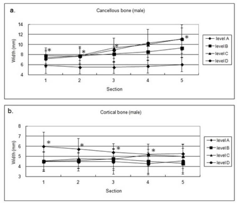
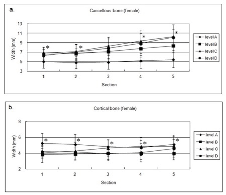
| The CAB and COB thicknesses of the mandibles are shown in Tables 2a,b, and 3a,b. The means and SD of the CAB and COB thicknesses ranged from 5.5 ± 1.3 mm to 11.1 ± 2.2 mm and 4.5 ± 1.0 mm to 5.3 ± 0.9 mm for males, respectively. For females, the CAB ranged from 4.8 ± 1.3 mm to 10.3 ± 1.5 mm and COB ranged from 3.8 ± 1.0 mm to 5.2 ± 1.2 mm. The CAB thickness was the greatest in sections 2 and 5, and the COB thickness was in sections 1 and 5 for both genders (Figure 2a,b). The CAB and COB thicknesses gradually increased from section 1 to section 5. The CAB and COB thicknesses in male patients were significantly greater than those in female patients (Figure 3a,b). |
| To investigate the correlation between patient age and the variability of the mandible CAB and COB thicknesses, coefficients of variation (CV) of these parameters were calculated. No tendency in age-related variability in thickness could be found. |
| Shape of the mandible |
| The shape of the mandible was classified into three types, as shown in Figure 4 (A, B, C) [23]. Lingual concavity, type B (62%-72%), was the most common in the anterior region for males, followed by type C (56%-58%) in the posterior region. For females, type C (25%-31%) in the posterior region and type B (18%-30%) were the most common types in the anterior region (Table 4a,b). |
| Location of the mandibular canal |
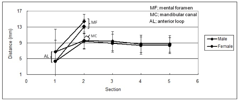
| The distances SAC, DOC, and IBM were measured in each section. The results are summarized in Table 5a,b. The SAC ranged from 10.8 ± 8.9 mm to 17.9 ± 2.8 mm for males and 6.2 ± 8.2 mm to 16.5 ± 3.5 mm for females. The SAC at the mental foramen were 13.3 ± 3.0 and 12.7 ± 3.3 mm for males and females, respectively (Figure 5). The inferior alveolar canal usually extends ahead of the mental foramen as the socalled anterior loop. In this study, the anterior loop was present at 57 sites (63%) for males and 18 sites (39%) for females, at a mean distance of 10.8 ± 8.9 mm for males and 6.2 ± 8.2 mm for females from the mental foramen (data not shown). The DOC ranged from 1.7 ± 1.4 mm to 2.9 ± 0.6 mm for males, and from 0.9 ± 1.2 mm to 3.0 ± 0.7 mm for females (Table 5). |
| Discussion |
| The objective of this study was to evaluate CAB and COB thicknesses and morphology using CT data for implant operation support. It is possible to estimate primary implant stability from CT diagnosis before surgery. Hence, various variables including CAB and COB, morphology of the mandible, and the location and diameter of the mandibular canal were measured on 688 CT cross-sectional images from 136 mandible sides. |
| Cancellous and cortical bone thickness |
| For a successful implant operation, the dental surgeon must consider not only the cortical bone thickness but also the cancellous bone thickness to ensure a good blood supply. The well-known implantologist Brånemark classified bone quality of class 2, thick cortical bone and a high density of cancellous bone, and class 3, thin cortical bone and a high density of cancellous bone, as being suitable for successful implant placement [4]. In the present study, in the anterior region (section 1), the CAB and COB thicknesses were almost the same for males (7.0 mm and 5.1 mm, respectively). For females, CAB thickness was 6.15 mm and COB thickness was 4.3 mm, but the thickness increased gradually through to the posterior region for both genders. The CAB and COB thicknesses peaked at 15 mm (11.1 ± 2.2 mm) and 5 mm (5.3 ± 0.9 mm) in the posterior region, above the inferior border of the mandible for males (level C and level A, respectively). For females, the CAB was greatest at level C (10.3 ± 1.5 mm) and the COB thickness (5.1 ± 1.2 mm) at level A. |
| The ratio of cortical to cancellous bone influences implant stability. Cortical bone has a high modulus of elasticity, is stronger and more resistant to deformation, and will bear more loads in clinical situations than cancellous bone [24,25]. Previous studies reported that there may be a strong relationship between cortical bone thickness and primary implant stability [3,25-28]. In the present study, the CAB and COB thicknesses increased gradually through the posterior region. On the other hand, above the mental foramen at levels C and D, the CAB thickness was slightly greater than that below the mental foramen. Furthermore, the COB thickness was wider below the foramen. This thickness measured from the inferior border of the mandible is more reliable than that measured from the superior border of the mandible. The measured CAB and COB thicknesses were significantly different between genders or sections. |
| Shape of the mandibular bone |
| The morphology of mandible bone has also been characterized and classified to facilitate predictable implant therapy. The cortical thickness of bone played a greater role in initial implant stability than the implant length [3]. A dental surgeon must know the relationship of CAB and COB thicknesses with the shape of the mandible to select the appropriate implant size for successful implantation. Cancellous bone was the most common (C level/15 mm above the inferior border of the mandible) in the posterior region, followed by cortical bone in the anterior region (B level/10 mm above the inferior border of the mandible) for both genders (Table 4). |
| The implant should be installed according to the shape of the mandible for preventing lingual or buccal strip perforation during drilling. Previous papers reported that a round shape was most frequently observed (58-74%), or that lingual concavity was observed (2.4-39%) [17,19]. In the present study, lingual concavity, type B (62- 72%), was the most common in the anterior region for males, followed by type C (56-58%) in the posterior region. As the buccal cortical bone (type B) has a higher risk of perforation, implant angulation should be toward the lingual side in such cases. For females, type C (25-31%) in the posterior region and type B (18-30%) were the most common types in the anterior region. The risk of perforation with type C is lower in males than that of females with the other types because of the sufficient lateral bone in the anterior and posterior regions. The risk of lingual cortical bone perforation is greater with type A. Therefore, the implant should be inserted accurately toward the buccal angle. |
| Location and diameter of the mandibular canal |
| Accurate determination of the exact location of the mandibular canal is imperative, to avoid interfering with the neurovascular bundle in the canal during implant placement [29,30]. The location of the mandibular canal is found by measuring the SAC distance using CT images, as shown in Figure 5. The SAC value was the greatest in section 2 (17.9 ± 2.8 mm and 16.5 ± 3.5 mm) and gradually decreased toward section 5 (15.7 ± 3.1 mm and 14.5 ± 3.1 mm for males and females, respectively). The SAC distance was greater in anterior and posterior regions for males (Figure 5). These data indicate that the SAC distance tends to decrease toward the posterior region in both genders. Previous studies reported that the superior aspect of the inferior alveolar canal was 16.0-17.4 mm from the alveolar crest in the area of the 1st molar, although they did not evaluate the size by gender [19,31]. Oral implants frequently perforate the canal to an unidentified extent in the interforaminal region [32]. Arzouman et al. reported that the inferior alveolar nerve bundle (IANB) may extend beyond the mental foramen as an anterior loop [33]. Solar et al. reported that a safe distance for implant installation is at least 6 mm anterior to the mental foramen [23]. Previous papers reported that, in 48-55% of the cases, an anterior loop was identified [19,34]. In this study, the anterior loop was observed in 63% and 39% of all sides in section 1 for males and females, respectively. The distances from the alveolar crest to the anterior loop were 10.8 ± 8.9 mm and 6.2 ± 8.2 mm, whereas the distances to the mental foramen were only 13.3 ± 3.0 mm and 12.7 ± 3.3 mm for males and females, respectively. Therefore, the surgeon should take care during implant insertion to avoid perforation of the canal in an anterior region in female patients as well as in males. Apostolakis et al. came to similar conclusions, finding the presence of an anterior loop and a safe distance of at least 6 mm (corresponding to our section 1) from the mental foramen (corresponding to section 2) [34]. |
| This finding indicates that, in the area around the mental foramen, the canal ascends step by step toward the mental foramen. Therefore, implant surgeons should take into account the high risk and inherent dangers of interfering with the IANB. The distances of DOC and IBM were greater in males than in females (Table 5a,b). Knowledge of the cancellous and cortical bone thicknesses in various areas can guide surgeons towards an appropriate implant installation protocol in the mandible. The purpose of this study was to evaluate the thicknesses of cancellous and cortical bone, and to identify and measure variation in the presence and extent of the anterior loop of the mandibular canal in the anterior and posterior regions to provide guidelines for implant operation support in the mandible. |
| Conclusion |
| For implant installation, careful evaluation and full knowledge of the thicknesses of cancellous and cortical bone, as well as the shape of a mandible, are necessary, providing anatomic guidelines to clinicians, which will be helpful for implant surgery. CAB thickness was from 5.5 to 11.1 mm and from 4.8 to 10.3 mm, and for COB, from 4.5 to 5.3 mm and from 3.8 to 5.2 mm, for males and females, respectively. Lingual concavity, type B (62%-72%), was the most common in the anterior region for males, followed by type C (56%-58%) in the posterior region. For females, type C (25%-31%) in the posterior region and type B (18%- 30%) in the anterior region were the most common types. The SAC was from 10.8 to 17.9 mm and 6.2 to 16.5 mm, and the DOC from 1.7 to 2.9 mm and from 0.9 to 3.0 mm for males and females, respectively. This study is the first to involve quantitative evaluation of the CAB and COB and the morphology of a mandible on the basis of cross-sectional CT images. |
References |
|
Tables and Figures at a glance
| Table 1 | Table 2 | Table 3 | Table 4 | Table 5 |
Figures at a glance
 |
 |
 |
 |
 |
| Figure 1 | Figure 2 | Figure 3 | Figure 4 | Figure 5 |
Relevant Topics
- Abdominal Radiology
- AI in Radiology
- Breast Imaging
- Cardiovascular Radiology
- Chest Radiology
- Clinical Radiology
- CT Imaging
- Diagnostic Radiology
- Emergency Radiology
- Fluoroscopy Radiology
- General Radiology
- Genitourinary Radiology
- Interventional Radiology Techniques
- Mammography
- Minimal Invasive surgery
- Musculoskeletal Radiology
- Neuroradiology
- Neuroradiology Advances
- Oral and Maxillofacial Radiology
- Radiography
- Radiology Imaging
- Surgical Radiology
- Tele Radiology
- Therapeutic Radiology
Recommended Journals
Article Tools
Article Usage
- Total views: 14874
- [From(publication date):
January-2014 - Jul 13, 2025] - Breakdown by view type
- HTML page views : 10208
- PDF downloads : 4666
