Make the best use of Scientific Research and information from our 700+ peer reviewed, Open Access Journals that operates with the help of 50,000+ Editorial Board Members and esteemed reviewers and 1000+ Scientific associations in Medical, Clinical, Pharmaceutical, Engineering, Technology and Management Fields.
Meet Inspiring Speakers and Experts at our 3000+ Global Conferenceseries Events with over 600+ Conferences, 1200+ Symposiums and 1200+ Workshops on Medical, Pharma, Engineering, Science, Technology and Business
Case Report Open Access
Pyrexia of Unknown Origin "Misleading First Impressions"
| Vijayan T* and Nagaraj Shetty I | |
| AJ Institute Of Medical Sciences Mangalore, Karnataka, India | |
| Corresponding Author : | Theertha Vijayan AJ Institute Of Medical Sciences Mangalore, Karnataka, India Tel: 8197589025 E-mail: theertha920@gmail.com |
| Received: June 21, 2015 Accepted: September 4, 2015 Published: September 10, 2015 | |
| Citation: Vijayan T, Shetty NI (2015) Pyrexia of Unknown Origin "Misleading First Impressions". J Infect Dis Ther 3:232. doi:10.4172/2332-0877.1000232 | |
| Copyright: © 2015 Vijayan T. This is an open-access article distributed under the terms of the Creative Commons Attribution License, which permits unrestricted use, distribution, and reproduction in any medium, provided the original author and source are credited. | |
| Related article at Pubmed, Scholar Google | |
Visit for more related articles at Journal of Infectious Diseases & Therapy
| Introduction |
| 48 year old foundry worker from Kumta district, Karnataka (India) presented with 3 weeks history of fever, body ache, cough, weight loss (10 kg) and anorexia. Recently detected to have type 2 DM. No significant illness in the past. No history of any recent travel outside the district. O/E Temp 101.F, PR 112/min, RR-22/min BP 100/70 mm Hg Toxic looking. No pallor, icterus, clubbing, lymphadenopathy, skin lesions. Sparse crepitations heard over right lung base. Mild hepatomegaly present. No focal neurological deficit. With the above findings provisional diagnosis of |
| • Pneumonia |
| • Tuberculosis |
| • Enteric fever |
| • Brucellosis was made. |
| Materials and Methods |
| Day-1 |
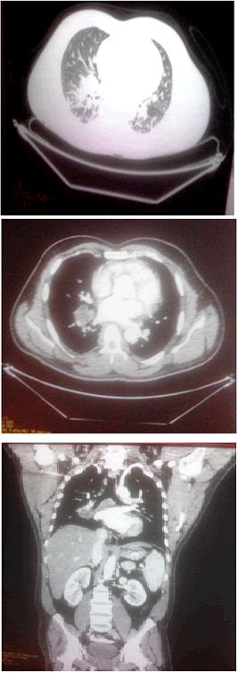
| TLC – 15,400/cu mm with neutrophilia, ESR – 140 mm/hr, RBS - 247 mg%, HIV, HbsAg, HCV status was negative. Tests for Dengue, Malaria, Typhoid, Leptospira, Brucella was negative. USG abdomen showed hepatomegaly and ECHO showed no vegetations.Blood and urine cultures were sent which were awaited, started on broad spectrum antibiotics in view of probable right lower lobe pneumonia (Figures 1 and 2). |
| Day-2 |
| Impression: tuberculosis/malignancy: Bronchoscopy showed only inflammatory cells, no endobronchial lesion, bronchial washings sent for gram stain, C&S, AFB. |
| Day-3 |
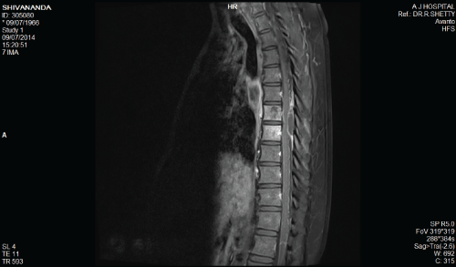
| He developed sudden onset of flaccid paraplegia with absent sensation below T8 level and urinary retention. Now the possibilties of acute myelitis, Spinal Abscess or Potts spine were considered (Figures 3 and 4). |
| Impression: epidural abscess |
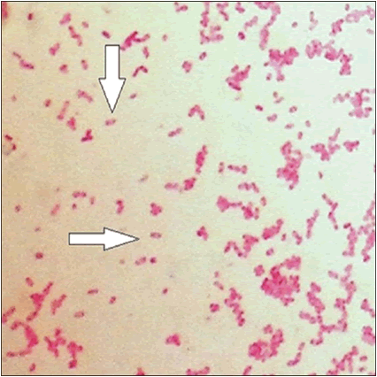
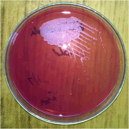
| He underwent an emergency T6-T10 laminectomy along with drainage of the epidural abscess, frank pus, sent for gram stain, C&S. Other tests: DNA PCR for Mycobacterium tuberculosis : -ve, Bronchial wash AFB : -ve. Meanwhile blood C&S, bronchial wash C&S and pus C&S were obtained (Figure 5). |
| The organism finally captured is Bukholderia pseudomallei . No other serology tests or immunological tests were done for confirmation as culture from blood, bronchial waasings and pus yeilded the organism. |
| Result |
| The was started on Inj Ceftazidime 2g iv Q6H for 2 weeks together with oral co-trimoxazole BD and Tab Doxy 100 mg BD given for six months. He gradually improved in motor power, sensations were regained. |
| Discussion |
| Melioidosis caused by Bukholderia pseudomallei , a widely distributed environmental saprophyte in soil and fresh surface water in endemic regions. Predominant modes of transmission are percutaneous inoculation and inhalation. Thailand has reported the largest number of cases,with an estimated 2000 to 3000 cases of melioidosis each year followed by Northern Territory of Australia, Increasingly seen in India and other Asian countries [1-3]. In India approximately 300-500 cases have been reported yearly. Central nervous system melioidosis is an unusual infection in humans. A similar case of thoracic epidural abscess was reported in Nizams Institute of Medical Sciences,Hyderabad, Andhra pradesh in 2011 [4]. |
| The retrospective melioidosis study at University Malaya Medical Centre has documented 3 cases of CNS melioidosis out of more than 160 cases of melioidosis since 1978. There were two patients with brain abscess and one with spinal epidural abscess [5]. Of 14 cases of spinal pyogenic infection reported by Nather et al. only one was caused by B. pseudomallei [6]. |
| Although healthy people may get melioidosis [7], the major risk factors are diabetes, excessive alcohol use, liver disease, chronic renal disease, chronic lung disease, urolithiasis, thalassemia, cancer or another immunosuppressing condition not related to human immunodeficiency virus (HIV) and occupational exposure. Morbidity and mortality of melioidosis are also higher in people with major risk factors. Isolates are generally susceptible to imipenem, piperacillin, amoxycillin-clavulanic acid, doxycycline, ceftazidime, aztreonam and chloramphenicol. These are found to be resistant to colistin and gentamicin. Chetchotisakd et al. [3] showed that combination of cefoperazone– sulbactam plus co-trimoxazole is an effective alternative to the use of ceftazidime and co-trimoxazole [8]. There is currently no effective vaccine against this disease. The usage of ceftazidime during the initial phase has been shown to reduce the mortality rate to almost half [9]. Generally, patients with melioidosis involving the CNS may have a good outcome if there is early diagnosis, early treatment, appropriate surgical intervention and 6 months of appropriate antibiotic therapy. If treated incorrectly, the mortality rate for melioidosis was 95%. |
| Conclusion |
| An increased awareness, high index of suspicion, early diagnosis and initiation of appropriate therapy is necessary for a favorable outcome. In all the reported cases, the abscess occurs in lower dorsal area, including in our case, reason for this needs evaluation. |
References
- White NJ (2003) Melioidosis. Lancet 361: 1715-1722.
- Cheng AC, Currie BJ (2005) Melioidosis: epidemiology, pathophysiology, and management. ClinMicrobiol Rev 18: 383-416.
- Wiersinga WJ, Currie BJ, Peacock SJ (2012) Melioidosis. N Engl J Med 367: 1035-1044.
- RavikanthVuppu, MudumbaVijayasaradhi, Barada Prasad Sahu (2011) A Thoracic Epidural Bukholderia Abscess- a case report, Pan Arab Journal.
- Muthusamy KA, Waran V, Puthucheary SD (2007) Spectra of central nervous system melioidosis. J ClinNeurosci 14: 1213-1215.
- Nather A, David V, Hee HT, Thambiah J (2005) Pyogenic vertebral osteomyelitis: a review of 14 cases. J OrthopSurg (Hong Kong) 13: 240-244.
- Lim MK, Tan EH, Soh CS, Chang TL (1997) Burkholderiapseudomallei infection in the Singapore Armed Forces from 1987 to 1994--an epidemiological review. Ann Acad Med Singapore 26: 13-17.
- Chetchotisakd P, Porramatikul S, Mootsikapun P, Anunnatsiri S, Thinkhamrop B (2001) Randomized, double-blind, controlled study of cefoperazone-sulbactam plus cotrimoxazole versus ceftazidime plus cotrimoxazole for the treatment of severe melioidosis. Clin Infect Dis 33: 29-34.
- Chaowagul W, Suputtamongkol Y, Dance DA, Rajchanuvong A, Pattara-arechachai J, et al. (1993) Relapse in melioidosis: incidence and risk factors. J Infect Dis 168: 1181-1185.
Figures at a glance
 |
 |
 |
 |
 |
| Figure 1 | Figure 2 | Figure 3 | Figure 4 | Figure 5 |
Post your comment
Relevant Topics
- Advanced Therapies
- Chicken Pox
- Ciprofloxacin
- Colon Infection
- Conjunctivitis
- Herpes Virus
- HIV and AIDS Research
- Human Papilloma Virus
- Infection
- Infection in Blood
- Infections Prevention
- Infectious Diseases in Children
- Influenza
- Liver Diseases
- Respiratory Tract Infections
- T Cell Lymphomatic Virus
- Treatment for Infectious Diseases
- Viral Encephalitis
- Yeast Infection
Recommended Journals
Article Tools
Article Usage
- Total views: 14506
- [From(publication date):
October-2015 - Jul 13, 2025] - Breakdown by view type
- HTML page views : 9881
- PDF downloads : 4625
