Pseudoangiomatous Stromal Hyperplasia (PASH) of the Breast
Received: 29-Dec-2017 / Accepted Date: 05-Feb-2018 / Published Date: 09-Feb-2018 DOI: 10.4172/2167-7964.1000291
Abstract
Pseudoangiomatous Stromal Hyperplasia (PASH) is one of the rare benign breast lesions. It typically occurs in women of reproductive age however it is reported in postmenopausal women under hormonal therapy. PASH is frequently incidental finding in breast biopsies but it may also manifest as palpable mass. We report a case of PASH which correlates with radiological and pathological findings. The knowledge of this rare lesion may be of importance to surgeons & radiologists for proper adequate management.
Keywords: Pseudoangiomatous stromal hyperplasia; PASH; Benign breast disease
Introduction
PASH of the breast is a benign myofibroblastic process, first described by Vuitchet et al. in 1986. The age of the diagnosis varies between 14 to 74 years. The size of PASH usually ranges between 0.6- 12 cm with most cases ranging from small to medium size [1]. The etiologic factors of PASH are unknown, but most investigators think that it represents a proliferative response of myofibroblasts, probably to hormonal stimuli [2]. Imaging features are not sufficiently specific to allow for a prospective diagnosis. Histological confirmation, preferably with core biopsy, should always be considered [3]. The following case report illustrates the radiographic abnormalities caused by this lesion.
Case Report
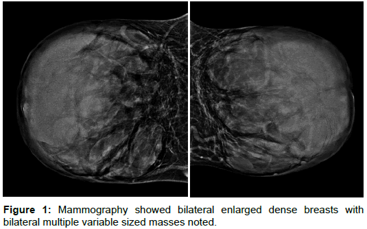
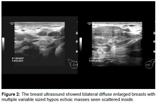
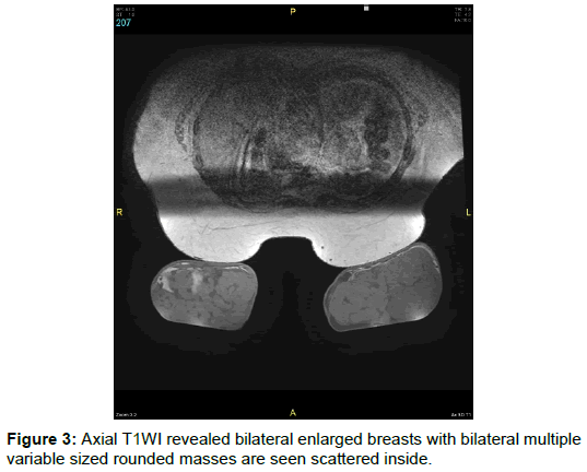
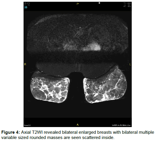
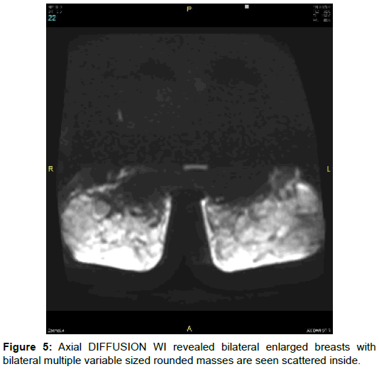
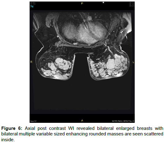
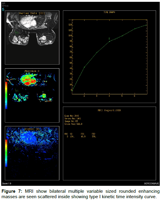
28 year-old female patient presented herself with enlarged both breasts. On clinical examination, the patient had large both breasts with bilateral palpable masses. The patient was initially examined with mammography which showed bilateral enlarged dense breasts with bilateral multiple variable sized masses however no speculation or calcification (Figure 1). Breast ultrasound was done with a high frequency (8-10 MHz) linear array head, which showed bilateral diffuse enlarged breasts with multiple variable sized hypoechoic masses seen scattered inside (Figure 2). The masses were well defined with smooth margin and posterior acoustic enhancement. No detected cystic degeneration. Magnetic Resonance Imaging (MRI) was performed with a 3 T MR-scanner and a dedicated breast coil. The examination protocol consisted of T1-weighted image (T1WI) (Figure 3), T2-weighted image (T2WI) (Figure 4), short-tau inversion recovery (STIR) and Diffusion weighted imaging were also obtained (Figure 5), as well as dynamic contrast-enhanced magnetic resonance imaging (DCE-MRI) after the administration of gadolinium (Figure 6). MRI show bilateral enlarged breasts with bilateral multiple variable sized rounded masses are seen scattered inside showing type I kinetic time intensity curve (Figure 7). Finally biopsy was taken and gross pathology revealed PASH.
Discussion
Pseudoangiomatous stromal hyperplasia (PASH) is an uncommon benign proliferative breast tumor composed of complex, anastomosing slit-like pseudovascular spaces, which are either acellular or lined by slender spindle-shaped stromal cells. Our case presents clinically as a bilateral multiple palpable breasts masses with the clinical and radiological assessment coping bilateral multiple fibroadenoma and also MRI findings cope with multiple bilateral lesions with benign criteria however the histo-pathological findings cope with PASH. Yoo et al. [4] had reported that clinical and radiological findings of PASH resemble those of fibrodenoma and all imaging modalities have no specific features to characterize PASH and distinguish it from other pathologic entities. In asymptomatic patients it usually presents as a breast imaging-reporting and data system (BI-RADS) type 3 lesion, suggesting a probably benign lesion [1]. In our case mammography revealed bilateral enlarged dense breasts with bilateral multiple variable sized masses noted and this agree with Solomou et al. [1] stated that say mammography of PASH can reveals single or multiple non-calcified masses, with well circumscribed margins, usually ranging from 1-10 cm, but it can also present as an asymmetric appearance of the gland whose size or density increases over time [1]. On our ultrasound they showed bilateral diffuse enlarged breasts with multiple variable sized hypo echoic masses seen scattered inside and this agree with Hargaden et al. [5] that say ,PASH has no characteristic appearance, including both its margins and echogenicity. It usually presents as a round or oval solid single or multiple masses, mainly hypoechoic and rarely hyperechoic with a central hypoechoic area. The presence of acoustic shadow is not very often [5]. On MRI images our case show bilateral enlarged breasts with bilateral multiple variable sized rounded masses are seen scattered inside showing type I kinetic time intensity curve and this agree with Mai et al. [6] that say nodular PASH has the same characteristics as a fibroadenoma, with rapid enhancement in the two minutes after contrast injection, followed by a slowly increasing enhancement during the rest of the examination. While diffuse PASH presents as a focal or segmental non-mass like clump of enhancement with a plateau or persistent phase [6]. PASH can be a relatively common incidental finding at breast biopsy. PASH radiologically mimics that of breast fibroadenoma, on histology, it is characterized by the presence of open slit-like spaces in dense collagenous stroma which is lined by a discontinuous layer of flat, spindle-shaped myofibroblasts with bland nuclei. It can also contain complex anastomosing spaces that may be confused with an angiosarcoma at histologic analysis [7].
Conclusion
Pseudoangiomatous stromal hyperplasia (PASH) is a rare benign breast lesion belonging to benign myofibroblastic process. PASH radiologically mimics that of breast fibroadenoma and histologically it may be confused with an angiosarcoma. PASH is considered to be a lesion of BI-RADS 3, which means probably benign. Proper diagnosis of this rare breast lesion is important for proper treatment.
Ethical Approval
Approval of the medical ethics committee was obtained for publication of this case report and accompanying images. The case was done in KAAH, KSA and ministry of health in October 2016.
References
- Solomou E, Kraniotis P, Patriarcheas G (2012) A case of a giant pseudoangiomatous stromal hyperplasia of the breast: Magnetic resonance imaging findings. Rare Tumors 4: 73-77.
- Jones KN, Glazebrook KN, Reynolds C (2010) Pseudoangiomatous stromal hyperplasia: Imaging findings with pathologic and clinical correlation. Am J Roentgenol 195: 1036-1042.
- Celliers L, Wong DD, Bourke A (2010) Pseudoangiomatous stromal hyperplasia: A study of the mammographic and sonographic features. Clin Rad 65: 145-149.
- Yoo K, Woo OH, Yong HS, Kim A, Ryu WS, et al. (2007) Fast-growing pseudoangiomatous stromal hyperplasia of the breast: Report of a case. Surg Today 37: 967-970.
- Hargaden GC, Yeh ED, Georgian-Smith D, Moore RH, Rafferty EA, et al. (2008) Analysis of the mammographic and sonographic features of pseudoangiomatous stromal hyperplasia. Am J Roentgenol 191: 359-363.
- Mai C, Rombaut B, Hertveldt K, Claikens B, Wettere P (2014) Diffuse pseudoangiomatous stromal hyperplasia of the breast: A case report and a review of the radiological characteristics. J Belg Soc Rad 97: 81.
- Cyrlak D, Carpenter PM (1999) Breast imaging case of the day: Pseudoangiomatous stromal hyperplasia. Radiographics 19: 1086-1088.
Citation: Alawi M, Harraz M, Kamr WH (2018) Pseudoangiomatous Stromal Hyperplasia (PASH) of the Breast. OMICS J Radiol 7: 291. DOI: 10.4172/2167-7964.1000291
Copyright: ©2018 Alawi M, et al. This is an open-access article distributed under the terms of the Creative Commons Attribution License, which permits unrestricted use, distribution, and reproduction in any medium, provided the original author and source are credited.
Share This Article
Open Access Journals
Article Tools
Article Usage
- Total views: 7779
- [From(publication date): 0-2018 - Mar 13, 2025]
- Breakdown by view type
- HTML page views: 6966
- PDF downloads: 813

