Case Report Open Access
Primary Cutaneous Ewing Sarcoma: Case Report and Review of Literature
Jlalia Z1*, Sboui I2, Bouhajja L2, Abid L2, Smida M2, Klibi F2 and Daghfous MS21Department of Anatomopathology, Kassab institute of orthopedic surgery, Ksar said, Tunisia
2Department of Kassab institute, University of Tunis, El Manar, Ksar said, Tunisia
- *Corresponding Author:
- Jlalia Z
Department of Orthopedic and traumatology
Kassab institute of orthopedic surgery
Ksar said 2010 Tunis, Tunisia
Tel: +00 216 95562337
E-mail: sboui_imedk@yahoo.fr
Received date: January 28, 2016; Accepted date: February 04, 2017; Published date: February 10, 2017
Citation: Jlalia Z, Sboui I, Bouhajja L, Abid L, Smida M, et al. (2017) Primary Cutaneous Ewing Sarcoma: Case Report and Review of Literature. J Orthop Oncol 3:115. doi: 10.4172/2472-016X.1000115
Copyright: © 2017 Jlalia Z, et al. This is an open-access article distributed under the terms of the Creative Commons Attribution License, which permits unrestricted use, distribution, and reproduction in any medium, provided the original author and source are credited.
Visit for more related articles at Journal of Orthopedic Oncology
Abstract
Ewing sarcoma (ES) is a primitive neuroectodermal tumor. It’s usually a primary bone tumor but rarely occurs in the skin and subcutaneous tissues. Current literature reports only a few isolated cases or small series. To date, less than 100 cases have been reported. The diagnosis is made by aspiration cytology, histochemical stains, immunohistochemistry, electron microscopy, cytogenetics and molecular genetics of translocations. Cutaneous ES has better prognosis than primary bone or soft tissue ES with a survival rate of 91% in 10 years. The presence of metastasis is really rare. Currently, no specific recommendations to primitively cutaneous Ewing tumors, these latter are treated as bone Ewing's sarcomas: neoadjuvant chemotherapy, surgery, adjuvant chemotherapy (+/− radiotherapy), and autologous bone marrow transplantation in high risk patients. We report a new case in a 20-yearold female with a lesion in the left elbow.
Keywords
Ewing Sarcoma; Skin; Chemotherapy; Radiotherapy
Introduction
Ewing sarcoma (ES) is a primitive neuroectodermal tumor. It’s usually a primary bone tumor but rarely occurs in the skin and subcutaneous tissues [1], and generally appears as a single small lesion, circumscribed mid to deep dermis or involving subcutis. Due to their rarity and morphological similarity to other cutaneous tumors, cutaneous Ewing sarcoma (CES) are subject to being clinically and pathologically sub-diagnosed [1]. We report a new case of a 20-yearold female with a lesion in the left elbow.
Case report
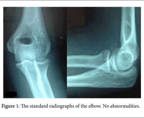
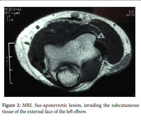
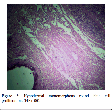
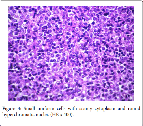
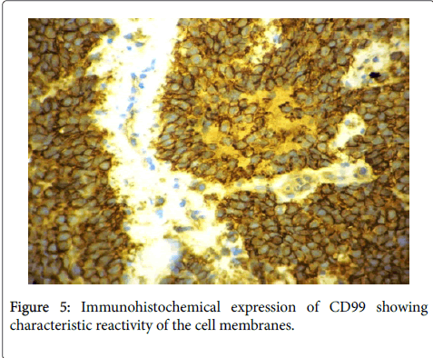
A 20-year-old female, with no past medical history, presented with a progressively increasing lump, of the external face of the left elbow for 7 months. He was asymptomatic with no fever or weight loss. On physical examination, the mass was superficial, firm, oval shaped, mobile, painless, and measuring 2 cm in greatest diameter. There was no lymphadenopathy detected. The standard radiographs of the elbow was normal (Figure 1). The magnetic Resonance Imaging (MRI) revealed a susaponevrotic lesion, invading the subcutaneous tissue of the external face of the left elbow (Figure 2). A surgical biopsy was done. Histological examination showed a subcutaneous (hypodermic and dermal) sarcomatous nodular proliferation (Figure 3) It is composed of monomorphic small round blue cells, with scanty cytoplasm, and round or ovoid hyperchromatic nuclei, mitotic Figures were rare (Figure 4). The epidermis was normal. The immunohistochemical study revealed a strong and diffuse membranous immunostaining for CD 99, and lack of stain for lymphoïd markers (CD3, CD 20) and muscular markers (desmin and myogenin) (Figure 5). The molecular study showed a chromosomal translocation t (11; 22). These features confirmed the diagnosis of extraskeletal Ewing’s sarcoma. Thoracic CT was normal. The patient received 6 courses of chemotherapy (Ewing 99 protocol). After chemotherapy, MRI was done and revealed a stable lesion. She underwent a surgical excision with negative margins. The skin fragment measured 6.5 × 4 × 1.5 cm. On the cut, the tumor was whitegrayish and measured 2 × 2 × 0.8 cm of greatest diameter. Histologically, tumour remnants corresponded to subcutaneous Ewing’s sarcoma. Afterwards, she received an adjuvant chemotherapy. At 2 years of follow-up, there was no recurrence and no metastasis.
Discussion
Current literature reports only a few isolated cases or small series. To date, less than 100 cases have been reported [2]. Extraskeletal Ewing’s sarcoma family of tumors (EESFT) most frequently occur in the deep soft tissues of children and young adults, such as paraspinal muscles, chest wall, and the lower extremities [2]. Nevertheless, it occasionally presents in a superficial location either as a primary tumor or a metastasis from osseous or deep seated EESFT. Those superficially located lesions, the so called Primary cutaneous EESFT are exceedingly rare and they are limited to the skin and generally present as a single small lesion, circumscribed mid to deep dermis or involving. superficial subcutis They were first described by Angerwall and Enzinger in 1975.
The diagnosis is made by aspiration cytology, histochemical stains, immunohistochemistry, electron microscopy, cytogenetics and molecular genetics of translocations [1,3,4]. The differencial diagnosis of ES include primary cutaneous small round cell tumors like Merckel cell carcinoma, eccrine spiradenoma, lymphoma, melanoma, clear cell sarcoma, rhabdomyosarcoma, malignant rhabdoïd tumor, malignant primitive neuroectodermal tumor and poorly differenciated adnexal tumors [5,6]. It includes also cutaneous metastases from osseous ES, large cell neuroendocrine carcinoma, small cell lung carcinoma and neuroblastoma [1,7]. The ES is composed of small round cells which express the CD99 and molecular study shows a specific chromosomal translocation t(11;22) involving gene EWSR1 in chromosome 22q12 or a fusion or combination between EWSR1 gene and gene of ETS family [1,3,8].
This study is made by FISH or real time polymerase chain reaction (RT-PCR) [8]. Cutaneous ES has better prognosis than primary bone or soft tissue ES with a survival rate of 91% in 10 years [1].
The less aggressive behavior is due to superficial location, small size and easy access [1,8]. One case of metastasis and one with local node involvement have been reported [9]. The presence of metastasis is really rare [4,10]. Currently, no specific recommendations to primitively cutaneous Ewing tumors, these latter are treated as bone Ewing's sarcomas: neoadjuvant chemotherapy, surgery, adjuvant chemotherapy (+/− radiotherapy), and autologous bone marrow transplantation in high risk patients [11]. This approach has been used successfully in bone tumors and deep tissue tumors, but there are data that supports the theory in the cutaneous and subcutaneous Ewing’s sarcoma, the intensity should be lowered [12].
Conclusion
Cutaneous Ewing sarcoma is a very rare entity, confined to the epidermis, dermis and subcutaneous tissue. It’s clinically and pathologically sub-diagnosed because of its rarity and morphological similarity to other cutaneous tumors. The molecular biology can lead us to the correct diagnosis. It seems to have better prognosis than osseous ES. The treatment consists on local surgery and chemotherapy and/or radiation.
References
- Oliveira Filho JD, Tebet AC, Oliveira AR, Nasser K (2014) Primary cutaneous Ewing sarcoma - Case report. An Bras Dermatol 89: 501-503.
- Yuste V , Sierra E , Ruano D Llamas-Velasco M Conde E et al. (2015) Primary Cutaneous Ewing Sarcoma: Report of a Case. Fetal PediatrPathol 34: 315-321.
- Luca Grassetti, MatteoTorresetti, Donatella Brancorsini, CorradoRubini, DavideLazzeri, et al. (2015) A peculiar case of large primary cutaneous ewing’s sarcoma of the foot: case report and review of the literature. Int J Surg Case Rep 15: 89-92.
- Delaplace M, Lhommet C, de Pinieux G, Vergier B, de Muret A (2012) Primary cutaneous Ewing sarcoma: a systematic review focused on treatment and outcome. Br J Dermatol 166: 721-726.
- Machado I, Llombart B, Calabuig-Fariñas S, Llombart-Bosch A (2011) Superficial Ewing's sarcoma family of tumors: a clinicopathological study with differential diagnoses. J CutanPathol 38: 636-643.
- Terrier-Lacombe MJ, Guillou L, Chibon F, Gallagher G, Benhattar J, et al. (2012) Superficial primitive Ewing's sarcoma: a clinicopathologic and molecular cytogenetic analysis of 14 cases 2012: 12-14.
- Shingde MV, Buckland M, Busam KJ, McCarthy SW, Wilmott J, et al. (2009) Primary cutaneous Ewing sarcoma/primitive neuroectodermaltumour: a clinicopathological analysis of seven cases highlighting diagnostic pitfalls and the role of FISH testing in diagnosis. J ClinPathol 62: 915-919.
- Delaplace M, Melard P, Perrinaud A, Gore C, Vergier B, et al. (2011) Primitive cutaneous Ewing's sarcoma: a diagnostic and therapeutic dilemma. Ann DermatolVenereol 38: 395-398.
- Collier AB, Simpson L, Monteleone P (2011) Cutaneous Ewing sarcoma: report of 2 cases and literature review of presentation, treatment and outcome of 76 other reported cases. J PediatrHematolOncol 33: 631-634.
- Mizawa M, Makino T, Norisugi O, Hara H, Shimizu K, et al. (2014) Primary cutaneous Ewing sarcoma following metastasis to the bone and lymph nodes. Br J Dermatol 171: 660-662.
- Angervall L, Enzinger FM (1975) Extraskeletal neoplasm resembling Ewing’s sarcoma. Cancer 36: 240-251.
- Chow E, Merchant TE, Pappo A (2000) Cutaneous and subcutaneous Ewing’s sarcoma: an indolent disease. Int J Radiation Oncology BiolPhys 46: 433-438.
Relevant Topics
- 3D Printing in Limb-Sparing Surgery
- Adamantinoma
- Aneurysmal Bone Cysts
- Chondrosarcoma
- Chordomas
- Cryosurgery
- Enchondroma
- Ewing’s Sarcoma
- Fibrous Dysplasia
- Giant Cell Tumor of Bone
- Immunotherapy for Osteosarcoma
- Liquid Biopsy in Orthopedic Oncology
- Malignant Osteoid
- Metastatic Bone Cancer
- Molecular Profiling of Bone Tumors
- Multilobular Tumour of Bone
- Orthopaedic Oncology
- Osteocartilaginous Exostosis
- Osteochondrodysplasia
- Osteoma
- Osteonecrosis
- Osteosarcoma
- Primary Bone Tumors
- Sarcoma
- Secondary Bone Tumours
- Targeted Therapy in Bone Sarcomas
- Tumours of Bone
Recommended Journals
Article Tools
Article Usage
- Total views: 3957
- [From(publication date):
March-2017 - Jan 15, 2025] - Breakdown by view type
- HTML page views : 3194
- PDF downloads : 763

