Primary Breast Liposarcoma
Received: 17-Nov-2017 / Accepted Date: 24-Nov-2017 / Published Date: 30-Nov-2017 DOI: 10.4172/2167-7964.1000282
Abstract
Primary Breast Sarcomas are rare, accounting for less than 1% of breast malignancies. The authors present a case of primary breast liposarcoma in a 58 year old female with a left palpable lump, present since four months. Following core needle biopsy, the nodule was diagnosed as well differentiated liposarcoma. She underwent conservative surgery of the left breast, without radiation therapy. The patient is currently free from any symptoms of local recurrence and metastases 9 months after surgery.
Keywords: Breast sarcoma; Primary breast liposarcoma; Breast cancer
Introduction
Primary Breast Sarcomas are rare, accounting for less than 1% of breast malignancies. These mesenchymal neoplasms begin in the connective tissue elements of the breast.
Subtypes of breast sarcomas are: angiosarcoma (most common), liposarcoma (0.3% of breast sarcomas), malignant fibrous histiocytoma, leiomyosarcoma, sarcomas with bone and cartilage differentiation and fibrosarcoma [1-3].
To our knowledge, Austin and Dupruee published in 1986 the largest series with 20 cases of breast liposarcomas [4].
We must know that sarcomas metastasize in first place to the lungs, then to the liver, brain and bones, respectively; lymphatic metastases are not common [5].
Case Report
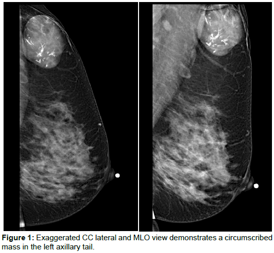
58 year old female with a painless palpable mass in the left breast presented to our hospital; a stiff nodule not attached to deep planes was found in the left axillary tail of Spence. The mammography (Figure 1) demonstrated an oval, circumscribed, equal density mass with dystrophic calcifications, no other associated features was present.
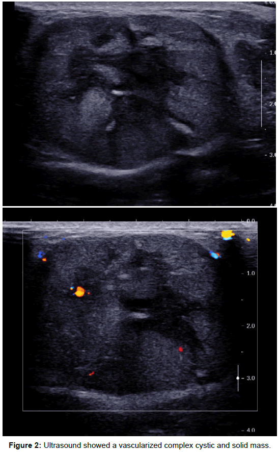
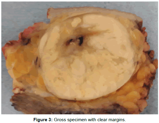
High frequency ultrasound (Figure 2) showed an oval, parallel, circumscribed, complex cystic and solid mass, with intranodal calcifications, peripheral and internal vascularity maximum diameter of 50 mm. A core needle biopsy was performed and the histopathological diagnosis was: well differentiated liposarcoma with a recommendation for excisional biopsy. The patient underwent conservative surgery, macroscopically (Figure 3) a well-defined nodule was identified, not encapsulated, solid, with yellow-brown areas; loose, medium consistency.
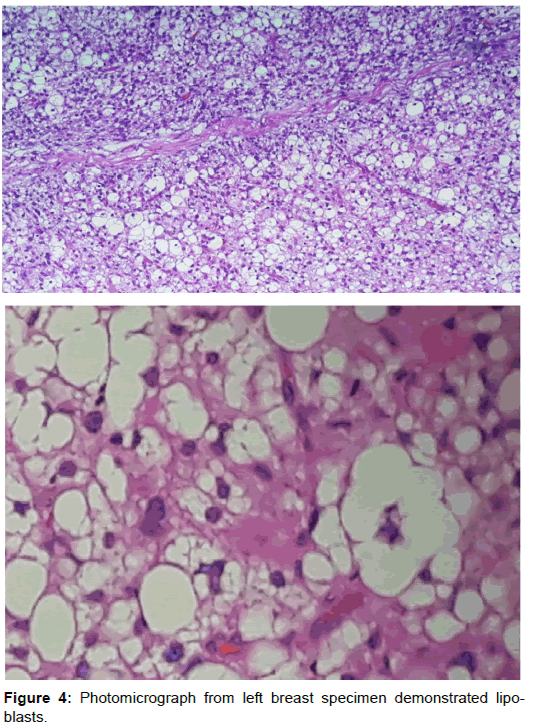
Histologically (Figure 4) a malignant mesenchymal neoplasm was identified, with lipoblasts of small to intermediate nuclei, hyperchromatics, multivacuolar cytoplasm, mitosis 1 by 10 high power fields, with hyaline focal areas and fusiform cells, Phyllodes or other tumor component was not identified in extensive sampling.
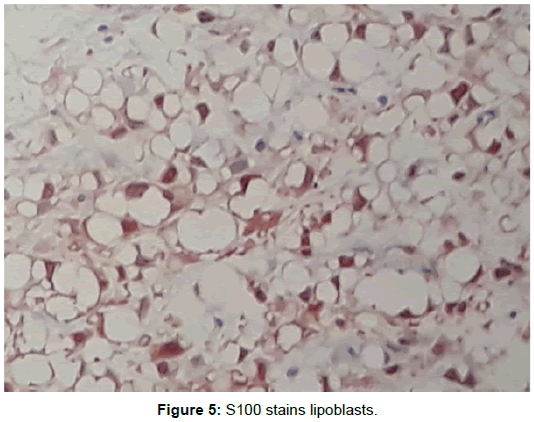
Immunohistochemistry (Figure 5) for S100 was positive in lipoblasts, negative for CD34 and BCL-2 and negative for P-63, which discarded phyllodes tumor and metaplastic carcinoma respectively.
No axillary dissection was performed, radiotherapy or chemotherapy was not administered and extension studies did not demonstrate metastasis.
The patient is currently free from any symptoms of local recurrence and metastases 9 months after surgery.
Discussion
In adults, liposarcoma is the most frequent soft tissue tumor, it is found predominantly in the extremities and retro peritoneum, it is rare to find it in the vulva, axilla and spermatic cord or in the breast.
The world health organization has classified the liposarcoma in to seven subtypes:
Atypical lipomatous tumor/well differentiated liposarcoma, dedifferentiated liposarcoma, myxoid liposarcoma, round cell liposarcoma, pleomorphic liposarcoma, mixed-type liposarcoma, liposarcoma not otherwise specified [5,6].
There is no consensus about imaging presentation of breast sarcomas, for example Berg et al. [1], Lamia Charfi et al. [7], describes that mammographically, these tumors often present as a well limited, round, oval or polycyclic opacity. So the mammographic impression in some cases may be of a benign fibroadenoma.
Smith et al. include twenty-four women with the final diagnosis of primary sarcoma of the breast in a study of the MD Anderson hospital and published the most frequent imaging findings: by mammography an oval nodule with indistinct margins and no calcifications by sonography an oval, indistinct, hypoechoic and hypervascular solid mass was found by MRI the most frequent form of presentation was an oval or round nodule with irregular margins, high signal intensity on T2 and heterogeneous enhancement [2].
The rarity of this disease prevents a consensus on the proper management of breast sarcomas, in the literature surgical treatments are reported through complete resection and free margins greater than one centimeter or mastectomy, there are no agreements about the administration of radiotherapy or chemotherapy in patients without metastatic disease.
To our knowledge, Yin M, et al. have published the biggest series of breast sarcomas patients and its outcomes with 785 women from the United States of America SEER database (Surveillance, Epidemiology and End Results Program), their major findings includes better survival rates with breast conserving surgery than with mastectomy and an improvement of survival outcomes with the use of adjuvant radiotherapy in neoplasm dimension over 50 mm [8].
Conclusion
The role of the breast cancer team on diagnosis, staging and treatment of rare breast cancer are highlighted with this case report of a primary breast liposarcoma.
References
- Berg W, Yang T (2013) Diagnostic Imaging: Breast. Amirsys Publishing Inc., USA.
- Smith TB, Gilcrease MZ, Santiago L, Hunt KK, Yang WT (2012) Imaging features of primary breast sarcoma. Am J Roentgenol 198: 386-393.
- Kanemoto K, Nakamura T, Matsuyama S, Arai M, Muratani M (1981) Liposarcoma of the breast, review of the literature and a report of a case. Japanese J Surg 11: 381.
- Austin RM, Dupree WB (1986) Liposarcoma of the breast: A clinicopathologic study of 20 cases. Human Pathol 17: 906-913.
- Akan A, Eryavuz Y, Akarsu C, Bademci R, Filiz F, et al. (2012) Primary liposarcoma of the breast. J Breast Heal 8: 89-91.
- Saito T, Ryu M, Fukumura Y, Asahina M, Arakawa A, et al. (2013) A case of myxoid liposarcoma of the breast. Int J Clin Exp Pathol 6: 1432.
- Charfi L, Driss M, Mrad K, Abbes I, Dhouib R, et al. (2009) Primary well differentiated liposarcoma: An unusual tumor in the breast. Breast J 15: 206-207.
- Yin M, Mackley HB, Drabick JJ, Harvey HA (2016) Primary female breast sarcoma: Clinicopathological features, treatment and prognosis. Sci Rep 6: 31497.
Citation: Rodríguez Suárez I, Moguel N, Espejo R, Ríos N, Bautista V (2017) Primary Breast Liposarcoma. OMICS J Radiol 6:282. DOI: 10.4172/2167-7964.1000282
Copyright: © 2017 Rodríguez Suárez I, et al. This is an open-access article distributed under the terms of the Creative Commons Attribution License, which permits unrestricted use, distribution, and reproduction in any medium, provided the original author and source are credited.
Share This Article
Open Access Journals
Article Tools
Article Usage
- Total views: 6682
- [From(publication date): 0-2017 - Apr 26, 2025]
- Breakdown by view type
- HTML page views: 5779
- PDF downloads: 903

