Prevalence of Staphylococcus aureus subsp. anaerobius in Sheep and Goats abscesses in Nyala, South Darfur State, Sudan
Received: 18-Nov-2019 / Accepted Date: 17-Jul-2020 / Published Date: 24-Jul-2020 DOI: 10.4172/2161-1165.1000380
Abstract
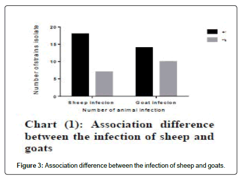
This study was conducted in Nyala, South Darfur State; samples were collected between August and November 2015 aiming at investigating the prevalence of Morel’s disease caused by Staphylococcus aureus subsp. anaerobius in sheep and goats at meat inspection in Nyala North abattoir. Out of 1050 slaughtered animals (441 sheep and 609 goats) 24 sheep (5.4%) and 25 goats (4.1%) had superficial lymph nodes abscesses, S. aureus subsp. anaerobius was isolated from 18 (72%), 14 (58.3%) of sheep and goats, respectively, giving prevalence rates of 4.1% in sheep and 2.3% in goats. Fisher’s test showed that there was no significant association between the prevalence of Morel’s disease between sheep and goats (p<0.05). The parotid (36%) and prescapular (20%) lymph nodes were more frequently affected by Morel’s disease in sheep and the prescapular (37.5%) and precrural (20.8%) in goats. The position of abscess was located close to the lymph node in goats and close to and/or within the lymph nodes in sheep. In one sheep, 3 abscesses were observed at the same time, and in one goat 2 abscesses were observed. The size of one abscess in sheep reached 12.5×14.5 cm. The resulting of this study revealed the high prevalence of Morel’s disease in sheep and goats and the great economic losses due to triming carcass and skin.
Keywords: Morel’s disease; Sheep; Goat; Staphylococcus aureus subsp. anaerobius; Sudan
Introduction
Morel’s disease (MD) is a bacterial disease caused by Staphylococcus aureus subsp anaerobius. It causes abscessation in superficial lymph nodes with high morbidity rate in small ruminant mainly in early ages and give food supplements, aged between 4-10 months and less in 1-5 years, abscess reached to 30-70 cm. The etiologic agent is S. aureus subsp. anaerobius. MD is prevailing in some African and Asian countries plus some reports of outbreaks in some European countries [1,2].
In the Sudan, it’s known as Aldamamil. Results of high prevalence rate of Morel’s disease conducted by investigators of Sudanese sheep abscesses illustrate poor vaccination against disease and observed the prevalence rate highest among Hamari and Kabbashi breed than other breeds and the incidence of disease was higher among feed lots animals when compared with natural cases. However, there have been vigorous efforts to produce the vaccine Musa. Many researchers worked in epidemiology of this disease in difference part of countries, so as to solve export sheep and goats problem to make control of the disease that causes problematic and negatively economic impact throughout the world. Speed of the disease from infected to susceptible animals is mainly through skin injuries and abrasion during rough gathering [3,4]. This study aimed at carrying out of prevalence study of Staphylococcus aureus subsp. anaerobius in sheep and goat abscesses.
Materials and Methods
Study area
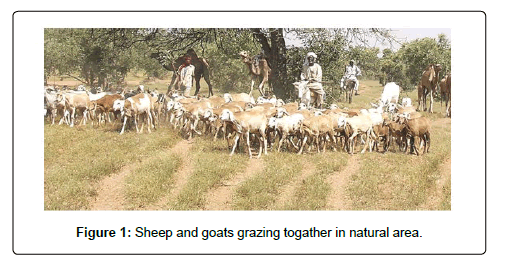
The study was conducted in Nyala town, South Darfur State. It is situated on the South West part of the Sudan; it is about 1446 km from Khartoum. The main activities of population are agriculture and animal breeding. There are about 3.7 million head of sheep and goats about 4.2 million head according to the Ministry of Animal Resources, South Darfur State report [5]. It has the biggest markets. Most of animal wealth in western Sudan is concentrated in South Darfur State and has become of great importance both locally and for exportation. Nomads migrate to the North during to the rainy season (August to October) and to the South during the dry season and remain there until rainfall ensuring in May to June in search of pasture and water. The climate is savannah type, which plant cover natural pasture comprised grasses, bushes shrubs and trees. In district the people breed goats often raised with sheep and mainly rearing together in open range system and rarely separated as shown in Figure 1, where they are expected to be subjected to various injures and so considered as predisposing factors for occurrence the disease.
Sampling
A total number of 1050 sheep and goats were slaughtered at Nyala North abattoir in South Darfur State between August to November, 2015 for domestic consumption. Samples were collected from enlarged superficial lymph nodes of those were that routinely palpated at meat inspection. The whole intact enlarged lymph nodes were incised and placed in sterile plastic bags in an ice box and sent to the bacteriology laboratory at the Institute of Molecular Biology, University of Nyala for bacterial culture.
Data analysis
Data were analysed using PRISM® version 6.07 (Graph Pad Software Inc., San Diego, California, USA). Fisher’s exact test was used to assess the significant difference between the prevalence of Staphylococcus aureus subsp. anaerobius in sheep compared with goats.
Descriptive statistics
Sample collection for identification of Staphylococcus aureus subsp. anaerobius associated with sheep and goats abscesses. A total 49 abscesses were collected, of which 18, 14 strains respectively were obtained
Procedures for identification of isolates
The organisms were examined for morphological, cultural and biochemical tests as shown in Table 1. The pure isolates were identified according to the scheme for identification of Staphylococci species modified by El-Sanousi et al. [7].
| Data analysed | positive | negative | Total Significance | |
|---|---|---|---|---|
| Sheep infection | 18 | 7 | 25 | |
| Goat infection | 14 | 10 | 24 | P = 0.3772 |
| Total | 32 | 17 | 49 | Not significant |
Table 1: Significance of sheep infection result in comparison with goats infection.
Results
Bacteriological isolation
Out of 1050 sheep and goats slaughtered, 49 abscesses were collected from sheep and goat (25, 24 respectively) as shown in Table 2.
| Species | No. of Slaughtered Animals | No of samples Collection | No of Isolation | Prevalence rate |
|---|---|---|---|---|
| Sheep | 441 | 25 | 18 | 4.1 |
| Goat | 609 | 24 | 14 | 2.3 |
| Total | 1050 | 49 | 32 | 6.4 |
Table 2: Isolation of Staph aureus subsp. anaerobius from slaughtered sheep and goats in Nyala north abattoir South Darfur State.
Statistical analysis
By using Fisher’s test there is no statistically (P<0.05) significant differences. Rate of infection in sheep and goats showed no significant difference (P<0.05) in detection of prevalence of sheep infection when compared with the goats.
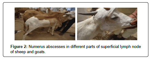
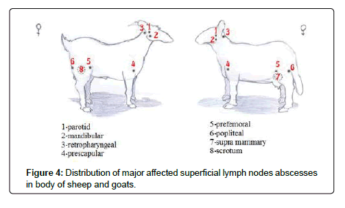
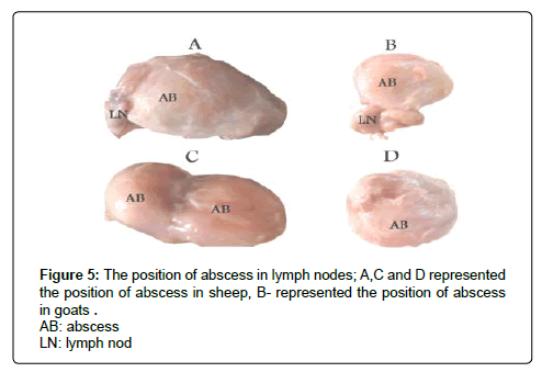
Data in the Figures 2 and 3 represent the prevalence and distributions of abscesses among slaughtered sheep and goats In Figure 4 showed variable shape of abscesses which contains of pus varied from greenish yellow creamy and thick consistency, whereas Figure 5 observe 2-3 abscesses in the same carcass and the big size was reached to 12.5×14.5 cm.
The formation of abscesses in difference part of the body located in superficial lymph nodes, close to lymph nodes in goats, while in sheep it was found in three forms: abscess developed close to, within lymph nodes and in both form (close to and adjacent) Figures 6 and 7.
Discussion
In study area, affected sheep and goat with abscesses passed after treaming abscesses during meat inspection in abattoir for human consumption. Previous studies have confirmed the goats are resistant to the disease, but it has spread widely.
In the present study showed the number of collected abscesses from superficial lymph nodes in different parts of the body in carcasses of sheep and goats were 25, 24 abscess respectively. These findings suggested that the sheep and goats in state are mainly rearing together in open range system where they are expected to be subjected to various injures and so considered as predisposing factors for occurrence the abscesses. The bacteriological culture result showed that the percentage of Staph aureus subsp. anaerobius in slaughtered sheep was 18(72%), while in goats was 14(58.3%). The results of prevalence rate of the disease in sheep (4.1%) while in goats (2.3%) as shown in Table 3.
| Lymph node | No of abscesses | |
|---|---|---|
| Sheep | Goats | |
| Parotid | 9(36%) | 3(12.5%) |
| Prescapular | 5(20%) | 9(37.5%) |
| Precrural | 4(16%) | 5(20.8%) |
| Popliteal | 2(8%) | 2(8.2%) |
| Supramammary | 1(4%) | 0(0%) |
| Mandibular | 3(12%) | 2(8.2) |
| Retropharyngeal | 1(4%) | 2(8.2) |
| Scrotum | 0(0%) | 1(4.1%) |
| Total | 25(100) | 24(100%) |
Table 3: Frequency and percentage of lymph node abscessses among slaughtered sheep and goats.
The current study revealed that the high percentage of Staphylococcus aureus subsp. anaerobius in sheep and goat was 72%, 58%; respectively; this percentage indicated that the incidence of Morel’s disease is higher in slaughtered sheep when compared with goats. Many studies have reported the occurrence of abscesses in sheep in Sudan and who stated that Staph aureus subsp. anaerobius is the common pathogen in sheep among bacterial isolation. The results obtained in sheep by Hamad et al. was 73.9%, Babiker 75%, Musa 63.3% and Radwan and Babiker et al. 65.95%, Nagah 53.3%, Karamala 26% and Bihary 24% [7-10]. Lower rate reported by Elgadal and Alharbi who reported that the Corynbacterium pseudotuberculosis was dominating 51.85%, 25.77% followed by Staph. aureus subsp. anaerobius 27.07%, 27.84 respectively. The prevalence rate of infection in sheep was found higher than that in goats (4.1%, 2.3% respectively). This contrast may be due to comparatively increase susceptibility of sheep to various adverse environmental conditions and disease in general. Higher prevalence rates recorded in sheep by Hamad, Hamad, Karamalla, Alharbi and Radwan and Babiker et al. 73%, 12.6%, 10%, 5-44.1% and 16.7% respectively [8-12]. Radwan found that the prevalence rate in natural rang area was 5.8%. Also Hassan recorded the prevalence rate of 8%. While the higher prevalence rate recorded in goat by Valenti and Bieler, 46.7%, De la Fuente et al., 71%, Szalus-Jordanow, et al. 93.6%, Al-Harbi, 2.2-6.5% [13,14]. Overall prevalence of Morel’s disease in sheep 4.1% was recorded in the current study, which is different with 1.21% reported by Elgadal, how investigated the aetiology of sheep abscess in postural area in the same district. This variability in our study could be attributed to difference in breeds and management of the herd, which the majority of the slaughtered animals submitted to the fattening process, in addition to mostly slaughtered animals at early ages due to consumer preferences so that the important factors to occurrence the disease. Thus incidence of abscesses high rate more probably in our study. In addition, higher prevalence rate of goat in Saudi Arabia 6.5-22% was recorded by Alharbi while Szalus -Jordanow recorded the highest prevalence rate 93.6%.
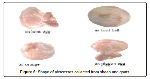
Earlier studies of Morel’s disease, described the size and shape of abscess as a pigeon and as orange, Bajmocy et al. described hen egg, Alhendi, El-sanousi and Moller et al. described the abscess as big as football. The same observations were found in this study [15-17].
In this study the contents of abscesses were creamy and greenish yellow in color and thick in consistency. Similar results were obtained among many studies. Concerning the formation of abscess in lymph node associated with, within, close and within (in lymph node and adjacent) lymph node in sheep. These observations disagree with earlier reporters they reported abscesses developed close to, but not within lymph nodes. Also Bajmocy et al. and Fuente and Suarez found abscesses inside the superficial lymph nodes and not around them.
Abscesses were located close with lymph nodes in goats [18]. Our observations agree with Valenti and Bieler, Moller et al., Koba et al., and Szalus -Jordanowet al. obcervations.
Morel’s investigations reported by Ayaund, Joubert, El-Sanousi and Szalus-Jordanow et al. found that the most commonly sites affected lymph nodes among slaughtered sheep were parotid lymph nodes and prescapular region in goats may be associated with behavior of sheep and goats that tend to scratch their jaw and shoulder in hard objects such as wall, fences and metallic feeding [19-25].
Our study is considered the first report of prevalence of Staphylococcus aureus subsp anaerobius in goats in Sudan.
Conclusion
This study provide the facts for prevalence of Staphylococcus aureus subsp. anaerobius in sheep and goats in Nyala, South Darfur State, Sudan, which will help to make information on the prevalence of the disease to be available and so put forward an appropriate control strategies for this economically important disease and their control method [26,27]. The very high prevalence of Morel’s disease during inspection of carcasses revealed the great economic losses due to reduction of wool and meat because terming skin and carcass. The resulting of this study warrants the need for strategic approach directed to the competent authorities for the disease risk, and control measures to achieve high animal health standards.
Acknowledgments
The author thanks to Institute of Molecular Biology university of Nyala for preservation of samples and bacterial culture in their laboratory. Also express special thanks to members of Microbiology Department for bacterial identification and acknowledge the Department of Preventive Medicine and veterinary public health for their collaboration during the study period. Members of the Nyala North abattoir in the collection samples are also very much appreciated for their cooperation in the field work.
References
- Rodwan K, Babiker A, Eltom H Musa O, Abbas B, El Sanousi S (2014) Abscess disease in pastoral and feedlot sheep. J Sci Tec 14: 45-53.
- Alhendi AB, Al-Sanhousi SM, al-Ghasnawi YA, Madawi M (1993) An outbreak of abscess disease in goats in Saudi Arabia. Zentralbl Veterinarmed A 40: 646-651.
- Ben Said MS, Ben Maitigue H, Benzarti M, Messadi L, Re- jeb A, Amara A (2002) Epidemiological and clinical studies of ovine caseous lymphadenitis. Arch Inst Pasteur Tunis 79: 51-57.
- Kaba J, Szalus O, Rzewuska M, Stefanska I, Binek M, et al. (2007) An outbreak of Morel’s disease in a goat flock (in Polish). Mag Weter 16: 46-48.
- Anon (2016) Annual report of Ministry of Animal Resources and Fisheries in South Darfur State.
- El-sanousi SM, Saeed KB, Gamal S, Awad S, Altom K (2016) Modified scheme for identification of Staphylococci ssp. Department of Microbiology, J.Vet. Science and animal production, Univ of Khrtoum 6: 93-97.
- Hamad AR, Shigiddi MT and El Sanousi SM (1992) Abscess disease of sheep in the Sudan. Sud J Vet Sc Anim Husb 31: 60-61.
- Babiker A (1996) Studies on Morel's disease of sheep. M. V. Sc. Thesis, University of Khartoum, Sudan.
- Karamalla NM (1997) Taxonomy, fatty acids pattern and protein profiles of Staphylococcus species isolated from abscess in sheep. Ph.D. Thesis, University of Khartoum, Sudan. Faculty of Vet Science.
- Bihary SO (2002) Pathogenicity of various species of Staphylococcus isolated from lymph nodes of sheep suspected for Morel s disease. M. V. Sc. Thesis. University of Khartoum.
- Al-Harbi KB (2011) Prevalence and etiology of abscess disease of sheep and goats at Qassim Region. Saudi Arabia. Vet World 4: 495-499
- El-Sanousi SM, Hamad AA and Gameel AA (1989) Abscess disease in goats in the Sudan. La maladie caseuse chez des chevres au Soudan. Revue Elev. Med Vet Pays Trop 42: 379-382.
- Valenti and Bieler (1984) Cultural and biochemical characteristics of strains of Morel’s coccus responsible for abscesses in goats in the valleys of the Cuneo area. Ann Fac Med Vet Torino 28: 286-307.
- De la Fuente R, Cid D, Sanz R and Ruiz-Santa-Quiteria JA (1997) An outbreak of abscess disease associated with shearing. Small Rumin Res 26: 283-286.
- Shirlaw JF and Ashford WA (1962) The occurrence of caseous lymphadenitis and Morel’s disease in a sheep flock in Kenya. Vet Rec 74: 1025-1026.
- Bajmocy E, Fazekas B and Tanyi J (1984) An outbreak of Morel’s disease (a contagious sheep disease accompanied by abscess formation) in Hungary. Act Vet Hung 32: 9-13.
- Moller Agerholm, Ahrens JS, Jensen P, and Nielsen TK (2000) Abscess disease, caseous lymphadenitis and pulmonary adenomatosis in imported sheep. J Vet Med 47: 55-62.
- Fuente R, De la and Suarez G (1985) Respiratory deficient Staphylococcus aureus as the aetiological agent of abscess disease. J Vet Med Ser B 32: 397-406.
- El-Sanousi SM (1989) Abscess disease in Najdi sheep. Saudia Arabia. El Yamama. 10: 23-24.
- Babiker A and El-Sanousi SM (2004) Effects of fattening on the occurrence of sheep abscess disease (Morel’s Disease). In: Peters, KJ, Kirschke D, Manig W, Burkert A, Schultze-Kraft R, Bharati L, Bonte-Friedheim C, Deininger A, Bhandari N, Weitkamp H (Eds.), Deutscher Tropentag Book of Abstracts. druckhausköthen GmbH, Kothen pp:s 135.
- De la Fuente R, Suarez G, and Shleifer KH (1985) Staphylococcus aureus subsp anaerobius. nov., the causal agent of abscess disease in sheep. Int J Syst Bacteriol 35: 99-102.
- Habrun B, Lestes E, Kompes G, Cvetnic Z, Mitak (2004) Caseous lymphadenitis in goats and sheep. Vet Stanica 35: 139-144.
- Musa NO (2012) Morel's Disease Vaccine: Evaluation of the Protective Effect in Sheep. Lambert Academic Publishing, Saarbrücken, Germany, ISBN: 978-3-659-14472-1.
- Pegram RG (1973) An unusual form of lymphadenitis in sheep and goats in the Somali Democratic Republic. Trop Anim Health Prod 5: 35-39
- Quiteria JA, Cid D, Sanz R, Garcia S, de la Fuente R (1996) Influence of age of the donor sheep on the phagocytosis of Staphylococcus aureus subspecies anaerobius and S. aureus by neutrophils. Res Vet Sci 61: 231-233.
- Rodwan K, Babiker A, El Sanousi S (2004) Studies on vaccination trials against Morel’s disease and monitoring of transfer of passive immunity from dams to lambs. Deut- scher Tropentag, October 5-7, Berlin.
- Szalus-Jordanow O, Kaba J, Czopowicz M, Witkowski L, Nowicki M, et al. (2010) Epidemiological features of Morel’s disease in goats. Pol. J Vet Sci 13: 437-445.
Citation: Khadeega YHS, El Sanousi SM, Mohamed AEM (2020) Prevalence of Staphylococcus aureus subsp. anaerobius in Sheep and Goats abscesses in Nyala, South Darfur State, Sudan. Epidemiol Sci 10: 380 DOI: 10.4172/2161-1165.1000380
Copyright: © 2020 Khadeega YHS, et al. This is an open-access article distributed under the terms of the Creative Commons Attribution License, which permits unrestricted use, distribution, and reproduction in any medium, provided the original author and source are credited.
Select your language of interest to view the total content in your interested language
Share This Article
Recommended Journals
Open Access Journals
Article Tools
Article Usage
- Total views: 6917
- [From(publication date): 0-2020 - Dec 19, 2025]
- Breakdown by view type
- HTML page views: 5884
- PDF downloads: 1033

