Preoperative Medication Co-ordinating all Aspects of Patient Care
Received: 19-May-2023 / Manuscript No. JPAR-23-103637 / Editor assigned: 22-May-2023 / PreQC No. JPAR-23-103637 / Reviewed: 05-Jun-2023 / QC No. JPAR-23-103637 / Revised: 10-Jun-2023 / Manuscript No. JPAR-23-103637 / Published Date: 17-Jun-2023 DOI: 10.4172/2167-0846.1000515 QI No. / JPAR-23-103637
Abstract
Beyond this technique’s clinical benefits, there is avoidance of the cost associated with preoperative anesthesia clearance, surgical supply costs, and avoidance of the costs associated with equipment and staff required for operating rooms. Moreover, patient’s stay in the healthcare facility is reduced and the overall financial burden to the already strained healthcare system is decreased. This led to a paradigm shift in the management of hand and wrist surgery cases which were done in the hand unit of the Department of Orthopaedics and our services adopted rapidly and expanded the use of WALANT. Despite limitations in the sensory examination, subjective numbness or paresthesias can be further explored independently with the use of a paper clip. Providing pictorial depictions of relevant sensory distributions can aid the patient in a subjective comparison of sensory function.
Keywords: Surgical supply; Patient visualization; Digitorium Pro-fundus; Lumbrical muscles; Telehealth; Radiographic studies
Keywords
Surgical supply; Patient visualization; Digitorium Profundus; Lumbrical muscles; Telehealth; Radiographic studies
Introduction
For the motor examination, begin by having the patient demonstrate passive ROM using the contralateral hand to demonstrate end range of flexion and extension of all digits and both wrists. Explore any deficits in greater detail by having the patient provide closer visualization so the provider can obtain a more accurate estimate of ROM of each joint. For active motor function, the provider should demonstrate each maneuver beforehand to help with patient compliance [1]. Begin with the extrinsic flexors. Ideal visualization for the provider is achieved with the patient’s hands in a neutral position, with the palms perpendicular to the camera or slightly pronated. Assess the flexor pollicis longus by having the patient block the meta-carpo-phalangeal joint of the thumb and flex through the inter-phalangeal joint. Assess the flexor digitorum profundus of each digit by having the patient block the proximal interphalangeal joint and flexing through the distal inter-phalangeal joint. Assess the flexor digitorum superficialis by blocking flexion of adjacent digits before flexing through the digit of interest. Ideal assessment of the extrinsic extensors involves a change in camera angle [2]. If possible, instruct the patient to lower the camera so that the palms can rest flat on the table in view of the camera. Assess the abductor pollicis longus and extensor pollicis brevis by having the patient extend and abduct the thumb away from the hand. Assess the extensor pollicis longus by having the patient lift the thumb off the table. Assess the extensor digitorum communis for each digit by independently extending and lifting it off the table, making sure that MCP joints are in extension.
Methodology
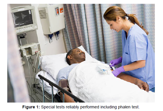
Have the patient make a fist and assess the extensor carpi radialis brevis and longus by demonstrating extension and radial deviation of the wrist, and the extensor carpi ulnaris by ulnar deviation of the wrist. Next, evaluate the function of the intrinsic muscles of the hand. Have the patient demonstrate MCP flexion and proximal interphalangeal joint extension to evaluate the function of the lumbrical muscles. Have the patient abduct, adduct, and then cross the fingers to evaluate interosseous muscle function, paying close attention for Wartenberg sign. Finally, have the patient demonstrate opposition of the thumb and little finger to evaluate the intrinsic thenar and hypothenar muscles [3]. The physical examination should conclude by using any special tests or provocative maneuvers that are warranted based on the patient’s history and physical examination thus far as shown in (Figure 1). In brief, special tests that can be reliably performed include Phalen test, elbow flexion test, and modified versions of Finklestein, Froment, Cozen, and TFCC load test [4]. To perform the Phalen test, have the patient oppose the dorsal aspect of each hand to achieve complete and forced flexion of both wrists. Have the patient hold this position for 30 to 60 seconds and assess for the presence of symptoms in the median nerve distribution. The elbow flexion test is performed by having the patient hold the elbow in full flexion for 1 minute to assess for the presence of symptoms in the ulnar distribution [5]. A modified Finklestein test can be performed independently by ulnarly deviating the wrist and using the contralateral hand to flex the thumb into the palm to assess for pain along the extensor pollicis brevise abductor pollicis longus tendons. A modified test for Froment sign for ulnar nerve palsy can be performed with the patient holding both ends of a piece of paper. A modified test for Cozen sign for lateral epicondylitis can be performed with the patient’s forearm and palm flat on the table. Have the patient make a fist, extend the wrist to elevate fist off the table, and use the contralateral hand to provide resistance to wrist extension [6]. A modified golfer’s elbow test can be performed in a similar fashion with wrist flexion. A modified TFCC load test can be performed with the patient loading the little finger with the contralateral hand. As part of a quality improvement initiative at Flinders Medical Centre, a review of antibiotics prescribed to all patients presenting to the Emergency Department with an open hand injury and who were subsequently admitted under the care of the Plastic and Reconstructive Surgery Department was undertaken [7]. Although these tests ideally should be performed in their unmodified form by a trained provider for proper interpretation, we believe that these modifications are adequate and reliable for the purposes of a telehealth encounter when in-person visitation is not possible. To maximize the usefulness of any telemedicine visit, it is vital for radiographic studies and other advanced diagnostic testing such as EMG results to be available for review [8].
Discussion
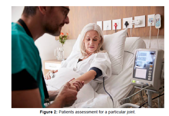
Special attention should be paid on the days leading up to a telemedicine visit to contact patients and provide them with instructions regarding how to make imaging and test results available to their provider for timely review. In addition, if radiographic studies are necessary for a visit but have not been completed, these should be ordered in advance. Because of the limitations of the remote patient encounter, access to all diagnostic studies is even more important [9]. Because of the limitations of the remote physical examination, it may be helpful and a more efficient use of time and resources to give scheduling preference to patients who have diagnostic studies ready for review for telemedicine visits. As mentioned, several studies outlined and validated appropriate use for telemedicine for specific types of patient encounters. Several recent studies commented on smartphone photography as a reliable alternative to goniometry, the reference standard for measuring joint ROM [10]. This has been shown to be effective for measuring ROM in the elbow, wrist, and fingers, especially in the setting of contracture as shown in (Figure 2). For patients for whom it is known that an assessment of ROM for a particular joint will be a necessary component of the physical examination, instructions can be provided regarding how to photograph themselves accurately [11]. Another specific patient encounter that was validated in the telemedicine setting is postoperative care for carpal tunnel release. As described by Tofte patients provided with instructions about removing surgical dressing and sutures, photographing the incision, and performing a basic neuromuscular examination were able to complete an effective 10- to 14-day postoperative visit remotely. However, suture removal proved to be the most difficult aspect. With this in mind, we believe that postoperative visits for minor hand surgery such as carpal tunnel release or trigger finger release can be readily managed through a telemedicine visit, especially if absorbable sutures were used. Many other postoperative visits can likely be managed remotely but should be evaluated on a case by case basis to determine the level of care and individual patient demands. Thought should also be given to specific patient encounters that are not appropriate to be conducted via telemedicine [12]. Clearly, any injuries that require manipulation, such as displaced fractures or dislocations, or other specialized care such as pin removal or cast removal or application, cannot be conducted remotely. In addition, patients for whom it is known with relative certainty that an injection needs to be administered, such as recurrent trigger finger or carpometacarpal arthritis, would benefit little from a telemedicine visit. A more complicated issue to tackle is the surgical decision-making process and informed consent. It has been shown that telemedicine encounters are not inferior to in-person encounters for patient comprehension during informed consent. However, there is uncertainty whether reliable risk and benefit assessment can be conveyed remotely [13]. For patients with a previous in-person encounter with documented appropriateness for surgical intervention, surgical decision-making and informed consent can proceed without issue. We also think that conditions with well-defined radiographic criteria for surgery are appropriate for surgical decision-making and informed consent via a remote visit. In these cases, the risks and benefits of intervention versus non-intervention, such as persistent deformity, posttraumatic arthritis, and recurrent joint instability, can be reliably assessed from certain radiographic studies and do not depend on physical examination. In cases in which indications for surgery are more complex and have not been discussed in previous detail, an adequate risk and benefit assessment might not be possible in a remote setting [14]. In such cases, patient comprehension of risks and benefits may require in-depth demonstration, patient education, or understanding of the patient’s own physical examination findings, which may necessitate in-person consultation. Thus, we advise providers to exercise caution before obtaining consent for surgery in these situations. The demand for telemedicine has been increasing over the past several years with the growth of technology and digital connectivity in our daily lives. With the impact of the global coronavirus disease 2019 pandemic, telemedicine implementation has become a necessity for many specialties because social distancing measures have greatly affected access to routine medical care. This article presents a detailed and systematic approach to conducting a hand physical examination during a video telemedicine encounter. Although the telemedicine physical examination has limitations, most components of the normal physical examination can be completed remotely with a systematic approach.
Conclusion
We enumerate modifications to maximize examination remotely and present considerations for improved delivery of telemedicine care. These methods may be beneficial to providers incorporating telemedicine into their practice.
Acknowledgement
None
Conflict of Interest
None
References
- Bidaisee S, Macpherson CNL (2014) Zoonoses and one health: a review of the literature. J Parasitol 2014:1-8.
- Cooper GS, Parks CG (2004) Occupational and environmental exposures as risk factors for systemic lupus erythematosus. Curr Rheumatol Rep EU 6:367-374.
- Parks CG, Santos ASE, Barbhaiya M, Costenbader KH (2017) Understanding the role of environmental factors in the development of systemic lupus erythematosus. Best Pract Res Clin Rheumatol EU 31:306-320.
- M Barbhaiya, KH Costenbader (2016) Environmental exposures and the development of systemic lupus erythematosus. Curr Opin Rheumatol US 28:497-505.
- Cohen SP, Mao J (2014) Neuropathic pain: mechanisms and their clinical implications. BMJ UK 348:1-6.
- Mello RD, Dickenson AH (2008) Spinal cord mechanisms of pain. BJA US 101:8-16.
- Bliddal H, Rosetzsky A, Schlichting P, Weidner MS, Andersen LA, et al. (2000) A randomized, placebo-controlled, cross-over study of ginger extracts and ibuprofen in osteoarthritis. Osteoarthr Cartil EU 8:9-12.
- Maroon JC, Bost JW, Borden MK, Lorenz KM, Ross NA, et al. (2006) Natural anti-inflammatory agents for pain relief in athletes. Neurosurg Focus US 21:1-13.
- Birnesser H, Oberbaum M, Klein P, Weiser M (2004) The Homeopathic Preparation Traumeel® S Compared With NSAIDs For Symptomatic Treatment Of Epicondylitis. J Musculoskelet Res EU 8:119-128.
- Ozgoli G, Goli M, Moattar F (2009) Comparison of effects of ginger, mefenamic acid, and ibuprofen on pain in women with primary dysmenorrhea. J Altern Complement Med US 15:129-132.
- Raeder J, Dahl V (2009) Clinical application of glucocorticoids, antineuropathics, and other analgesic adjuvants for acute pain management. CUP UK: 398-731.
- Świeboda P, Filip R, Prystupa A, Drozd M (2013) Assessment of pain: types, mechanism and treatment. Ann Agric Environ Med EU 1:2-7.
- Nadler SF, Weingand K, Kruse RJ (2004) The physiologic basis and clinical applications of cryotherapy and thermotherapy for the pain practitioner. Pain Physician US 7:395-399.
- Trout KK (2004) The neuromatrix theory of pain: implications for selected non-pharmacologic methods of pain relief for labor. J Midwifery Wom Heal US 49:482-488.
Indexed at, Google Scholar, Crossref
Indexed at, Google Scholar, Crossref
Indexed at, Google Scholar, Crossref
Indexed at, Google Scholar, Crossref
Indexed at, Google Scholar, Crossref
Indexed at, Google Scholar, Crossref
Indexed at, Google Scholar, Crossref
Indexed at, Google Scholar, Crossref
Indexed at, Google Scholar, Crossref
Indexed at, Google Scholar, Crossref
Citation: Cain J (2023) Preoperative Medication Co-ordinating all Aspects ofPatient Care. J Pain Relief 12: 515. DOI: 10.4172/2167-0846.1000515
Copyright: © 2023 Cain J. This is an open-access article distributed under theterms of the Creative Commons Attribution License, which permits unrestricteduse, distribution, and reproduction in any medium, provided the original author andsource are credited.
Share This Article
Recommended Conferences
42nd Global Conference on Nursing Care & Patient Safety
Toronto, CanadaRecommended Journals
Open Access Journals
Article Tools
Article Usage
- Total views: 464
- [From(publication date): 0-2023 - Dec 23, 2024]
- Breakdown by view type
- HTML page views: 401
- PDF downloads: 63


