Post-Traumatic Femoral Epiphysiodesis in Children
Received: 06-Oct-2022 / Manuscript No. roa-22-76767 / Editor assigned: 08-Oct-2022 / PreQC No. roa-22-76767 (PQ) / Reviewed: 21-Oct-2022 / QC No. roa-22- 76767 / Revised: 24-Oct-2022 / Manuscript No. roa-22-76767 (R) / Published Date: 31-Oct-2022 DOI: 10.4172/2167-7964.1000405
Image Article
The growth cartilage is a complex zone, found at the Epiphysometaphyseal junction, in the secondary ossification centers and in the growing apophysis. It allows the growth in length of long bones.
During a trauma, if an abnormal solution of continuity exists in the growth cartilage, a definitive communication is formed between the metaphyseal bone and the epiphyseal bone which constitutes an epiphysiodesis bridge thus hindering growth [1].
Post-traumatic epiphysiodesis must absolutely be detected and feared, especially since the residual growth potential is great. When the bridge is central, the consequence is the cessation of growth. When the bridge is lateral, the consequence is a misalignment of the type of varus, valgus, flessum. It is the consequence of an incomplete epiphysiodesis [1].
MRI is the ideal examination in the exploration of the growth cartilage, it is important in locating the bridges of epiphysiodesis, and their nature and knowing the functional quality of the remaining growth cartilage [2].
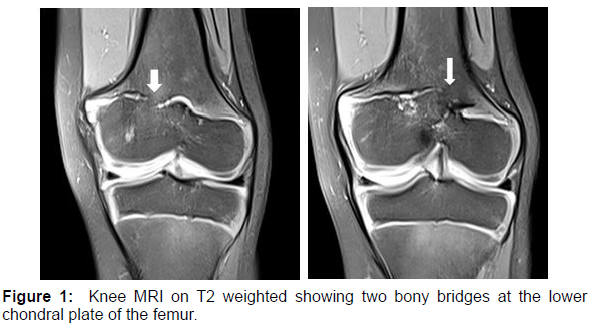
CT can also locate the epiphysiodesis bone bridge (Figure 1).
Figure 1 shows Knee MRI of 5 years-old child after severe knee trauma, on T2 weighted sequence showing a hypo signal interrupting the normal hyper signal of the growth plate, related to areas of lower femoral epiphysiodesis.
MRI makes the diagnosis by showing on the T2-weighted sequences, a hypo signal interrupting the normal hyper signal of the growth plate. The T1-weighted sequences will find a hypo signal or a discreet hyper signal relative to the cartilage, associated with the indirect sign represented by the convergence of the metaphyseal lines in hypo signal towards the bridge.
References
- Jouve JL, Bollini G, Launay F, Glard Y, Craviari T, et al. (2010) Growth cartilage and growth in orthopedics. Encycl Méd Chir 23: 1-15.
- Panuel M, Petit P, Chaumoitre K, Portier F, Bourlière B, et al. (2001) Magnetic resonance imaging of musculoskeletal trauma in children and adolescents. Encycl Méd Chir 31: 25.
Citation: Kabila B, Drissi A, Lrhorfi N, Allali N, Chat L (2022) Post-Traumatic Femoral Epiphysiodesis in Children. OMICS J Radiol 11: 405. DOI: 10.4172/2167-7964.1000405
Copyright: © 2022 Kabila B. This is an open-access article distributed under the terms of the Creative Commons Attribution License, which permits unrestricted use, distribution, and reproduction in any medium, provided the original author and source are credited.
Share This Article
Open Access Journals
Article Tools
Article Usage
- Total views: 1632
- [From(publication date): 0-2022 - Mar 13, 2025]
- Breakdown by view type
- HTML page views: 1390
- PDF downloads: 242

