Postpartum HELLP Syndrome with Atypical Features: A Case Study
Received: 20-May-2019 / Accepted Date: 11-Sep-2019 / Published Date: 18-Sep-2019
Abstract
Introduction: HELLP syndrome is a severe and potentially life-threatening variant of pre-eclampsia, consisting of a triad of Hemolysis (H), Elevated Liver enzymes (EL) and Low Platelet count (LP). The majority of preeclampsia and HELLP syndrome cases develops in the last part of pregnancy and rarely establish postpartum.
Case presentation: A 28-year-old primiparous woman, without any known risk factors for developing preeclampsia, developed jaundice and general discomfort 1 day postpartum.
The patient was diagnosed with HELLP syndrome on the basis of laboratory and urine findings, despite a normal blood pressure, lack of hemolysis and an absence of typical symptoms. The patient showed kidney involvement, which is a rare and serious complication of HELLP syndrome. The patient rapidly and fully recovered, without any permanent kidney damage. Due to the short administration time of intravenous magnesium sulphate, there is reason to believe that the recovery occurred spontaneously.
Conclusion: This case reports what may be an atypical presentation of postpartum HELLP syndrome.
Keywords: HELLP syndrome; Pregnancy; Hemolysis; Preeclampsia
Introduction
Preeclampsia (PE) is a pregnancy-specific disorder affecting 2-8% of all pregnancies [1]. Diagnosed as new-onset hypertension after 20 weeks of gestation, in addition to proteinuria, distinctive symptoms and/or affected blood test showing organ involvement, it remains a leading cause of maternal and perinatal morbidity and mortality worldwide. In the US, PE is responsible for approximately 15.9% of all maternal mortalities [2].
While the causative or initiating event in PE is not entirely understood, it has been postulated to involve a reduced placental perfusion leading to widespread dysfunction of the maternal vascular endothelium [3]. With progression this may lead to severe PE, a multisystemic disease which can result in placental abruption, liver- and kidney failure, disseminated intravascular coagulation or cardiovascular- and cerebrovascular disease [4]. Cerebrovascular deterioration may cause generalized seizures, known as eclampsia [5].
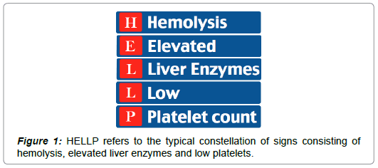
The so-called HELLP syndrome is considered to be a variant of severe PE and is a potentially life-threatening condition [6]. HELLP refers to the typical constellation of signs consisting of hemolysis (H), elevated liver enzymes (EL) and low platelets (LP) and it is believed to occur in 0.5-1% of all pregnancies [7]. Although symptoms of PE generally accompany the HELLP syndrome, approximately 20% of cases develop without any sign of PE, which can make the diagnosis challenging (Figure 1) [8].
The majority of PE cases are seen between 27 and 37 weeks of gestation and patients exhibiting severe features and/or HELLP syndrome are usually induced order to prevent maternal and fetal complications [8,9]. PE typically resolves with the birth of the infant, but is not always a definite cure [10].
Infrequently, PE, eclampsia or HELLP syndrome develops postpartum. In such cases, the majority occur within 48 h of childbirth. However, postpartum preeclampsia has been seen to develop up to 6 weeks after delivery or even later, termed late postpartum preeclampsia [11].
Case Report
A 28-year-old Asian woman, gravida 1, para 0, was admitted to our clinic at 39 weeks and 2 days of gestation. Upon arrival, the patients’ water had broken and contractions had started. The patient gave birth to a healthy baby boy after receiving an episiotomy.
The total bleeding was estimated to be around 700-800 ml, mostly due to bleeding after the episiotomy, which was sutured, as well as placental separation.
Directly after childbirth, the patient felt unwell and dizzy, tachycardia with a pulse between 120-130 beats per minute (BPM), expected to result from the blood loss. The patient’s blood pressure, temperature and other vital parameters were normal.
On the first day postpartum, over the course of a few hours the patient suddenly developed jaundiced sclera and palms of the hands. Furthermore, it was revealed that the patient had experienced an extreme sense of thirst over the preceding days, with a fluid intake of 4-6 liters daily.
Laboratory findings showed altered kidney parameters with increased serum creatinine and decreased estimated glomerular filtration rate, despite the high fluid intake and similarly high urine output. Furthermore, alanine aminotransferase, amylase, basic phosphatase, bilirubin, bile salts, gamma-glutamyl transferase and lactate dehydrogenase all showed markedly increased levels, while the thrombocytes were below normal level (thrombocytopenia). The urine dipstick test was positive for glucose, blood, protein and leukocytes.
Laboratory tests and clinical signs raised suspicion of postpartum HELLP syndrome and the Patient was moved to the intensive care unit for treatment with intravenous magnesium sulphate (MgSO4) infusion. During infusion, the patient developed massive nausea and the MgSO4 was paused and later stopped. The patient’s blood pressure was normal throughout the admission and the pulse 120-130 BPM.
After 12 h in the intensive care unit, the patient’s lab parameters started to improve. At two days, the patient subjectively felt better and was discharged from the hospital for outpatient follow-ups. The patient’s blood tests normalized completely within a few weeks.
During the period of admission, hereditary haemochromatosis and undiscovered Gestational Diabetes Mellitus (GDM) were suspected as differential diagnoses. GDM was ruled out due to normal fasting blood sugar levels as well as normal hemoglobin 1Ac. Hereditary haemochromatosis was excluded due to normal levels of serum ferritin, iron and transferrin.
Discussion
In this case study, we report the atypical development of the HELLP syndrome in a patient without any identified risk factors for developing PE, except null parity. Risk factors for PE include antiphospholipid syndrome, insulin dependent diabetes, preexisting hypertension, however this patient was a young woman with normal Body Mass Index (BMI), no known diseases and was expecting a singleton [10]. As the patient was adopted from Vietnam during childhood, her family history with regards to PE was unknown.
Cases of HELLP syndrome usually occur with preeclampsia, typically seen with a rise in blood pressure and proteinuria, however, there have been described cases in which signs are absent or only slight. In the present case, the blood pressure was normal throughout the whole course of the disease, including every routine check-up during pregnancy and no protein was found in the urine prior to the time of delivery. Proteinuria appears to have only developed postpartum.
The patient experienced a general malaise and discomfort but did not suffer from any of the typical symptoms for severe PE (e.g. headache, liver or epigastric pain, edema or dyspnea). Furthermore, the hemolysis was missing, which is otherwise an essential part of the HELLP syndrome (“H”-Hemolysis). The rapidly developing jaundice, resulting from the liver involvement (“EL”-Elevated Liver enzymes), in combination with thrombocytopenia (“LP”-Low Platelet), on the other hand, gives a clear suspicion towards the diagnosis of HELLP syndrome. Kidney involvement, as occurred in this case, is a rare, but serious complication, occurring only in 2-3% of cases of HELLP syndrome [8].
Conclusion
MgSO4 infusion was given only for a few hours but withdrawn due to extreme nausea. Nevertheless, the patient’s parameters and subjective well-being improved after a few days, suggesting the course of spontaneous regression. The patient was diagnosed with HELLP on the basis the laboratory and urine findings, despite a normal blood pressure and lack of hemolysis and an absence of typical symptoms. Differential diagnoses of undiscovered GDM and hemochromatosis were ruled out. Infection was not suspected due to a normal body temperature. No other cause of the patient’s symptoms was found.
In the event of any future pregnancy this patient is to be considered as having a high-risk pregnancy.
Conflicts of Interest
None.
References
- Jeyabalan A (2013) Epidemiology of preeclampsia: Impact of obesity. Nutr Rev 71: S18-25.
- Rasouli M, Pourheidari M, Gardesh ZH (2019) Effect of self-care before and during pregnancy to prevention and control preeclampsia in high-risk women. Int J Prev Med 10:21.
- LaMarca BD, Alexander BT, Gilbert JS, Ryan MJ, Sedeek M, et al. (2008) Pathophysiology of hypertension in response to placental ischemia during pregnancy: A central role for endothelin? Gend Med 5: S133-138.
- Kongwattanakul K, Saksiriwuttho P, Chaiyarach S, Thepsuthammarat K (2018) Incidence, characteristics, maternal complications and perinatal outcomes associated with preeclampsia with severe features and HELLP syndrome. Int J Womens Health 10: 371-377.
- Naljayan MV, Karumanchi SA (2013) New developments in the pathogenesis of preeclampsia. Adv Chronic Kidney Dis 2013 20: 265-270.
- Sharma RM, Sandhu GS (2006) HELLP syndrome: Report of two cases. Med J Armed Forces India 62: 373-374.
- Mai C, Wang B, Chen R, Duan D, Lv L, et al. (2018) HELLP syndrome complicated by pulmonary edema: A case report. Open Med 13: 509-511.
- Pop-Trajković S, Antić V, Kopitović V, Popović J, Trenkić M, et al. (2013) Postpartum HELLP syndrome--the case of lost battle. Ups J Med Sci 118: 51-53.
- Rimaitis K, Grauslyte L, Zavackiene A, Baliuliene V, Nadisauskiene R, et al. (2019) Diagnosis of HELLP syndrome: A 10-year survey in a perinatology centre. Int J Environ Res Public Health 16: 109.
- English FA, Kenny LC, McCarthy FP (2015) Risk factors and effective management of preeclampsia. Integr Blood Press Control 8: 7-12.
- https://www.mayoclinic.org/diseases-conditions/postpartum-preeclampsia/symptoms-causes/syc-20376646
Citation: Hansen IR, Khalil MR (2019) Postpartum HELLP Syndrome with Atypical Features: A Case Study. J Preg Child Health 6:420.
Copyright: © 2019 Hansen IR, et al. This is an open-access article distributed under the terms of the Creative Commons Attribution License, which permits unrestricted use, distribution, and reproduction in any medium, provided the original author and source are credited.
Select your language of interest to view the total content in your interested language
Share This Article
Recommended Journals
Open Access Journals
Article Usage
- Total views: 5360
- [From(publication date): 0-2019 - Feb 03, 2026]
- Breakdown by view type
- HTML page views: 4387
- PDF downloads: 973

