Pneumomediastinum
Received: 29-Jul-2015 / Accepted Date: 31-Jul-2015 / Published Date: 31-Jul-2015 DOI: 10.4172/2161-119X.1000i001
251255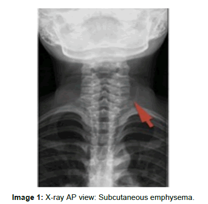
Image 1: X-ray AP view: Subcutaneous emphysema.
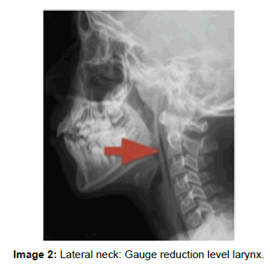
Image 2: Lateral neck: Gauge reduction level larynx.
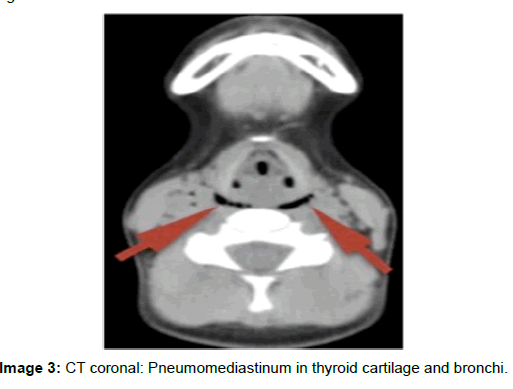
Image 3: CT coronal: Pneumomediastinum in thyroid cartilage and bronchi.
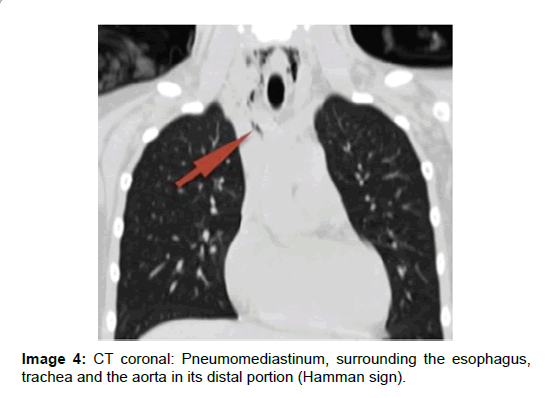
Image 4: CT coronal: Pneumomediastinum, surrounding the esophagus, trachea and the aorta in its distal portion (Hamman sign).
Citation: Lomeli JRL (2015) Pneumomediastinum. Otolaryngology 5: i001. DOI: 10.4172/2161-119X.1000i001
Copyright: © 2015 Lomeli JRL. This is an open-access article distributed under the terms of the Creative Commons Attribution License, which permits unrestricted use, distribution, and reproduction in any medium, provided the original author and source are credited.
Share This Article
Recommended Journals
Open Access Journals
Article Tools
Article Usage
- Total views: 13675
- [From(publication date): 7-2015 - Apr 07, 2025]
- Breakdown by view type
- HTML page views: 9166
- PDF downloads: 4509