Pleomorphic Adenoma of the Larynx
Received: 02-Dec-2022 / Manuscript No. asoa-23-88068 / Editor assigned: 05-Dec-2022 / PreQC No. asoa-23-88068 (PR) / Reviewed: 19-Dec-2022 / QC No. asoa-23-88068 / Revised: 21-Dec-2022 / Manuscript No. asoa-23-88068 (R) / Published Date: 28-Dec-2022 DOI: 10.4172/asoa.1000194
Abstract
Pleomorphic adenomas (PA) also called benign mixed tumors are most common tumors of salivary gland. Pleomorphic adenoma arising from the larynx is very rare. We report a 28 years old male patient who presented with shortness of breath and dry cough since 2 years, treated as asthma. The last crisis the patient warrants an intubation which failed and warrants an ENT consultation. ENT examination showed a mass in the subglottic region. After excision the pathology was in favor of pleomorphic adenoma.
Keywords
Pleormorphic adenoma; Larynx; Airway obstruction
Introduction
Pleomorphic adenomas (PA) also called benign mixed tumor are most common tumors of salivary glands. They occur mainly in major salivary glands but may also arise from minor salivary glands of upper respiratory and alimentary tract [1]. Histologically, these tumors are encapsulated and consist of benign epithelial and /or myoepithelial and stromal elements [2]. About 80% of PAs arise in the parotid gland, 10% in the submandibular gland, and 10% in the minor salivary glands of the oral cavity, paranasal sinuses, and upper respiratory and alimentary tract [3]. Those tumors have the capacity to transform into malignancy when they stay long without treatment [4].
Pleomorphic adenoma arising from the larynx are very rare, the literature report less than 30 cases[5]. Pleomorphic adenoma may occur at any age, but mainly they affect patients in the 4-6 decades with a male to female ration 2:1 [6]. When the larynx is involved, the most common site of involvement is the epiglottis and subglottis in some cases [5].
Case Report
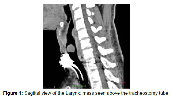
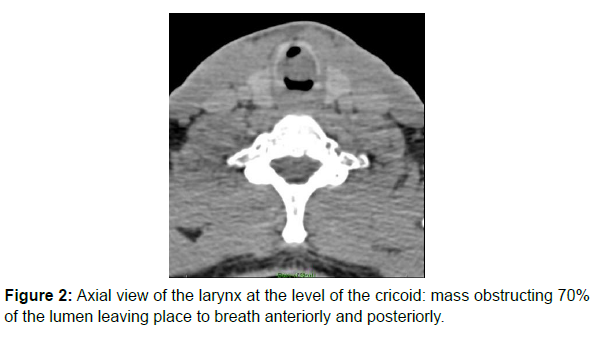
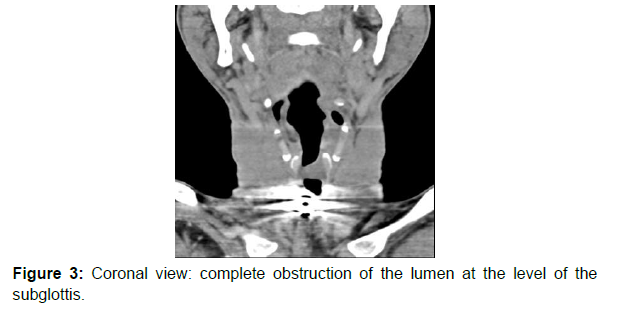
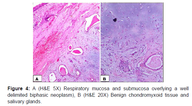
A 28 years old male patient with a 2-year history of shortness of breath, and dry cough. During those 2 years he presented to the accident and emergency department, where he was treated as a patient with asthma crisis. Initially he was improving on asthma treatment by bronchodilators and steroids. However over the course of time the symptoms were increasing in frequency and severity. Lastly the patient presents with severe l respiration distress that has been alleviated by a difficult intubation using a number 6.0 endotracleal tube as the biggest tube to pass in the subglottis and this warranted an ENT consultation. Meanwhile the patient had an accidental ex-tubation and re-intubation was difficult hence an emergent tracheostomy was done. CT scan was performed and showed (Figures 1-4).
A direct laryngoscopy was done and revealed a submucosal mass arising from the right posterolateral subglottic region extending into the lumen with occlusion of up to 70% with normal vocal cord mobility. According to the history and physical examination the mass was in favor of a benign mass. Endoscopic surgery of the mass was done with complete excision. Macroscopically the mass was submitted as 2 fragments, the biggest was 2x1x0.5 cm and the smallest 0.5x0.5x0.2cm.
Microscopy showed respiratory mucosa overlying a well circumscribed, biphasic neoplasm composed of proliferative benign salivary glands into a benign chondromyxoid stroma.
Discussion
For our case there has been a delay in the diagnosis of around 2 years. The literature also reports that usually there is a delay in the diagnosis of those uncommon tumors due to their non-specific clinical presentation [5]. Initially our patient was treated as asthmatic till he warranted an emergency intubation. Therefore, a complete head and neck examination including indirect or direct laryngoscopy with regard to the benign tumors in this organ is necessary for patients with respiratory problems. Current approaches in use for the excision of laryngeal pleomorphic adenomas include lateral pharyngotomy, laryngofissure, and use of laser [5]. As lack of experienced surgeons in the above procedure we did complete endoscopic resection which is usually done for small tumors. At the time of surgery the patient was having a tracheostomy: the tube has been kept for 7 months after surgery with a monthly evaluation of the patency of airway by transstomal flexible nasopharyngoscopy and finally a complete decanulation was done [7-10]. Since then the patient has been reviewed every month for a period of 6 months to exclude any reccurence. At the time of our report the patient had no complication [11, 12].
Conclusion
We recommend that every patient with signs of upper respiratory tract obstruction must be examined by a head and neck surgeon. The signs of obstruction may include hoarseness of voice, stridor and shortness of breath. This facilitates early diagnosis and prevents morbidity and mortality which may occur if the disease is not recognized early.
References
- Shafi S, Ansari HR, Bahitham W, Aouabdi S (2019) The Impact of Natural Antioxidants on the Regenerative Potential of Vascular Cells. Front Cardiovascu Med 6: 1-28.
- Ala-Korpela M (2019) The culprit is the carrier, not the loads: cholesterol, triglycerides and Apo lipoprotein B in atherosclerosis and coronary heart disease. Int J Epidemiol 48: 1389-1392.
- Esper RJ, Nordaby RA (2019) Cardiovascular events, diabetes and guidelines: the virtue of simplicity. Cardiovasc Diabetol 18: 42.
- Wityk RJ, Lehman D, Klag M, Coresh J, Ahn H, et al. (1996) Race and sex differences in the distribution of cerebral atherosclerosis. Stroke 27: 1974-1980.
- Qureshi AI, Caplan LR (2014) Intracranial atherosclerosis. Lancet 383: 984-998.
- Kay JE (2020) Early climate models successfully predicted global warming. Nature 578: 45-46.
- Traill LW, Lim LMM, Sodhi NS, Bradshaw CJA (2010) Mechanisms driving change: altered species interactions and ecosystem function through global warming. J Anim Ecol 79: 937-947.
- Kajinami K, Akao H, Polisecki E, Schaefer EJ (2005) Pharmacogenomics of statin responsiveness. Am J Cardiol 96: 65-70.
- Zavodni AE, Wasserman BA, McClelland RL, Gomes AS, Folsom AR, et al. (2014) Carotid artery plaque morphology and composition in relation to incident cardiovascular events: the Multi‐Ethnic Study of Atherosclerosis (MESA). Radiol 271: 381-389.
- Polonsky TS, McClelland RL, Jorgensen NW, Bild DE, Burke GL, et al. (2010) Coronary artery calcium score and risk classification for coronary heart disease prediction. JAMA 303: 1610-1616.
- Arad Y, Goodman KJ, Roth M, Newstein D, Guerci AD (2005) Coronary calcification, coronary disease risk factors, C‐reactive protein, and atherosclerotic cardiovascular disease events: the St. Francis Heart Study. J Am Coll Cardiol 46: 158-165.
- Dichgans M, Pulit SL, Rosand J (2019) Stroke genetics: discovery, biology, and clinical applications. Lancet Neurol 18: 587-599.
Indexed at, Google Scholar, Crossref
Indexed at, Google Scholar, Crossref
Indexed at, Google Scholar, Crossref
Indexed at, Google Scholar, Crossref
Indexed at, Google Scholar, Cross Ref
Indexed at, Google Scholar, Crossref
Indexed at, Google Scholar, Crossref
Indexed at, Google Scholar, Crossref
Indexed at, Google Scholar, Crossref
Indexed at, Google Scholar, Crossref
Indexed at, Google Scholar, Crossref
Citation: Jackson S, Parhami F, Xi XP, Berliner J, Hsueh W, et al. (2023) Pleomorphic Adenoma of the Larynx. Atheroscler Open Access 8: 194. DOI: 10.4172/asoa.1000194
Copyright: © 2023 Jackson S, et al. This is an open-access article distributed under the terms of the Creative Commons Attribution License, which permits unrestricted use, distribution, and reproduction in any medium, provided the original author and source are credited.
Share This Article
Open Access Journals
Article Tools
Article Usage
- Total views: 1224
- [From(publication date): 0-2023 - Mar 31, 2025]
- Breakdown by view type
- HTML page views: 877
- PDF downloads: 347


