Editorial Open Access
Pilon Fractures of Middle Phalanx Managed with Lag Screw and Early Mobilisation
Chandan Jadhav N*
Department of Plastic Surgery, Royal Perth Hospital, Perth, WA
- *Corresponding Author:
- Chandan Jadhav N
MS, MCh., Department of Plastic Surgery
Royal Perth Hospital
Wellington Street, WA 6000
Tel: +61 449925922
E-mail: chandansurgery@gmail.com
Received date: November 20, 2015; Accepted date: November 24, 2015; Published date: November 28, 2015
Citation: Chandan Jadhav N (2015) Pilon Fractures of Middle Phalanx Managed with Lag Screw and Early Mobilisation. Transplant Rep 1:e102.
Copyright: © 2015 Chandan Jadhav N. This is an open-access article distributed under the terms of the Creative Commons Attribution License, which permits unrestricted use, distribution, and reproduction in any medium, provided the original author and source are credited.
Visit for more related articles at Transplant Reports : Open Access
Keywords
Pilon fracture of middle phalanx; Open reduction internal fixation (ORIF); Lag screw
Level of Evidence
V, Therapeutic study
Introduction
Proximal interphalangeal (PIP) joint injuries are commonly seen in athletes and especially the pilon fractures are quite common due to axial loading injuries. It is associated with volar lip fragment with dorsal subluxation of middle phalanx with varying degree of comminution of the base of middle phalanx. Varied treatment options have been described ranging from extension block splint, external frame finger distraction and open reduction internal fixation [1-3]. The key is in restoring the joint congruity with early controlled mobilisation thereby preventing joint stiffness or deformity.
Case Series
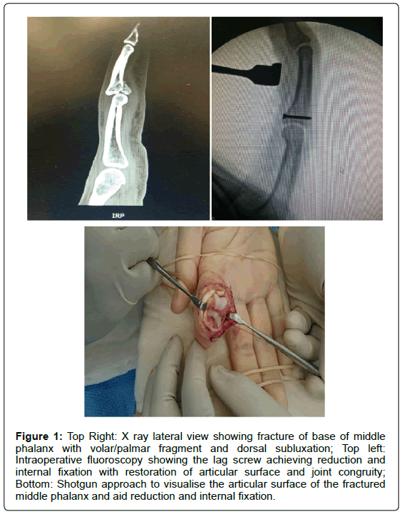
Five patients presented between Jan to June 2015 with pilon fracture of the middle phalanx. Age ranged from 21 to 29 yrs with an average age of 25.2 years. All were males. Mechanism of injury was axial loading onto to the proximal interphalangeal joint, while playing Australian football. Radiological evaluation revealed all of them had fracture of the volar lip of the base of the middle phalanx with dorsal subluxation as seen in Figure 1. Additional CT scan was done to assess the degree of comminution of the base of middle phalanx and also to plan accordingly. Average size of the largest volar fragment was 3.5 mm on CT scan. 3 out of 5 patients had marked comminution of base of middle phalanx classical of true pilon fractures. At presentation, active range of flexion at the PIP joint was limited to 0 - 25 degrees and passive movements were upto 35 degree. All of them underwent surgery with aim to restore the joint congruity and achieve optimal functional outcome and joint stability.
Figure 1: Top Right: X ray lateral view showing fracture of base of middle phalanx with volar/palmar fragment and dorsal subluxation; Top left: Intraoperative fluoroscopy showing the lag screw achieving reduction and internal fixation with restoration of articular surface and joint congruity; Bottom: Shotgun approach to visualise the articular surface of the fractured middle phalanx and aid reduction and internal fixation.
Surgical Technique
Regional block anaesthesia was administered. Brachial tourniquet was applied. Approach was through hemi- Bruner incision centred over the flexion crease of the PIP joint. The skin flaps were elevated, and care was taken to protect the radial and ulnar digital neurovascular bundles. The flexor tendon sheath was entered between the A2 and A4 pulleys. The proximal aspect of the volar plate was reflected from the proximal phalanx. Care was taken to maintain the volar plate’s distal attachment on the middle phalanx, which was repaired at the end. The collateral ligaments were then mobilised from the proximal phalanx and repaired at the end of the procedure. At this point, with the volar plate and collateral ligaments released, the joint could be shotgun opened to visualise the articular surface as shown in Figure 1. Fracture segment was reduced and articular surface alignment confirmed by direct visualisation. 1.3 mm screw is used for internal fixation in all patients. Traditional technique of lag screwing by drilling the near cortex with 1.3 mm drill bit and the far cortex with 1.0 mm drill bit was employed. So when 1.3 mm screw was used, it provided the necessary lag screw effect with adequate compression. 3 out of 5 patients had significant comminution with impacted articular surface of the base of middle phalanx. They required additional bone grafting to restore and lift back the impacted articular surface. The bone graft was harvested from the Lister`s tubercle of distal radius of the same limb, permissible as the procedures were being done under regional block. Tendon sheath was repaired and wound closed in layers. Passive flexion was done at the end of the procedure to assess the degree of improvement in the range of flexion and assess the joint alignment. All joints were stable at the end of the procedure.
Postoperative protocol:
All patients underwent early active mobilisation of the PIP joint within the splint which was maintained at 30 degree flexion at the PIP joint in the first week. The degree of flexion in the PIP joint splint age was gradually reduced at the rate of 10 degree ever week. So at the end of four weeks the PIP joint was in extension. The patient underwent early active mobilisation in the form of flexion at PIP joint right from postoperative day 1.
Results
All patients at the end of six weeks had 0-90 degree of active flexion at the injured PIP joint and 0-100 degree of passive flexion at the same joint. Proper reduction and congruency of the joint was noted on anteroposterior and lateral radiographs at 6 weeks post op follow up. Final evaluation was done at 3 months showed, 0-100 degree of active flexion and 0-110 of passive flexion at PIP joint without any joint instability. No patients had complaints of pain during post-operative mobilisation.
Discussion
Fracture dislocation of the PIP joint of the hand, although relatively uncommon, is a potentially disabling injury which leads to persistent pain and stiffness [1,2].
Unstable palmar lip fractures involve greater than 50% of the palmar articular surface of the middle phalanx base and those involving 30– 50% require more than 30° of flexion to maintain concentric reduction of the PIP joint. Treatment is aimed at restoring or stabilising the cupshaped geometry of the base of the middle phalanx by reconstructing its palmar deficiency, which is responsible for the instability of these fractures. Ruland et al. [3] used a dynamic distraction external fixator device for the unstable fracture of PIP joint in 26 patients. This procedure relies on ligamentotaxis for indirect fracture reduction which requires a pin that can be problematic from an infection or functional point of view. Dynamic distraction procedure has been viewed as a less invasive treatment option, but its effectiveness on reduction of joint and aligning the depressed fracture fragments remains doubtful. Patient with single fragment fractures amenable to open reduction and internal fixation with early post-operative mobilisation in a protected volar plate protocol offers the best possible functional outcome [4]. Our case series describes patients with similar mechanism of injury and presentation. It emphasises on the importance of anatomical reduction and articular surface realignment to establish the joint congruity, which is the key factor determining successful treatment of these injuries. Although one might argue as the procedure being too invasive as we have to divide the volar plate and collaterals to shot gun the joint. The authors feel this is the only approach through which one can directly visualise the joint articular surface and realign it to best possible position. Also the fear of joint stiffness or instability is overcome by, early active controlled mobilisation and protected volar plate protocol.
Conclusion
Early intervention in the form of open reduction and internal fixation in cases of pilon fractures restores the joint congruity. Significant comminution with impaction of the articular surface of base of middle phalanx warrants the need of additional bone grafting. Early postoperative mobilisation in a controlled volar plate protocol prevents the development of joint stiffness which is commonly associated with these kind of injuries.
Compliance with Ethical Standards
Disclosure of potential conflicts of interest – All authors declare that there are no conflicts of interest.
• Research involving Human Participants - All procedures performed in studies were in accordance with the ethical standards of the institutional research committee and with the 1964 Helsinki declaration and its later amendments or comparable ethical standards.
• Informed consent - Informed consent was obtained from all individual participants included in the study.
References
- McElfresh EC, Dobyns JH, O’Brien ET (1972) Management of fracture-dislocation of the proximal interphalangeal joints by extension-block splinting.J Bone Joint Surg Am 54:1705-1711
- Wilson JN, Rowland SA(1966) Fracture-dislocation of the proximal interphalangeal joint of the finger: Treatment by open reduction and internal fixation.J Bone Joint Surg Am.48:493-502
- Ruland RT, Hogan CJ, Cannon DL, Slade JF(2008)Use of dynamic distraction external fixation for unstable fracture-dislocations of the proximal interphalangeal joint.J Hand Surg Am33:19-25
- Williams RM, Kiefhaber TR, Sommerkamp TG, Stern PJ(2003)Treatment of unstable dorsal proximal interphalangeal fracture/dislocations using a hemi-hamate autograft.J Hand Surg Am.28:856-865
Relevant Topics
- Allografts Transplant Reports
- Bone Marrow Transplant Reports
- Brain Transplant Reports
- Corneal Transplant Reports
- Eye Transplant Reports
- Eyebrow Transplant Reports
- Hair Transplant Reports
- Head Transplant Reports
- Heart Transplant Reports
- Individual Organ Transplants
- Kidney Transplant Reports
- Liver Transplant Reports
- Lung Transplant Reports
- Pancreatic Transplantation
- Stem Cell Transplant Reports
Recommended Journals
Article Tools
Article Usage
- Total views: 12392
- [From(publication date):
December-2015 - Apr 03, 2025] - Breakdown by view type
- HTML page views : 11529
- PDF downloads : 863

