Pilomatrixoma-A Case Report
Received: 07-Jul-2016 / Accepted Date: 26-Jul-2016 / Published Date: 02-Aug-2016 DOI: 10.4172/2161-119X.1000251
Abstract
A 27 year old female attended our ENT out patient department with swelling in the left preauricular region. Excision biopsy was done and was reported as pilomatrixoma. Pilomatrixoma is a rare tumor in head and neck region. It is commonly misdiagnosed and often missed while making a differential diagnosis. It is a benign tumour but occasionally it may become malignant. For this reason surgical excision is the treatment of choice.
Keywords: Pilomatrixoma; Tumor; Epithelioma
252286Introduction
Pilomatrixoma (calcifying epithelioma of Malherbe), was first described by Malherbe and Chenantais in 1880 as a calcified tumor, originating from the sebaceous glands [1]. With further study Forbis and Helwig discovered that the cell of origin for pilomatrixoma is the outer sheath cell of the hair follicle root [2]. Multiple lesions are found in 3.5% of the reported cases [3]. Familial pilomatrixomas are reported but the incidence is very rare [4]. Patients presenting with multiple pilomatrixoma are usually associated with other diseases and/or syndromes. Pilomatrixomas usually present in the first 20 years of life [5]. There appears to be a 3:2 female:male incidence ratio, with Caucasians being the most affected group [3]. Head and neck region is the most common location for the tumour. The prognosis is typically good, and the treatment of choice is surgical removal.
Case Report
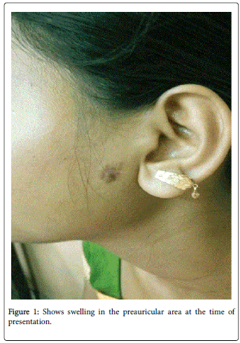
A 27 year old female patient attended our ENT outdoor with a mass in the left preauricular area for last 1 year. The swelling was insidious in onset and gradual on progession. On inspection the swelling was smooth surfaced with well-defined margin having dimension of 1.5 cm*1 cm. On palpation it was stony hard and free from skin and underlying structures. On ultrasonography it showed an ovoid mass with 1.2*0.8 cm dimension at the junction of dermis and subcutaneous fat having well defined margin, heterogenous echotexture, hypoechoic rim and calcification. There was no cervical lymph node enlargement or facial muscle weakness. Excision biopsy was planned under local anaesthesia. Macroscopically it was a grayish mass with 1.2*0.8*0.5 cm dimension. Histopathology report revealed multiple islands of epithelial cells composed mainly of eosinophilic shadow cells, which are pathognomonic for pilomatrixoma (Figures 1 and 2).
Discussion
Pilomatrixoma, or calcifying epithelioma of Malherbe, is a benign epithelial neoplasm. When Malherbe and Chenantais first mentioned this tumor in 1880, they described this as a calcifying epithelioma arising from sebaceous glands [1,6]. Forbis and Helwig later discovered that the origin of pilomatrixoma is the outer sheath of the hair follicle root [2,7]. These tumours are commonly present in the head and neck region but occurrence in other parts of the body has also been reported. These tumors are most common in 0-20 years of age group [6,9,10]. Pilomatrixoma presents usually as a single asymptomatic nodule. The skin over the tumour is mostly normal but occasionally may have a reddish or bluish discoloration [10]. These tumours are well circumscribed ovoid or spherical in shape and sometimes it may be encapsulated [9,10]. Multiple tumours are associated with Gardner syndrome, Turner syndrome, myotonic dystrophy,sarcoidosis and Steinert disease [6]. Malignant transformation of pilomatrixoma though reported are rare [8,12]. The histopathological characteristics of a pilomatrixoma is a well circumscribed tumor which is surrounded by a connective tissue capsule. Usually pilomatixoma is situated in the subcutaneous layer or dermis of skin and composed of islands of epithelial cells which are made up of varying amounts of basaloid matrical cells and often shows some cystic changes [6,9]. As the tumor matures, there is a a central degeneration of these basaloid cells.This degeneration forms the ghost (anucleated shadow) cells that is the cental unstained area which is a histopathological feature of pilomatrixoma [6,9]. As the tumour matures the basaloid cells decrease in number and the ghost cells predominates [11]. But these ghost cells, are not pathognomonic histopathological feature of pilomatrixomas. The presence of inflammatory reaction, central calcifications,foreign body giant cells and keratin debris is also characteristic. With the use of von Kossa stain, cal 75% of the tumors calcium deposits are found [11]. Trichilemmal cysts which keratinize losing their nuclei and undergo calcification should be histopathologically differentiated from pilomatrixoma.There is a palisading pattern of the peripheral basophilic cells in the trichilemmal cysts which are not found in pilomatrixoma [6,9,12]. The clinical differential diagnosis of these tumours should include sebaceous cyst, dermoid, epidermoid cysts, metaplastic bone formation, foreign body reaction, trichoepithelioma, heamatoma and basal cell epithelioma [8,12]. Surgical excision is the treatment of choice.After adequate exicision recurrence of the tumour is rare.After surgical excision of the tumor dermatological evaluation and long-term follow-up is mandatory [6]. This case is being reported because this is a rare tumour and it should be differentiated from other soft tissue tumours.
References
- Malherbe A, Chenantais J (1880) Note sur l’ epitheliomacalcifie des glandes sebacees. Prog Med 8:826-828.
- Forbis R Jr, Helwig EB (1961) Pilomatrixoma (calcifying epithelioma).Arch Dermatol 83: 606-618.
- Moehlenbeck FW (1973) Pilomatrixoma (calcifying epithelioma). A statistical study.Arch Dermatol 108: 532-534.
- Hills RJ, Ive FA (1992) Familial multiple pilomatrixomas.Br J Dermatol 127: 194-195.
- Knight PJ, Reiner CB (1983) Superficial lumps in children: what, when, and why?Pediatrics 72: 147-153.
- Birman MV, McHugh JB, Hayden RJ, Jebson PJ (2009) Pilomatrixoma of the forearm: a case report.Iowa Orthop J 29: 121-123.
- Marrogi AJ, Wick MR, Dehner LP (1992) Pilomatrical neoplasms in children and young adults.Am J Dermatopathol 14: 87-94.
- Zaman S, Majeed S, Rehman F (2009) Pilomatricoma-study on 27 cases and review of literature. D:/Biomedica25: 69–72.
- Kaddu S, Soyer HP, Cerroni L, Salmhofer W, Hödl S (1994) Clinical and histopathologic spectrum of pilomatricomas in adults.Int J Dermatol 33: 705-708.
- Schweitzer WJ, Goldin HM, Bronson DM, Brody PE (1989) Solitary hard nodule on the forearm. Pilomatricoma.Arch Dermatol 125: 828-829, 832.
- Peterson WcJr, Hult Am (1964) Calcifying Epithelioma of Malherbe.Arch Dermatol 90: 404-410.
- Chuang CC, Lin HC (2004) Pilomatrixoma of the head and neck.J Chin Med Assoc 67: 633-636.
Citation: Sannigrahi R, Saha J, Ghosh D, Biswas D, Basu SK, et al. (2016) Pilomatrixoma-A Case Report. Otolaryngol (Sunnyvale) 6:251. DOI: 10.4172/2161-119X.1000251
Copyright: © 2016 Sannigrahi R, et al. This is an open-access article distributed under the terms of the Creative Commons Attribution License, which permits unrestricted use, distribution, and reproduction in any medium, provided the original author and source are credited.
Share This Article
Recommended Journals
Open Access Journals
Article Tools
Article Usage
- Total views: 12377
- [From(publication date): 8-2016 - Apr 07, 2025]
- Breakdown by view type
- HTML page views: 11481
- PDF downloads: 896


