Case Report Open Access
Physiotherapy Management of Peripheral Unilateral Isolated Hypoglossal Nerve Palsy in a 53 Year Old Woman: A Case Report
| Maduagwu Stanley M1*, Kaidal Amina2, Aremu Bukuola J1, Kodiya Aliyu M3, Adetoyeje Y Oyeyemi4, Adewale Oyeyemi L4, Jaiyeola Olabode A1 and Mungonu Mustapha1 |
|
| 1Department of Physiotherapy, University of Maiduguri Teaching Hospital, Nigeria | |
| 2Department of Physical and Health Education, University of Maiduguri, Nigeria | |
| 3Department of Ear, Nose and Throat, University of Maiduguri Teaching Hospital, Nigeria | |
| 4Department of Physiotherapy, University of Maiduguri, Nigeria | |
| Corresponding Author : | Maduagwu Stanley M Department of Physiotherapy University of Maiduguri Teaching Hospital Bama Road, Maiduguri, Borno State, Nigeria Tel: +2348034998207 E-mail: madusta@yahoo.com |
| Received February 21, 2014; Accepted July 14, 2014; Published July 16, 2014 | |
| Citation: Stanley MM, Amina K, Bukuola AJ, Aliyu KM, Oyeyemi AY, et al. (2014) Physiotherapy Management of Peripheral Unilateral Isolated Hypoglossal Nerve Palsy in a 53 Year Old Woman: A Case Report. J Nov Physiother 4:218. doi: 10.4172/2165-7025.1000218 | |
| Copyright: © 2014 Stanley MM, et al. This is an open-access article distributed under the terms of the Creative Commons Attribution License, which permits unrestricted use, distribution, and reproduction in any medium, provided the original author and source are credited. | |
Visit for more related articles at Journal of Novel Physiotherapies
Abstract
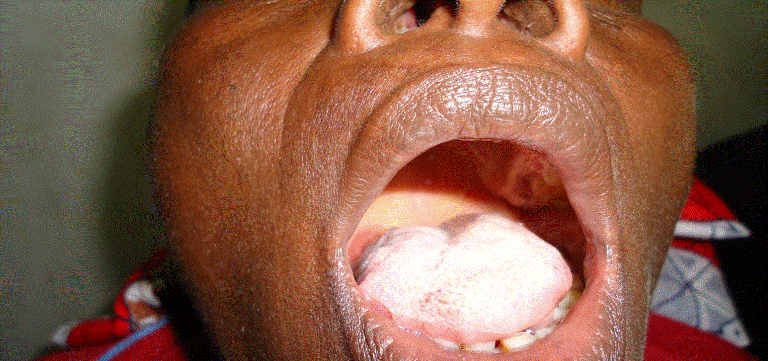
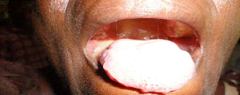
Hypoglossal nerve palsy is not common in literature, especially its physiotherapy assessment and management. We described a case of a 53 year old woman who presented clinically with right sided atrophy of the tongue, ipsilateral deviation and fasciculation, a speech defect, inability to swallow solids, and difficulty in swallowing fluid as a result of one year history of hypoglossal nerve palsy after a traditional uvulectomy. She was fully assessed and received physiotherapy management in the form of electrical stimulation and a home programme in combination with spiritual therapy. After 11 weeks of treatment there was improvement in terms of her initial clinical presentation.
The tongue (Latin. lingua; Greek. glossa) is a highly mobile muscular organ that varies greatly in shape and size. Its main functions include mastication, taste, deglutition (swallowing), articulation (speech), and oral cleansing. The tongue is divided into two halves; each half has four extrinsic and four intrinsic muscles. The extrinsic muscles comprise the genioglossus, hyoglossus, styloglossus and palatoglossus. The superior and inferior longitudinal, transverse and vertical musculatures are the intrinsic muscles of the tongue [2]. Motor innervations of the tongue are as documented above [1]. Sensory innervations depend on the part of the tongue, and can be either general sensory or special sensory or both. The general sensory innervations of the anterior two-third of the tongue is by the lingual nerve, a branch of the 5th cranial nerve, i.e. the trigeminal nerve, while the special sensory innervations come from the chorda tympani nerve, a branch of the facial nerve (7th cranial nerve). For the posterior one-third of the tongue, the glossopharyngeal nerve, the 9th cranial nerve, is responsible for both the general and special innervations [2].
The hypoglossal nerve palsy unlike the facial nerve palsy is seldom reported in literature. Most available studies focus mainly on aetiology, diagnosis, medical and surgical management, and prognosis and most are primarily case reports. To our knowledge, no study has been published reporting on the physiotherapy management of this condition, probably due to its rare occurrence. The purpose of this case report was therefore, to illustrate the outcome of physiotherapy management of hypoglossal nerve palsy as presented in a 53 year old woman who underwent a traditional uvulectomy two weeks prior to the clinical presentation. Our client indicated her consent for this publication by signing a written informed consent.
History
She was a known hypertensive, diagnosed in 2006, and claimed to take antihypertensive drugs as prescribed by a physician, but had no history of diabetes or rheumatoid arthritis. A full time house wife in a monogamous home with six children, she did not smoke cigarettes or drink any alcoholic beverages. She was a choir leader at her church. She has no formal education.
She was referred by the ENT Surgery department with right hypoglossal nerve palsy on the 5th June 2012 for physiotherapy at the University of Maiduguri Teaching Hospital, Maiduguri, Nigeria.
Laboratory and radiological investigations before her referral as documented in her case note were as follows: Complete blood cell count, erythrocyte sedimentation rate (ESR), urinalysis and other biochemical findings (e.g. fasting blood glucose, electrolyte, urea and creatinine) and thyroid function test were normal. Herpes simplex virus, retroviral screening and auto-antibodies tests were negative. Chest X-ray, computed tomography scanning and Magnetic Resonance Imaging (MRI) were unremarkable. Cranial and cervical MRI did not show any brain tumor or ischaemic lesion. On the MRI, other cranial nerves were normal.
We planned the following treatment: (1) spiritual therapy, (2) electrical stimulation, and (3) a home programme. The essence of spiritual therapy was to enable the client to cope with the psychological aspect of her condition. She was quite depressed and anxious. As a result, we instructed her to be prayerful and have faith in God. Evidence has suggested that certain spiritual beliefs and the practice of prayer are associated with improved coping and better health outcomes [5-7].
To commence electrical stimulation, the client laid comfortably on a couch. The right cheek and the ipsilateral lower jaw were thoroughly cleaned with sterilized water. Two pieces of lint cloths were soaked in saline water and gently squeezed to get rid of excess water. Passive and active electrodes were inserted inside each of the lint and placed at the cervical and right buccinator regions respectively. The salty water was to increase the conductivity of the electrodes. The parameters for the electrical stimulation were mono-rectangular waveform with 280 ms phase duration and 1.5 ms phase interval. The frequency, intensity, and duration of treatment were 0.80 Hz, 20 mA, and 10 minutes respectively. After this phase, the active electrode was transferred to the right lower plathysma region. The same parameters were repeated and frequency of treatment was 3 times per week. We used Sonoplus 992, 2600 AV Delft, Netherland, for the electrical stimulation. She was instructed to move the tongue as far as she could and as frequently as possible as her home programme.
Our patient may have resolved naturally with time; however unresolved cases of hypoglossal nerve palsy treated only with drugs have been previously reported [3,9,10]. Fernandes [9] presented a middle aged woman with a history of five months unresolved hypoglossal nerve palsy who was later considered for radiotherapy due to background metatastic disease. Loro and Owens [10] described a patient who was treated with drugs and followed up by a neurologist at 7 months post injury and the patient reported no real change or improvement from his 2-month post injury visit. Marwah et al. [3] reported a case of a patient with hypoglossal nerve palsy who was placed on drugs and examined periodically every other week, and showed no recovery at six months post lesion, although the authors asserted that the patient was more comfortable while speaking after receiving speech therapy. The spontaneous recovery following hypoglossal nerve palsy does not appear common.
Mathey et al. [11] posited that 12% of patients with carotid artery dissection present with hypoglossal nerve palsy and most dissections heal spontaneously. The authors reported that the tongue movement of their patient returned to normal within a few weeks. This complete resolution, according to the authors was due to spontaneously healing of the carotid artery dissection which initially was a source of neurapraxia in the hypoglossal nerve. Unfortunately ��?a few weeks� was not defined by the authors. Cheong et al. [12] described a 32 year old man who presented with hypoglossal nerve palsy due to compression by a pulsating normal vertebral artery and underwent microvascular decompression. After 3-month post-surgery, the patient reported marked improvement of deviation of the tongue, but not total resolution.
When a nerve��?s function is impaired, the impulses it sends to the muscle it supplies is diminished or totally ceased depending on the type and severity of lesion that causes the impairment. This nerve lesion could be neurapraxia, axonotmesis or neurotmesis [13], and each result to temporary or permanent denervation of the muscle. Based on this knowledge, the parameters we chose were therefore specifically to stimulate the denervated tongue muscles. The highest possible intensity of the electrical stimulating machine that was utilized is 100 mA; we applied 20% (20 mA) of this intensity based on our patient��?s tolerance level. We would have adjusted the parameters to suit the patient in case of any discomfort. The authors believe that the method utilized in managing this patient was a novel idea, since there is dearth of literature on physiotherapy management of hypoglossal nerve palsy. Future scholars may improve on our adopted method as well as validate what we had done.
The rarity of this condition means that the development of guidelines for physiotherapy management need to be developed by meticulous and consistent reporting of cases.
Electrical stimulation may be one possible physiotherapy intervention with which to manage hypoglossal nerve palsy.
References
- Prives M, Lysenkov N and Bushkovich V (1989) Human Anatomy (3rd edn): The science of the vessels, the science of the nervous system, the science of the sensory organs. USSR: Mir Publishers, pp 281, 293, 296.
- Wilson-Pauwels L, Akesson EJ and Stewart PA (1998) Cranial Nerves ��? Anatomy and Clinical Comments. Philadelphia: B.C. Decker, Inc.
- Marwah N, Agnihotri A and Goel M (2008) Idiopathic unilateral isolated hypoglossal nerve palsy: a case report. Journal of Oral Health and Community Dentistry 2(3):62-64.
- Arnheim D and Prentice W (1997) Management of Cerebral Concussion in Sports. The Athletic Trainer��?s Perspective. Principles of Athletic Training (8th edn). McGraw-Hill: St Louis, MO.
- Ellison CG, Levin JS (1998) The religion-health connection: evidence, theory, and future directions. Health EducBehav 25: 700-720.
- Levin JS (2001) God, faith, and health: exploring the spirituality��?healing connection. New York: John Wiley and Sons.
- Powell LH, Shahabi L, Thoresen CE (2003) Religion and spirituality. Linkages to physical health. Am Psychol 58: 36-52.
- Shin DS, Bae HG, Shim JJ, Yoon SM, Kim RS, et al. (2012) Morphometric study of hypoglossal nerve and facial nerve on the submandibular region in korean. J Korean NeurosurgSoc 51: 253-261.
- Fernandes R (2010) Unusual presentation of more common disease/injury: Metastatic disease causing unilateral isolated hypoglossal nerve palsy. British Medical Journal doi:10.1136/bcr.05.2010.2998.
- Loro WA, Owens B (2009) Unilateral hypoglossal nerve injury in a collegiate wrestler: a case report. J Athl Train 44: 534-537.
- Mathey DG, Wandler A, Rosenkranz M (2010) Images in cardiovascular medicine. Hypoglossal-nerve palsy caused by carotid dissection. Circulation 121: 457.
- Cheong JH, Kim JM, Yang MS and Kim CH (2011) Resolution of isolated unilateral hypoglossal nerve palsy following microvascular decompression of the intracranial vertebral artery. Journal of Korean Neurosurgery Society. 49: 167-170.
- Seddon HJ (1943) Three types of nerve injury. Brain 66: 237- 288 .
Figures at a glance
 |
 |
| Figure 1 | Figure 2 |
Relevant Topics
- Electrical stimulation
- High Intensity Exercise
- Muscle Movements
- Musculoskeletal Physical Therapy
- Musculoskeletal Physiotherapy
- Neurophysiotherapy
- Neuroplasticity
- Neuropsychiatric drugs
- Physical Activity
- Physical Fitness
- Physical Medicine
- Physical Therapy
- Precision Rehabilitation
- Scapular Mobilization
- Sleep Disorders
- Sports and Physical Activity
- Sports Physical Therapy
Recommended Journals
Article Tools
Article Usage
- Total views: 18684
- [From(publication date):
September-2014 - Apr 02, 2025] - Breakdown by view type
- HTML page views : 13936
- PDF downloads : 4748
