Phantom Tumor: A Source of Diagnostic Error
Received: 06-Jun-2023 / Manuscript No. roa-23-106682 / Editor assigned: 08-Jul-2023 / PreQC No. roa-23-106682 (PQ) / Reviewed: 22-Jul-2023 / QC No. roa-23-106682 / Revised: 24-Jul-2023 / Manuscript No. roa-23-106682 (R) / Published Date: 31-Jul-2023 DOI: 10.4172/2167-7964.1000467
Abstract
Phantom tumors are usually found in the minor fissure, but they can also be found, although rarely, in the oblique fissure. Interlobar Pseudo-tumor also known as phantom tumor, or vanishing tumor, is a localized collection of fluid that occurs between two lobes of the lung. While phantom tumors are relatively rare, they are important to recognize as they can be mistaken for pleural-based diseases, including neoplasia.
Keywords
Phantom tumor; Chest RX; TDM
Case History
60-year-old man, chronic weaned smoker, with a history of hypertension, ischemic heart disease and mitral valve replacement, presented with recurrent dyspnea and dry cough enduring five days.
physical examination revealed a dyspneic patient (NYHA 4) with swollen legs, auscultation reveals crackling noises, laboratory tests showed an increase in white blood cells (16000).
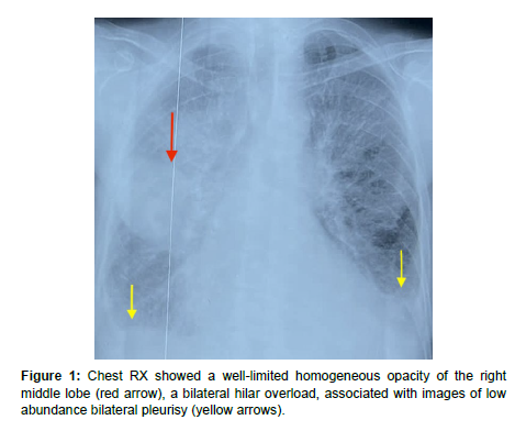
The chest X-ray (Figure 1) showed a well-limited homogeneous opacity of the right middle lobe (red arrow), a bilateral hilar overload, associated with images of low abundance bilateral pleurisy (yellow arrows).
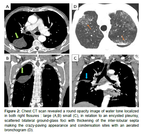
In addition to these lesions the chest CT scan (Figure 2) revealed a round opacity image of water tone localized in both right fissures: large (A,B) small (C), in relation to an encysted pleurisy, scattered bilateral ground-glass foci with thickening of the inter-lobular septa making the crazy-paving appearance and condensation sites with an aerated bronchogram (D).
Figure 2: Chest CT scan revealed a round opacity image of water tone localized in both right fissures : large (A,B) small (C), in relation to an encysted pleurisy, scattered bilateral ground-glass foci with thickening of the inter-lobular septa making the crazy-paving appearance and condensation sites with an aerated bronchogram (D).
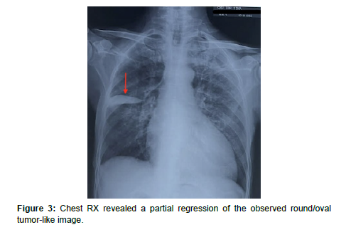
The diagnosis of cardiac decompensation due to infectious pneumonia was made and the parenteral antibiotic + diuretic therapy was immediately initiated with reduction of fluid intake. Clinical improvement has been noted and three days later, a control C-XR (Figure 3) revealed a partial regression of the observed round/oval tumor-like image.
Commentary
Scissural effusion in congestive heart failure is rare uncommon but well known entities. It resembles a mass and resolves rapidly, and is also known as a disappearing tumor. It is difficult to estimate the incidence because of the small number of cases reported. Phantom tumour is most common on the right hemithorax in men. It is mainly seen in the large fissure, less frequently in the small fissure and rarely in both fissures [2].
Pleuritis causing adhesions and obliteration in the pleural space also plays a key role in the pathogenesis. Treatment is diuretics along with treatment of the underlying aetiology [2].
Characteristic radiographic phantom lung tumor finding was discovered: right sided, well delimited pulmonary mass with smooth margins. Rapid resolution of the pseudo tumor after management of the left heart failure provided additional evidence for the diagnosis [1].
The differential diagnosis of loculated pleural effusions within the fissure includes the following: transudates (left ventricular failure or renal failure), exudates (parapneumonic pleural effusions, malignant pleural effusions, and benign asbestos-related pleural effusions), and hemothorax, chylothorax and fibrous tumors originating from the visceral pleura of the interlobar fissure [1]. Treatment is diuretics along with treatment of the underlying etiology.
References
- Sandal R, Jandial A, Mishra K, Malhotra P (2018) Phantom tumour and heart failure. BMJ Case Rep 2018: bcr2018227364.
- Argan O, Ural D (2017) Phantom tumor of the lung in heart failure patient. Turk J Emerg Med 17: 121-122.
Indexed at, Google Scholar, Crossref
Citation: Rostoum S, Zhim M, Chorfa SH, Naggar A, Fenni JE, et al. (2023) Phantom Tumor: A Source of Diagnostic Error. OMICS J Radiol 12: 467. DOI: 10.4172/2167-7964.1000467
Copyright: © 2023 Rostoum S, et al. This is an open-access article distributed under the terms of the Creative Commons Attribution License, which permits unrestricted use, distribution, and reproduction in any medium, provided the original author and source are credited.
Share This Article
Open Access Journals
Article Tools
Article Usage
- Total views: 1141
- [From(publication date): 0-2023 - Mar 29, 2025]
- Breakdown by view type
- HTML page views: 917
- PDF downloads: 224


