Case Report Open Access
Percutaneous Vertebroplasty Painful Malignant Involvement of the Second Cervical Vertebra (C2): Case Report
| Rispoli R1*, Zofrea G1, Bartolini N2, Caputo N2 and Carletti S1 | |
| 1Department of Neurosurgery, S.Maria Hospital, Terni, Italy | |
| 2Department of Neuroradiology, S.Maria Hospital, Terni, Italy | |
| Corresponding Author : | Rispoli R Department of Neurosurgery, “S. Maria” Hospital, Terni, Italy Tel: 00393331163632 E-mail: rossella.rispoli@libero.it |
| Received June 06, 2014; Accepted July 11, 2014; Published July 13, 2014 | |
| Citation: Rispoli R, Zofrea G, Bartolini N, Caputo N, Carletti S (2014) Percutaneous Vertebroplasty Painful Malignant Involvement of the Second Cervical Vertebra (C2): Case Report. J Pain Relief 3: 151. doi: 10.4172/2167-0846.1000151 | |
| Copyright: © 2014 Rispoli R, et al. This is an open-access article distributed under the terms of the Creative Commons Attribution License, which permits unrestricted use, distribution, and reproduction in any medium, provided the original author and source are credited. | |
Visit for more related articles at Journal of Pain & Relief
Abstract
The axis is an important element of the musculoskeletal complex in the upper cervical spine. It is surrounded by a number of delicate neurological and vascular structures and controls a wide range of movements. Thus, a pathological C2 fracture is a threatening condition. Multiple myeloma and osteolytic metastases are the most frequent malignant lesions affecting the spine. However, the cervical spine, especially the C1 and C2 region, seems to be involved less often. In this study, we report a case of unstable C2 fracture treated with the percutaneous vertebroplasty. Vertebroplasty is a well-established procedure for pain control and stabilization of vertebral pathology including metastasis, hemangioma, and multiple myeloma. The PVP procedure allows the option of preserving the mobility of the upper cervical spine
In this study, we report a case of unstable C2 fracture treated with the percutaneous vertebroplasty. Vertebroplasty is a well-established procedure for pain control and stabilization of vertebral pathology including metastasis, hemangioma, and multiple myeloma.
The PVP procedure allows the option of preserving the mobility of the upper cervical spine.
The patient sought help from his primary care physician who prescribed prescription analgesics, anti-inflammatory medication, muscle relaxants and physical therapy
Despite ten days of therapy, he had inadequate pain relief and he presented to the Emergency room to our Hospital.
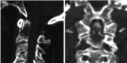
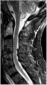
The X-ray images demonstrated a fracture in C2; a CT of the cervical spine was positive for a lytic lesion involving the C2 vertebral body and odontoid process with an underlying pathologic fracture (Figure 1a,1b); magnetic resonance imaging (MRI) demonstrated a focal area of signal abnormality with a pre-dominantly hypointense complex lesion on T1 weighted images, and hyperintense lesion on T2 weighted images. This MRI confirmed the presence of a lytic/complex cystic lesion at the base of dens with extension into the C2 vertebral body (Figure 2). At this point, the patient was transferred to Neurosurgery Unit. Total-body CT was performed and demonstrated a mass in the right upper lobe of the lung. We recommended biopsy and possible vertebroplasty. The patient provided informed consent after an explanation of the risks/benefits and indications for the procedure. The procedure was performed in the X-ray suite with a single-plane Angiography C-arm system. The patient was in the supine position and following the routines for percutaneous vertebroplasty (PVP) procedures at the cervical spine at our unit, under general anaesthesia. A fibrotic nasal intubation was performed to minimize movement of the neck. The patient was given a prophylactic dose of an antibiotic.
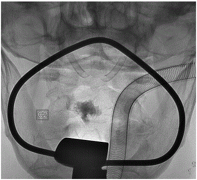
The otolaryngology service placed an oropharyngeal retractor (Crowe–Davis mouth gag) within the patient’s oral cavity in order to adequately expose the oropharynx. The soft palate was tethered superiorly using red rubber catheters. The oral cavity and oropharynx were then prepped using Beta dine swabs, and a 22-gauge, 5 in. spinal needle was used to localize the best approach toward the left aspect of the C2 vertebral body, in the location of the lytic lesion seen on the CT scan and avoiding the retropharyngeal right internal carotid artery. 10-gauge guide needle was then placed into the C2 vertebral body over an introducer stylet under fluoroscopic guidance. A 12-gauge biopsy needle was then advanced through the kyphoplasty guide needle into the C2 vertebral body beyond the anterior aspects of the guide needle. The biopsy needle was retracted under gentle suction, and specimens were obtained for histology. A trocar needle filled with high viscosity radiopaque poly-methyl-methacrylate (PMMA) was then placed within the guide needle. Each batch of PMMA was mixed with 500 mg vancomycin powder. Under intermittent fluoroscopic visualization, 2 cc of PMMA was slowly hand injected into the C2 vertebral body (Figure 3). The guide catheter was then removed.
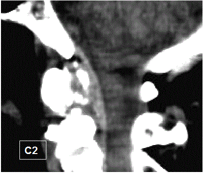
The patient tolerated the vertebroplasty of C2 without complications and he had adequate pain relief. Follow-up CT images of the upper cervical spine 3 months after the PVP procedure showed that he cement cast is unchanged. A clear osteosclerotic reaction occurred around the cement cast (Figure 4).
Otolaryngologists and neurosurgeons have extensively used the transoral approach to access the upper cervical spine. This is a well-established access route in the otolaryngological and neurosurgical literature [3,4] a thin layer of fascia and muscles separates the upper cervical spine from the oral mucosa. One of the potential downsides of the transoral cervical spine surgical approach is a reported risk of wound infection up to 2% and a risk of meningitis up to 4.5%. Extension of infection into facial planes can cause a retropharyngeal abscess and invasion of the meningeal layers leading to meningitis and encephalitis. The rate of infection has dropped significantly in recent years, due to the improvement in aseptic techniques, perioperative antibiotics, and thorough cleansing of the surgical bed prior to the procedure.
Percutaneous vertebroplasty (PVP) of the axis is a challenging procedure which may be performed by a percutaneous or a transoral approach [5,6]. There are few reports of PVP at the C2 level. Transoral vertebroplasty is postulated to have a lower risk of infection given the minimal tissue disruption by the needle. The addition of peri-operative intravenous antibiotics, antibiotics within the PMMA mixture, and post-procedural antibiotics should further reduce the infection risk.
Severe neck and back pain and reduced mobility are the most common symptoms in patients with vertebral metastases. The treatment is basically conservative, with analgesics, cytostatic, radiotherapy, reinforced corset and/or neck brace.
In patients with osteolytic lesions in the cervical spine and refractory pain, with or without fracture dislocations, different types of surgical stabilization methods have traditionally been used. The least invasive method consists of the halo-vest treatment, which can safely be performed concurrently with the medical treatment. This form of treatment may lead to bone reconstitution and stability. Posterior osteosynthesis with or without decompression involves permanent fixation from occipital bone to C4 with restriction of flexion, extension, bending and rotation. The procedure has a low risk of complications, provides immediate stabilization of the C2 lesion and allows safe mobilization of the patient with a neck brace.
During recent years an increasing number of patients with C2 metastases have been treated with PVP. This has offered rapid and long-lasting pain relief in up to 85% of cases [7].
Vertebroplasty is a well-established procedure for pain control and stabilization of vertebral pathology including metastasis, hemangioma, and multiple myeloma.
The PVP procedure allows the option of preserving the mobility of the upper cervical spine. On the other hand, posterior stabilization leads to fixation from the occipital bone to the C4 vertebra, resulting in a considerable decrease in mobility. If sufficient stabilization is not achieved with PVP, the surgical option is still available.
We think that PVP is a less aggressive procedure than any surgical stabilizing procedure in the upper cervical spine and does not restrict the mobility of the occipito-cervical junction [8]. The patient can usually walk a couple of hours after treatment, and can be discharged from hospital within 24 h of the procedure. Considering the higher risk of complications of PVP in this region, it is highly recommended that the treatment be performed by an experienced specialist at a centre where many PVP procedures are carried out and which has excellent radiological equipment.
References
- Fung KY, Law SW (2005) Management of malignant atlanto-axial tumours. J OrthopSurg 13: 232–239.
- Jenis LG, Dunn EJ, An HS (1999) Metastatic disease of the cervical spine. A review ClinOrthopRelat Res 359: 89–103.
- Bonney G, Williams JP (1985) Trans-oral approach to the upper cervical spine. A report of 16 cases. J Bone Joint Surg Br 67: 691- 698.
- Spetzler RF, Hadley MN, Sonntag V (1988) the transoral approach to the anterior supe- rior cervical spine. A review of 29 cases. ActaNeurochirSuppl 43: 69-74.
- Guglielmi G, Andreula C, Muto M, Gilula LA (2005) percutaneous vertebroplasty: indications, contraindications, technique, and complications. ActaRadiol 46: 256–268
- Levine SA, Perin LA, Hayes D, Hayes WS (2000) an evidence-based evaluation of percuta- neousvertebroplasty. Manag Care. 9: 56-60.
- Cotten A, Boutry N, Cortet B, Assaker R, Demondion X, et al. (1998) Percutane- ousvertebroplasty: state of the art. Radio- graphics. 18: 311-323.
- Martin JB, Gailloud P, Dietrich P-Y, Luciani ME, Somon T (2002) Direct transoral approach to C2 for percutaneous vertebroplasty. CardiovascInterventRadiol. 25: 517–519
Figures at a glance
 |
 |
 |
 |
| Figure 1 | Figure 2 | Figure 3 | Figure 4 |
Relevant Topics
- Acupuncture
- Acute Pain
- Analgesics
- Anesthesia
- Arthroscopy
- Chronic Back Pain
- Chronic Pain
- Hypnosis
- Low Back Pain
- Meditation
- Musculoskeletal pain
- Natural Pain Relievers
- Nociceptive Pain
- Opioid
- Orthopedics
- Pain and Mental Health
- Pain killer drugs
- Pain Mechanisms and Pathophysiology
- Pain Medication
- Pain Medicine
- Pain Relief and Traditional Medicine
- Pain Sensation
- Pain Tolerance
- Post-Operative Pain
- Reaction to Pain
Recommended Journals
Article Tools
Article Usage
- Total views: 13491
- [From(publication date):
September-2014 - Jul 06, 2025] - Breakdown by view type
- HTML page views : 8965
- PDF downloads : 4526
