Research Article Open Access
Decreased Radiation Exposure with Percutaneous Shielded Lead Compared to Unshielded Implant for Spinal Cord Stimulation
| Nathalie Zaidman1, Brice Constant2, Cristo Chaskis1 and Laurence Abeloos1,2* | |
| 1Department of Neurosurgery, CHU Charleroi, Belgium | |
| 2Pain Clinic, CHU Charleroi, Belgium | |
| Corresponding Author : | Laurence A Department of Neurosurgery, CHU Charleroi Chaussée de Bruxelles 140 6042 Charleroi, Belgium Tel: +32.71.92.23.63 Fax: +32.71.92.23.67 E-mail: laurenceabeloos@gmail.com |
| Received November 11, 2014; Accepted December 22, 2014;; Published December 24, 2014 | |
| Citation:Zaidman N, Constant B, Chaskis C, Abeloos L (2014) Decreased Radiation Exposure with Percutaneous Shielded Lead Compared to Unshielded Implant for Spinal Cord Stimulation. J Pain Relief 3: 166. doi: 10.4172/2167-0846.1000166 | |
| Copyright: ©2014 Zaidman N, et al. This is an open-access article distributed under the terms of the Creative Commons Attribution License, which permits unrestricted use, distribution and reproduction in any medium, provided the original author and source are credited. | |
Visit for more related articles at Journal of Pain & Relief
Abstract
Background: Spinal cord stimulation (SCS) is a safe and effective treatment for refractory failed back surgery syndrome (FBSS). Two implantation procedures exist depending on the use of a surgical versus percutaneous lead. Percutaneous leads implantation is less invasive but the placement procedure requires a higher X-ray exposure. The new MRI compatible shielded percutaneous leads (Vectris®) appear to be stiffer and therefore seem to be easier to implant compared to unshielded percutaneous electrodes (Octad®). The aim of this study is to compare the radiation exposure between percutaneous shielded leads and percutaneous unshielded leads in SCS implantation.
Setting: Retrospective study.
Material and methods: 20 patients successively underwent SCS for FBSS. The first 10 patients were implanted with an Octad ® lead and the 10 following with a shielded Vectris ® lead. All patients were operated on by the same surgical team. In all cases, the same intra-operative X-rays device (Ziehm Vision 8000®) was used, with identical parameters aiming at a minimal X-rays exposure (pulse mode with 2 impulses/sec, and automatic mode for kVp and mAs).
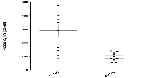
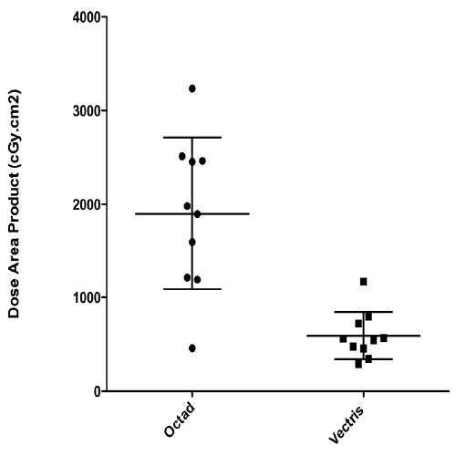
Results: Fluoroscopy time was significantly lower in patients implanted with the Vectris® lead (mean: 99 seconds) compared to the Octad® lead (mean: 291 seconds) (p=0.0035). Subsequently, total patient X-rays exposure in the Vectris group (mean 592 cGycm2) was significantly lower than in the Octad® group (mean: 1899 cGycm2) (p=0.0005).
Conclusion: The use of MRI compatible shielded percutaneous leads significantly reduces the fluoroscopy time and the total X-ray exposure in SCS implantation.
The new MRI compatible shielded percutaneous leads (Vectris®) appear to be stiffer and therefore seem to be easier to implant compared to unshielded percutaneous electrodes (Octad®).
The aim of this study is to compare the radiation exposure between percutaneous shielded leads and percutaneous unshielded leads in SCS implantation.
Setting: Retrospective study.
Material and methods: 20 patients successively underwent SCS for FBSS. The first 10 patients were implanted with an Octad ® lead and the 10 following with a shielded Vectris ® lead. All patients were operated on by the same surgical team. In all cases, the same intra-operative X-rays device (Ziehm Vision 8000®) was used, with identical parameters aiming at a minimal X-rays exposure (pulse mode with 2 impulses/sec, and automatic mode for kVp and mAs).
Results: Fluoroscopy time was significantly lower in patients implanted with the Vectris® lead (mean: 99 seconds) compared to the Octad® lead (mean: 291 seconds) (p=0.0035).
Subsequently, total patient X-rays exposure in the Vectris group (mean 592 cGycm2) was significantly lower than in the Octad® group (mean: 1899 cGycm2) (p=0.0005).
Conclusion: The use of MRI compatible shielded percutaneous leads significantly reduces the fluoroscopy time and the total X-ray exposure in SCS implantation.
Neurosurgeons generally favor the surgical approach, while interventional pain clinicians are more comfortable with the percutaneous approach. But putting personal preferences aside, both approaches come with advantages and disadvantages. The implantation of surgical paddle lead is more invasive [4] and therefore tends to be performed under general anesthesia without the ability to test the coverage of the pain area [5,6]. However, due to the shape of the lead and the greater number of stimulation contacts, surgical electrodes are more efficient in the treatment of lower back pain [6,7]. Another advantage of surgical lead is that the paddle placement is performed under visual control, thereby requiring less fluoroscopy [4]. Alternatively to the surgical approach, SCS electrodes can be implanted percutaneous. This less invasive procedure is done under local anesthesia; the patient is awake and can guide the surgeon in the exact positioning of the electrode, to insure coverage of pain area [8]. Because this procedure is less invasive, postoperative pain is reduced, as is the length of hospitalization. The main concerns with the use of percutaneous leads are the difficulty to cover lower back pain and the X-ray exposure for lead placement [9,10]. X-rays are known to be the cause of various harmful biological effects due to the production of free radicals, involved in carcinogenesis [11]. The biological deterministic effects of radiation from fluoroscopy depend on the dose, duration and distance of X-ray source [11,12]. Part of the energy released by the X-ray is deflected and scattered from its original path with less energy. This secondary scattered radiation does not contribute to the radiographic image and can be hazardous to the operator. It is therefore important to protect patients from such effects, especially given that radiation dose is cumulative [12]. One of the main concerns regarding percutaneous lead implantation is that the technique relies essentially on the use of X-ray fluoroscopy to guide needle insertion and lead placement, with an X-ray exposure significantly higher than in surgical electrode implantation. Recently, a newly developed, MRI compatible, percutaneous SCS electrode has been developed (Vectris®, Medtronic Neuromodulation, Minneapolis, MN, USA). The structure of this lead is shielded and therefore more rigid. Due to this property, the Vectris® electrode seems to be easier to control and to implant for SCS. The aim of this study is to compare X-ray exposure in patients implanted with a percutaneous unshielded (Octad®) versus shielded lead (Vectris®).
Eligible patients suffered from FBSS with neuropathic pain predominantly in the legs, of an intensity of at least 50 mm on a visual analogue scale (VAS: 0 equaling no pain, to 100 mm representing the worst possible pain). The pain was present for at least 6 months after a minimum of one anatomically successful lumbar surgery (i.e., lumbar disc surgery, laminectomy with or without foraminotomy and/or spinal fusion).
The neuropathic nature of pain was checked as per routine practice at the center (i.e. by clinical investigation of pain distribution, DN4 questionnaire, examination of sensory/motor/reflex change, with supporting tests such as X-ray, MRI and EMG).
Conservative treatments including opioids, anti-depressant drugs, gabapentin, multiple infiltrations, physiotherapy, TENS, and others were tried but were not efficient.
All cases were discussed during a multidisciplinary meeting comprising of neurosurgeon, pain clinician and psychiatrist, before a spinal cord stimulation trial was decided. All patients then underwent a four week screening trial with a percutaneous lead.
The needle was then removed and the lead fixed to the muscular fascia with an Injex® Anchoring System (Models 97791 from Medtronic Inc., Minneapolis, MN, USA). To avoid migration of the lead, a loop was formed and the lead connected to an external extension for the 4-weeks trial period.
A pulsed mode fixed at 2-pulses per second was used in all procedures. The applied constructs of the radiation safety program are described here. Fluoroscopic imaging was properly used, in that the fluoroscope was only activated when localizing, adjusting, or advancing a needle/lead. The low dose and automatic brightness control features were used with a 23 cm field of view (FoV) (i.e., the normal magnification mode). The C-arm anti-scatter grid was not removed. Automatic kilovolt peak/kilovolt potential (kVp) and milliampers (mA) determine the best images on the screen with lower radiation doses. Also, an electronic collimation was used and no zoom function was applied.
The fluoroscopy system automatically tabulated and stored total fluoroscopy time (in seconds) and DAP (in cGycm2) per case. Analysis of fluoroscopy time and DAP was conducted by means of summary statistics (GraphPad Prism version 6.00 for Mac OS X, GraphPad Software, La Jolla California USA). An unpaired t-test with Welch’s correction was used for statistical analysis because the two groups do not have equal standard deviation.
Multiple clinical studies have demonstrated the efficiency and safety of spinal cord stimulation in that indication [2,14]. Two types of leads can be used for the procedure: a surgical multicolumn paddle lead or a cylindrical percutaneous lead. Though the multicolumn surgical paddle electrode seems to be more efficient in alleviating the low back pain component [6,7] both types of leads are equivalent in the treatment of radicular neuropathic leg pain. In this study, the neuropathic radicular component was more important than low back pain, which is why percutaneous electrodes were implanted. Percutaneous leads are advantageous since they require a less invasive implantation procedure, which can be performed under local anesthesia, with feedback while the patient is awake. However, fluoroscopic guidance is required and with it a non-negligible radiation exposure for the physician, the patient, as well as the operating room staff. In the past few years, the assessment of radiation dose has received more attention. Especially in the demonstration of deterministic effects of X-ray correlated to the dose and for which dose thresholds have been fixed [9,15,16]. Radiation exposure can also be associated to biological modifications leading to stochastic adverse effects. Multiple dose reduction techniques exist, but the material used is also an important factor in terms of ease of placement. The shielded MRI compatible electrode is more rigid, making it easier to place, without relying so much on fluoroscopic guidance.
So far, only one article has been published in regards to X-ray exposure in SCS percutaneous lead implantation [10]. In this retrospective study, 110 SCS trailing procedures were performed by the same interventional spine team in an outpatient interventional pain management practice. The aim of the study was to report mean fluoroscopy time in the placement of percutaneous SCS lead, thereby providing a dosimetric reference level for such procedures. The mean total fluoroscopy time was 133.4 seconds (SD: 84.8 seconds). The authors concluded that fluoroscopy time associated with SCS may be considerable, compared to others interventional pain treatments. They suggested that deterministic effects exist but however, remain rare [10].
In our preliminary study, we compared radiation exposure between the implantation of unshielded versus the shielded percutaneous leads. We have found the mean fluoroscopy time as well as patient X-ray exposure to be significantly lower in patients implanted with the shielded electrode compared to the unshielded one. Those results reinforce the argument that the more rigid structure of the new lead allows for a better guidance, therefore decreasing radiation needs for the lead placement. Nevertheless, our study has its limitations among which its small sample size and the retrospective aspect. Further prospective studies with larger samples of patients still need to be performed. Those encouraging results must of course be associated with the knowledge of fundamental technical means to maximally reduce X-ray exposure, for patients as well as for practitioners. First of all, the fluoroscopy system has to comply with all federal and state rules/regulations, as well as manufacturer calibrations and physics acceptance testing. The fundamental principles of radiation protection include: 1) a perfect positioning of the C-arm fluoroscope in order to maximize the distance between the patient and operator from the radiation dose, 2) minimizing exposure time by using fluoroscopy only when necessary to check needle position and lead placement, and by avoiding the use of the C-arm by anyone else than the operator.
Multiple dose reduction techniques must been applied such as: 1) using pulse/boost fluoroscopy mode, 2) adjusting the beam quality, 3) maximally reduce the number of pulses per seconds when working on automatic mode, 4) avoiding the use of the zoom and using the collimation, 5) removing the grid, when possible, 6) last image holding. Finally, the application of the ALARA principle (as low as reasonably achievable) will keep radiation within the recommended and safe limits [16,17].
References
- Shealy CN, Mortimer JT, Reswick JB (1967) Electrical inhibition of pain by stimulation of the dorsal columns: preliminary clinical report. AnesthAnalg 46: 489-491.
- Frey ME, Manchikanti L, Benyamin RM, Schultz DM, Smith HS, et al. (2009) Spinal cord stimulation for patients with failed back surgery syndrome: a systematic review. Pain physician 12: 379-397.
- North RB, Kumar K, Wallace MS, Henderson JM, Shipley J (2011) Spinal cord stimulation versus re-operation in patients with failed back surgery syndrome: an international multicenter randomized controlled trial (EVIDENCE study). Neuromodulation : journal of the International Neuromodulation Society 14: 330-335.
- Pahapill PA (2013) Surgical Paddle-Lead Placement for Screening Trials of Spinal Cord Stimulation. Neuromodulation: journal of the International Neuromodulation Society.
- North RB, Kidd DH, Olin J, Sieracki JN, Petrucci L (2006) Spinal cord stimulation for axial low back pain: a prospective controlled trial comparing 16-contact insulated electrodes with 4-contact percutaneous electrodes. Neuromodulation: journal of the International Neuromodulation Society 9: 56-67.
- Rigoard P, Desai MJ, North RB, Taylor RS, Annemans L (2013) Spinal cord stimulation for predominant low back pain in failed back surgery syndrome: study protocol for an international multicenter randomized controlled trial (PROMISE study). Trials 14: 376.
- Song JJ, Popescu A, Bell RL (2014) Present and potential use of spinal cord stimulation to control chronic pain. Pain physician 17: 235-246.
- Kinfe TM, Schu S, Quack FJ, Wille C, Vesper J (2012) Percutaneous implanted paddle lead for spinal cord stimulation: technical considerations and long-term follow-up. Neuromodulation : journal of the International Neuromodulation Society 15: 402-407.
- Wininger KL (2012) Benefits of inferential statistical methods in radiation exposure studies: another look at percutaneous spinal cord stimulation mapping [trialing] procedures. Pain physician 15: 161-170.
- Wininger KL, Deshpande KK, Deshpande KK(2010) Radiation exposure in percutaneous spinal cord stimulation mapping: a preliminary report. Pain physician 13: 7-18.
- Wagner LK, Eifel PJ, Geise RA (1994) Potential biological effects following high X-ray dose interventional procedures. Journal of vascular and interventional radiology : JVIR 5: 71-84.
- Hall EJ (1994)Radiobiology for the radiologist. Philadeplphia.
- Hoy D, Brooks P, Blyth F, Buchbinder R (2010) The Epidemiology of low back pain. Best Pract Res ClinRheumatol 24: 769-781.
- Dario A, Fortini G, Bertollo D, Bacuzzi A, Grizzetti C, et al. (2001) Treatment of failed back surgery syndrome. Neuromodulation: journal of the International Neuromodulation Society 4: 105-110.
- Geleijns J, Wondergem J (2005) X-ray imaging and the skin: radiation biology, patient dosimetry and observed effects. Radiation protection dosimetry 114: 121-125.
- Mahesh M(2001) Fluoroscopy: patient radiation exposure issues. Radiographics: a review publication of the Radiological Society of North America, Inc 21: 1033-1045.
- Manchikanti L, Datta S, Smith HS, Hirsch JA (2009) Evidence-based medicine, systematic reviews, and guidelines in interventional pain management: part 6. Systematic reviews and meta-analyses of observational studies. Pain physician 12: 819-850.
Tables and Figures at a glance
| Table 1 |
Figures at a glance
 |
 |
| Figure 1 | Figure 2 |
Relevant Topics
- Acupuncture
- Acute Pain
- Analgesics
- Anesthesia
- Arthroscopy
- Chronic Back Pain
- Chronic Pain
- Hypnosis
- Low Back Pain
- Meditation
- Musculoskeletal pain
- Natural Pain Relievers
- Nociceptive Pain
- Opioid
- Orthopedics
- Pain and Mental Health
- Pain killer drugs
- Pain Mechanisms and Pathophysiology
- Pain Medication
- Pain Medicine
- Pain Relief and Traditional Medicine
- Pain Sensation
- Pain Tolerance
- Post-Operative Pain
- Reaction to Pain
Recommended Journals
Article Tools
Article Usage
- Total views: 13770
- [From(publication date):
January-2015 - Jul 13, 2025] - Breakdown by view type
- HTML page views : 9240
- PDF downloads : 4530
