Pellegrini-Stieda Syndrome: A Rare Complication of Knee Entorse
Received: 04-Jun-2022 / Manuscript No. roa-22-66819 / Editor assigned: 06-Jun-2022 / PreQC No. roa-22-66819 (PQ) / Reviewed: 20-Jun-2022 / QC No. roa-22-66819 / Revised: 23-Jun-2022 / Manuscript No. roa-22-66819 (R) / Published Date: 30-Jun-2022 DOI: 10.4172/2167-7964.1000389
Abstract
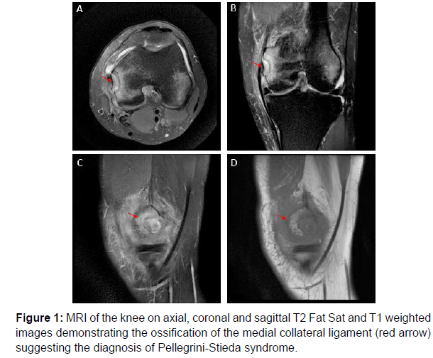
Axial, coronal and sagittal views of knee MRI of a 56 years old man with recurrent knee pain following a knee sprain, showing a calcification in the soft tissue situated mesial to the medial femoral condyle (red arrows) corresponding to the ossification of the medial collateral ligament, suggesting the diagnosis of Pellegrini-Stieda syndrome.
Keywords
Pellegrini-Stieda; Posttraumatic; MRI; Ossification; Medial collateral ligament
Introduction
The Pellegrini-Stieda syndrome is relatively infrequent and is commonly associated with sporting injuries [1, 2]. It occurs within 11 days to 6 weeks after trauma and patients may be asymptomatic or present a local pain and swelling in the medial aspect of the knee (Figure1).
The diagnosis can be suggested on the X-ray, and confirmed by the MRI which also delineates the extent of adhesion of the calcified mass to the MCL and the remaining ligamentous volume.
References
- Mendes LFA, Pretterklieber ML, Cho JH, Garcia GM, Resnick DL, et al. (2006) Pellegrini-Stieda disease: a heterogeneous disorder not synonymous with ossification/calcification of the tibial collateral ligament-anatomic and imaging investigation. Skeletal Radiol 35: 916-922.
- Miller TT (2009) Imaging of the medial and lateral ligaments of the knee. Semin Musculoskelet Radiol 13: 340-352.
Indexed at, Google Scholar, Crossref
Citation: Boularab J, Sahli H, Izi Z, Edderai M (2022) A Rare Complication of Knee Entorse: Pellegrini-Stieda Syndrome. OMICS J Radiol 11: 389. DOI: 10.4172/2167-7964.1000389
Copyright: © 2022 Boularab J, et al. This is an open-access article distributed under the terms of the Creative Commons Attribution License, which permits unrestricted use, distribution, and reproduction in any medium, provided the original author and source are credited.
Share This Article
Open Access Journals
Article Tools
Article Usage
- Total views: 1681
- [From(publication date): 0-2022 - Mar 29, 2025]
- Breakdown by view type
- HTML page views: 1281
- PDF downloads: 400

