Pediatric Renal Trauma : A Case Report and Imaging Finding
Received: 04-Mar-2024 / Manuscript No. roa-24-131433 / Editor assigned: 06-Mar-2024 / PreQC No. roa-24-131433 / Reviewed: 20-Mar-2024 / QC No. roa-24-131433 / Revised: 25-Mar-2024 / Manuscript No. roa-24-131433 / Published Date: 29-Apr-2024
Abstract
Pediatric abdominal trauma is a frequent occurrence. Post traumatic renal injuries rarely occur in isolation; instead, they often coincide with multiorgan injuries. We present the case of a 9-year-old child, victim of a road traffic accident. A full body CT was performed showing a right renal hematoma associated with this is a large adjacent hemoperitoneum.
Keywords
Renal trauma; CT; Renal hematoma; Hemoperitoneum
Image Article
Pediatric abdominal trauma is a frequent occurrence, with approximately 5% to 20% of children experiencing blunt abdominal trauma also suffering from renal trauma [1]. Unlike adults, children’s kidneys are less secured, being fixed primarily by the vascular pedicle and the ureter, specifically the pelviureteric junction. They are enveloped by Gerota’s fascia, their capsule, and a thinner, more pliable layer of perirenal fat. Additionally, due to incompletely ossified lower ribs and the natural disposition of renal lobulations, injury forces may propagate along these planes [2].
Post traumatic renal injuries rarely occur in isolation; instead, they often coincide with multiorgan injuries involving the liver, spleen, closed head, and orthopedic fractures [3].
Renal imaging is typically indicated in scenarios such as penetrating trauma, blunt trauma accompanied by hematuria or hypotension, flank hematoma, or rib or lumbar spine fractures [4]. Furthermore, computed tomography (CT) stands as the preferred modality for assessing hemodynamically stable patients following blunt abdominal trauma [2].
We present the case of a 9-year-old child, with no particular pathological history, who suffered a closed trauma with cranial and abdominal point of impact following a road traffic accident. Clinically, the patient was conscious, well-oriented in time and space, severe abdominal pain without any external bleeding.
Upon inspection, there were skin bruises on the right flank, and upon palpation, there was generalized abdominal rigidity.
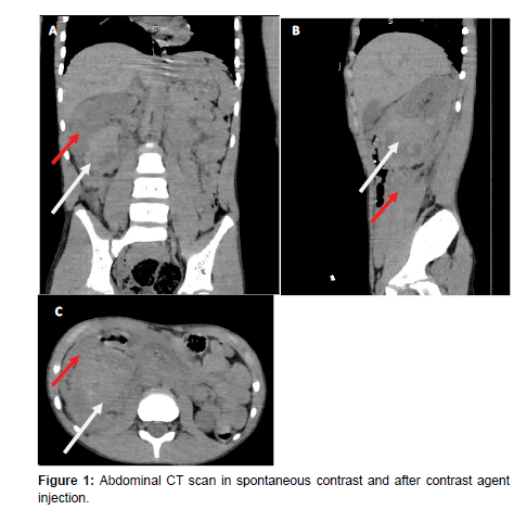
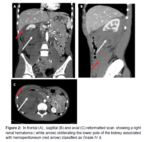
A full body CT was performed showing a right renal formation, oval-shaped, well-defined, obliterating the lower pole of the kidney and reaching the renal pelvis with hematoma density, measuring 58x55x64mm (Figure 1 and Figure 2).
Associated with this is a large adjacent peritoneal effusion of hematic density, in the right peri-renal area, extending to the perihepatic, right GPC and pelvic regions (Figure 1 and Figure 2). There was no urinary extravasation or vascular involvement (Figure 1).
References
- Fraser JD, Aguayo P, Ostlie DJ, St Peter SD (2009) Review of the evidence on the management of blunt renal trauma in pediatric patients. Pediatr Surg Int 25: 125–132.
- Martínez-Piñeiro L, Djakovic N, Plas E, Mor Y, Santucci RA, et al. (2010) EAU Guidelines on Urethral Trauma. Eur Urol 57: 791–803.
- Fernández-Ibieta M (2018) Renal Trauma in Pediatrics: A Current Review. Urology 113: 171–178.
- Salama H, Elshahawy A, Maboud Noha MA, Mashaly E (2020) Multidetector computed tomography in the diagnosis and staging of renal trauma. Tanta Med J 48: 152.
Indexed at, Google Scholar, Crossref
Indexed at, Google Scholar, Crossref
Indexed at, Google Scholar, Crossref
Citation: Loubna M, Sara Z, Nouha B, Lina B, Nazik A, et al. (2024) PediatricRenal Trauma: A Case Report and Imaging Finding. OMICS J Radiol 13: 548.
Copyright: © 2024 Loubna M, et al. This is an open-access article distributed underthe terms of the Creative Commons Attribution License, which permits unrestricteduse, distribution, and reproduction in any medium, provided the original author andsource are credited.
Share This Article
Open Access Journals
Article Usage
- Total views: 620
- [From(publication date): 0-2024 - Apr 04, 2025]
- Breakdown by view type
- HTML page views: 435
- PDF downloads: 185


