Paraurethral Cyst in Newborn
Received: 10-Apr-2018 / Accepted Date: 06-Jun-2018 / Published Date: 13-Jun-2018 DOI: 10.4172/2376-127X.1000381
Abstract
A girl was born at term. Four days old, an interlabial cyst appeared, located anterior to the vaginal introitus. The urethral meatus was normally positioned and there was no voiding difficulty. Interlabial cysts in newborn girls are rare and may have different origins. This girl had a so-called paraurethral cyst, which is often possible to recognize by clinical examination. This sort of cyst is generally completely asymptomatic and has a high rate of spontaneous involution. Without any treatment, the cyst had disappeared at examination two months later.
Keywords: Interlabial Cyst; Paraurethral Cyst; Renal Disorders; Gestational Diabetes
Introduction
Interlabial cysts in newborn girls are rare and prevalence are given from 1:1000 to 1:7000 but it may be higher than described in literature [1,2]. An interlabial cyst in a newborn girl may histologically be presented as a paraurethral cyst (skene duct cyst), Gartner duct cyst (a mesonephric ductal remnant) or a covered ectopic ureter. Paraurethral cysts usually have fine capillary vessels on the cyst surface. It is presumed that this sort of cyst appears when the outflow from the paraurethral gland is obstructed. If uncertain of the origin of the interlabial cyst, an abdominal ultrasound is needed in order to rule out renal disorders [3]. Differential diagnoses include imperforated hymen, urethral prolapse, urethral diverticulum, rhabdomyosarcoma of the vagina and prolapsed ectopic ureterocele [4]. The natural history of a congenital paraurethral cyst is one of spontaneous resolution within a few weeks after birth or rupture [5,6]. Surgical excision may be an option in a few cases with obstruction of urinary outflow, if the cyst recurs, or if it fails to resolve within a few months [7].
Case
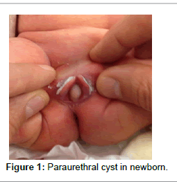
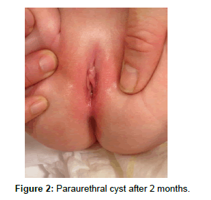
A girl was born at term, at gestational age 37 weeks and 3 days. Birthweight was 3415 g and Apgar scores were 10/1, 10/5 and 10/10. Pregnancy was only complicated by gestational diabetes. 4 days old, an interlabial cyst appeared located anterior to the vaginal introitus. The cyst was spherical, yellowish and with dilated blood vessels on the anterior surface (image A). There was no apparent voiding difficulty. The urethral meatus was normally positioned and not displaced. Consequently, it was found unnecessary to conduct further urologic or ultrasound examination. There was neither surgical intervention nor needle aspiration carried out for treatment. Spontaneous involution had occurred by examination two months later (Figures 1 and 2).
Discussion
Paraurethral glands and ducts are known to form as an outpouching of the urethra during the third trimester [8]. They are rudimentary homologues of the male prostate gland and empty into female urethra [4,8]. Paraurethral cysts are benign thin-walled, golden or whitish simple cysts. In general, there is no obstruction of the urinary tract and they are self-resolving. The largest of the paraurethral glands may have 6-30 ducts merging in the distal urethra [4]. The cause of ductal obstruction in newborns is unknown, whereas in adults, ductal obstruction is usually caused by inflammation [8]. One hypothesis suggests that dislocation of the urothelium into an adjacent area results in the blocking of skene’s ducts, leading to paraurethral cyst formation [8]. Others postulate that maternal estrogen exposure provokes glandular secretion in the newborn girl and stimulates the development of the cyst. The maternal hormones are eliminated during the newborn period, allowing spontaneous involution. In concordance with similar cases described in literature, the girl in the present case was born after a full-term pregnancy and by an uncomplicated vaginal delivery, with birth weight and Apgar scores within normal limits [8,9]. This might support the theory that the maternal hormones at or around, the term are important for the development of this condition. However, the true etiology remains unknown [4]. Paraurethral cysts are generally completely asymptomatic and have a high rate of spontaneous involution [9]. The diagnosis of paraurethral cysts can be made by clinical examination, and often further investigation is unnecessary. It is important to bear in mind that these cysts are generally benign and self-resolving, and there is normally no need for needle aspiration, drainage or marsupialization. There is no long-term sequalae from a paraurethral cyst.
Differential diagnosis must be kept in mind. Other types of interlabial masses, such as those of embryologic origin, ectopic tissue, prolapse, urologic anomalies and neoplasia, can superficially resemble simple cysts. These include prolapsed urethra, neonatal prolapsed ectopic ureterocele, prolapsed vagina or uterus. Most of these anomalies require some medical or surgical intervention [1]. Ultrasound examination and further urologic investigation are recommended in cases of urinary outflow obstruction.
References
- Shimizu M, Imai T (2013) Vaginal cyst in a newborn. J Pediatr 163: 1790.
- Badalyan V, Burgula S, Schwartz RH (2012) Congenital paraurethral cysts in two newborn girls: Differential diagnosis, management strategies and spontaneous resolution. J Pediatr Adolesc Gynecol 25: e1-e4.
- Tiwari C, Shah H, Desale J, Waghmare M (2017) Neonatal Gartner duct cyst: Two case reports and literature review. Dev Period Med 21: 35-37.
- Costantino E, Ganesan GS (2016) Skene's gland cyst as an interlabial mass in a newborn girl. BMJ Case Rep.
- Wright JE (1996) Paraurethral (Skene's duct) cysts in the newborn resolve spontaneously. Pediatr Surg Int 11: 191-192.
- Merlob P, Bahari C, Liban E, Reisner SH (1978) Cysts of the female external genitalia in the newborn infant. Am J Obstet Gynecol 132: 607-610.
- Ceylan H, Karakök M, Ozokutan BH, Sarsu SB (2002) Paraurethral cyst: Is conservative management always appropriate? Eur J Pediatr Surg 12: 212-214.
- Soyer T, Aydemir E, Atmaca E (2007) Paraurethral cysts in female newborns: Role of maternal estrogens. J Pediatr Adolesc Gynecol 20: 249-251.
- Fujimoto T, Suwa T, Ishii N, Kabe K (2007) Paraurethral cyst in female newborn: Is surgery always advocated? J Pediatr Surg 42: 400-403.
Citation: Moser LK, Fenger-Gron J, Khalil MR (2018) Paraurethral Cyst in Newborn. J Preg Child Health 5: 381. DOI: 10.4172/2376-127X.1000381
Copyright: © 2018 Moser LK, et al. This is an open-access article distributed under the terms of the Creative Commons Attribution License, which permits unrestricted use, distribution, and reproduction in any medium, provided the original author and source are credited.
Share This Article
Recommended Journals
Open Access Journals
Article Tools
Article Usage
- Total views: 17630
- [From(publication date): 0-2018 - Apr 29, 2025]
- Breakdown by view type
- HTML page views: 16556
- PDF downloads: 1074


