Papillon-Lefevre Syndrome: A Rare Case Report in Mongolia
Received: 04-Jun-2022 / Manuscript No. roa-22-66429 / Editor assigned: 06-Jun-2022 / PreQC No. roa-22-66429 (PQ) / Reviewed: 20-Jun-2022 / QC No. roa-22- 66429 / Revised: 23-Jun-2022 / Manuscript No. roa-22-66429 (R) / Published Date: 30-Jun-2022 DOI: 10.4172/2167-7964.1000386
Abstract
Papillon-Lefèvre Syndrome (PLS) is a rare genetic disorder, characterized by Palmoplantar keratosis, aggressive periodontitis and premature edentulous primary and permanent dentition at a very young age. Additional symptoms and findings associated with PLS may include frequent pyogenic skin infections, abnormalities of the nails, and excessive perspiration. Papillon-Lefèvre syndrome is inherited in an autosomal recessive pattern. The various etiological and pathogenic factors are associated with the syndrome: immunologic alterations, genetic mutation and the role of the pathogenic microorganism. Dentists, especially pediatric dentist is very important specialist to diagnosis and management of PLS, there were the periodontal destruction at early childhood. We introduce the clinical presentation and management options of 5 years old boy with PLS in Mongolia.
Keywords
Edentulous; Hyperkeratosis; Alveolar bone resorption; Genetic disorder; Immunoglobulin A
Introduction
Papillon-Lefèvre syndrome is an infrequent genodermal syndrome characterized initially by two French Physicians Papillon and Lefevre in 1924, in a brother and sister with palmoplantar hyperkeratosis accompanying with early onset periodontitis and premature loss of primary as well as permanent teeth [1, 2] Papillon-Lefevre Syndrome (PLS) is a rare autosomal recessive disorder featured by destructive periodontitis beginning in childhood, diffuse, transgradient palmoplantar keratoderma, premature loss of permanent teeth, and frequent cutaneous and systemic pyogenic infections [3-5].
Variety of systemic conditions increases patient susceptibility to periodontal disease, leading to more rapid and aggressive attachment loss. The fundamental factors are chiefly related to changes in endocrine, immune, and connective tissue status [6, 7]. These modifications can be related to diverse pathologies and syndromes that produce periodontal disease either as a principal manifestation or by provoking a preexisting condition related to local factors [4, 5]. This article describes a clinical presentation of boy diagnosed with PLS.
Case report
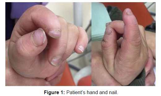
A 4-year-old male patient from Ulaanbaatar city of Mongolia reported to the Department of Pediatric and Preventive Dentistry, School of Dentistry, Mongolian National University of Medical Sciences, with a complaint of tongue ulcer and exposing root of deciduous teeth (Figure 1).
According to the patient’s parents, his deciduous teeth erupted normally, but exfoliated at the age of 3. He had suffered congenital immune deficiency and sensory neuropathy.
On physical examination, he is skinny and weak, hobble walking and hold to his mother occasionally, close narrow eyes, hand finger not stretched out completely, abnormal finger nails, the hyperkeratotic lesions on the palm were observed (Figure 1). His parents and relatives were not similar complications on his family history.
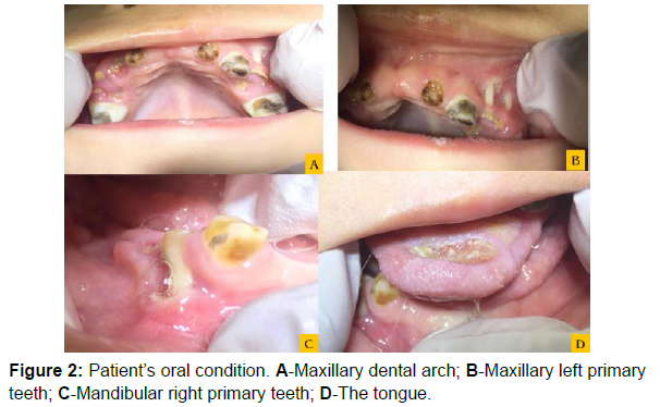
Intraoral examination had shown absence of primary maxillary central incisors and presence of maxillary molars, canine and the lateral incisors. In the mandible presence of first primary premolars and right primary canine and also other mandibular primary teeth had missing (Figure 2). The anterior third of tongue were traumatic ulcer by the maxillary left canine and lateral incisor (Figure 2).
Panoramic radiograph displayed the lower first permanent molars in the lower dental arch and the all permanent tooth germs, except #45 in mandible bone, when he was 4.5 years old. In the maxilla there were all permanent tooth germs, which are different stages of tooth development (Figure 3). The radiopaque image in the mesial side of the crown of mandibular right first permanent molar and alveolar bone was local horizontal resorption around this tooth (Figure 3).
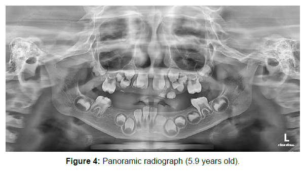
Panoramic radiograph, when he was 5.9 years old, displayed the lower first permanent molars and central incisors in the lower dental arch and the all permanent tooth germs were in next developmental stage than 4.5 years old. The new radiopaque image in the coronal occlusal side of mandibular right first permanent molar and alveolar bone was restored. The first permanent molar in the upper dental arch and the tooth germs of permanent premolar, canine and incisors were overlapped (Figure 4).
Diagnosis and Treatment
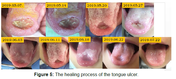
We offered “Papillon-Lefevre syndrome” based on patient’s clinical sign, radiographic image, palm hyperkeratosis, edentulous and the alveolar bone resorption. The treatment plan included the oral hygiene modification, the tongue ulcer treatment and the removable denture. During the oral hygiene modification some deciduous teeth fall prematurely by itself. We removed the traumatic agent, which was incisors enamel sharp edge for tongue ulcer, after two months full recovered (Figure 5). The patient goes to immune support treatment every two week by the 18 years old, we will be considered nonremovable denture (Figure 5).
Discussion
Papillon-Lefèvre syndrome is an infrequent genodermal syndrome characterized initially by two French Physicians Papillon and Lefevre in 1924, in a brother and sister with palmoplantar hyperkeratosis accompanying with early onset periodontitis and premature loss of primary as well as permanent teeth. Gorlin et al in 1964 added a third section of dural calcification to the diagnosis of this syndrome [1, 8].
Papillon-Lefèvre syndrome has been categorized into three groups: diffuse, focal, and punctuated [4]. The hyperkeratosis of the plantar surface often spreads to the edges of the soles and in some cases onto the skin overlying the Achilles tendon and the external malleoli. We observed from patient the only palmoplantar hyperkeratosis, nail shape abnormal and transverse grooving [9, 10].
The intraoral features that the premature loss of permanent and deciduous teeth mobility, aggressive periodontal destruction was reported by Nickles et al., [11]. Vassilopoulou and Laskaris [12]. The above mentioned clinical features were same our patient’s some clinical symptom, which were appeared tooth root on the gingiva and the alveolar bone resorption on the panoramic radiograph.
Haneke advised to make differential diagnosis of PLS the following three criteria (palmoplantar hyperkeratosis, autosomal recessive inheritance, loss of primary and permanent teeth) from prepubertal periodontitis and Haim-Munk syndrome [4, 12]. We observed in the case above 3 criteria.
In the press review the prevalence of PLS is about 1 to 4 per million people and with both parents as recessive carriers, there is a 25% chance of generating offspring with this syndrome [13, 14]. The fathers of patient had full lip and palate cleft and he is chromosomal disorder. However, the mother has not yet shown any symptoms the mother may have number of teeth that looked like she had been bitten by a source and had been cut.
The pathogenesis of PLS is still controversial, but two hypothesis is more prominent nowadays. First, some patients suffering from PLS display a cellular immune fault with reduced chemotactic and phagocytic function of neutrophils and other granulocytes. Second, some pathogenic microorganisms like Capnocytophaga gingivalis, Porphyromonas gingivalis, Actinobacillus actinomycetemcomitans, Peptostreptococcus micros, Fusobacterium nucleatum, and spirochetes have been concerned as the causal agents for periodontal problems in PLS. We had no possible to identify the pathogenic microorganisms in the periodontal pockets in this case. The patient’s organism is not able to produce immunoglobulin A, which is same features in the first hypothesis about PLS pathogenesis [2, 5, 15].
Identification of PLS at an early age and a multidisciplinary approach can improve the prognosis of these patients. Skin lesions are commonly treated with emollients, salicylic acid, and urea. Oral retinoids including acitretin and isotretinoin have also been reported for the treatment of keratoderma [11, 16]. Effective treatment of periodontal disease includes prompt institution of antibiotics with nonsurgical therapy, the modification of the patient’s oral hygiene, extraction of primary teeth, and periodontal maintenance visit every 3 months. There is no consensus regarding the use of antibiotics; however, the prescription of antibiotics in combination with periodontal therapy has shown some benefit [16, 17]. This case treatment consisted of the oral health modification and antibiotic therapy based on the result of the antibiotic susceptibility test of tongue ulcer. The patient is always admitted the recall system by pediatric dentist.
Conclusion
Papillon-Lefèvre syndrome can badly affect the psychological, social, and esthetic well-being of the patient at early age, as it is a devastating disease process associated with cutaneous involvement and partial or complete edentulism. The dentist is usually the first to diagnose this syndrome due to the involvement of the periodontium.
References
- Papillon MM, Lefevre P (1924) Deux cas de keratoderma palmaire et plantaire symmetrique familial (maladie de Meleda) chez le frère et la soeur: coexistence dans les deux cas d’alterations dentaire graves. Bull Soc Fr Dermatol Syph 31: 82-87.
- Kaur B (2013) Papillon Lefevre syndrome: a case report with review. Dentistry 3:1-4.
- Krol AL (2008) Keratodermas. Dermatology (2nd edn). Spain: Mosby pp: 777-789.
- Tasli L, Kacar N, Erdogan BS, Ergin S (2009) Successful treatment of Papillon Lefevre syndrome with a combination of acitretin and topical-PUVA; a four year follow up. J Turk Acad Dermatol 3: 1-4.
- Woo I, Brunner DP, Yamashita DD, Le BT (2003) Dental implants in a young patient with Papillon-Lefèvre syndrome: a case report. Implant Dent 12: 140-144.
- Galanter DR, Bradford S (1969) Case report. Hyperkeratosis palmoplantaris and periodontosis: the Papillon-Lefèvre syndrome. J Periodontol40: 40-47.
- Ingle JL (1959) Papillon-Lefèvre syndrome: precocious periodontosis with associated epidermal lesions. J Periodontol.30: 230-237.
- Gorlin RJ, Sedano H, Anderson VE (1964). The syndrome of palmar-plantar hyperkeratosis and premature periodontal destruction of the teeth. J Pediatr 65: 895-908.
- Dorairaj J, Selvaraj S, Sadique M, Shaw M, Jain SK (2012) Papillon Lefevre syndrome: a periodontist approach. Int J Case Rep Images3: 1-5.
- Pavankumar K (2010) Papillon-Lefèvre syndrome: a case report. Saudi Dent J22: 95-98.
- Nickles K, Schacher B, Schuster G, Valesky E, Eickholz P (2011) Evaluation of two siblings with Papillon-Lefèvre syndrome 5 years after treatment of periodontitis in primary and mixed dentition. J Periodontol 82: 1536-1547.
- Vassilopoulou A, Laskaris G (1989) Papillon-Lefèvre syndrome: report of two brothers. ASDC J Dent Child56: 388-391.
- Kaya FA, Polat ZS, Baran EA, Tekin GG (2008) Papillon-Lefèvre syndrome-3 years follow up: a case report. Int Dent Med Disord 1: 24-28.
- Griffiths WAD, Judge MR, Leigh IM (1998) Disorders of keratinization. Dermatology (6th edn), Oxford: Blackwell Scientific Publications pp: 1569-1571.
- Toomes C, James J, Wood AJ, Wu CL, McCormick D et al. (1999) Loss-of-function mutations in the cathepsin C gene result in periodontal disease and palmoplantar keratosis. Nat Genet 23: 421-424.
- Jain V, Gupta R, Parkash H (2005) Prosthodontic rehabilitation in Papillon Lefevre syndrome: a case report. J Indian Soc Pedod Prev Dent 23: 96-98.
- Ahmad M, Hassan I, Masood Q (2009) Papillon-Lefèvre syndrome. J Dermatol Case Rep 3: 53-55.
Indexed at, Google Scholar, Crossref
Indexed at, Google Scholar, Crossref
Indexed at, Google Scholar, Crossref
Indexed at, Google Scholar, Crossref
Indexed at, Google Scholar, Crossref
Indexed at, Google Scholar, Crossref
Citation: Jargaltsogt D, Rashsuren O, Bayarsaikhan O, Altannamar M (2022) Papillon-Lefèvre Syndrome: A Rare Case Report in Mongolia. OMICS J Radiol 11: 386. DOI: 10.4172/2167-7964.1000386
Copyright: © 2022 Jargaltsogt D, et al. This is an open-access article distributed under the terms of the Creative Commons Attribution License, which permits unrestricted use, distribution, and reproduction in any medium, provided the original author and source are credited.


