Like us on:
Research Article Open Access
Optimal Concentration of 2,2,2-Trichloroacetic Acid for Protein Precipitation Based on Response Surface Methodology
Albert N Ngo, Miezan JM Ezoulin, Ibrahima Youm and Bi-Botti C Youan*Laboratory of Future Nanomedicines and Theoretical Chronopharmaceutics, Division of Pharmaceutical Sciences, University of Missouri-Kansas City, Kansas City, Mo 64108, USA
- *Corresponding Author:
- Bi-Botti C Youan
Laboratory of Future Nanomedicines and Theoretical Chronopharmaceutics
Division of Pharmaceutical Sciences
University of Missouri-Kansas City
Kansas City, Mo 64108, USA
Tel: +816 2352410
Fax: 816 235 5779
E-mail: youanb@umkc.edu
Received date: June 25, 2014; Accepted date: August 05, 2014; Published date: August 08, 2014
Citation: Ngo AN, Ezoulin MJM, Youm I, Youan BC (2014) Optimal Concentration of 2,2,2-Trichloroacetic Acid for Protein Precipitation Based on Response Surface Methodology. J Anal Bioanal Tech 5:198 doi: 10.4172/2155-9872.1000198
Copyright: 2014 Ngo AN, et al. This is an open-access article distributed under the terms of the Creative Commons Attribution License, which permits unrestricted use, distribution, and reproduction in any medium, provided the original author and source are credited.
Visit for more related articles at Journal of Analytical & Bioanalytical Techniques
Abstract
For low protein concentrations containing biological samples (in proteomics) and for non proteinaceous compound assays (in bioanalysis), there is a critical need for a simple, fast, and cost-effective protein enrichment or precipitation method. However, 2,2,2-trichloroacetic acid (TCA) is traditionally used for protein precipitation at ineffective concentrations for very low protein containing samples. It is hypothesized that response surface methodology, can be used to systematically identify the optimal TCA concentration for protein precipitation in a wider concentration range. To test this hypothesis, a central composite design is used to assess the effects of two factors (X1 = volume of aqueous solution of protein, and X2 = volume of TCA solution 6.1N) on the optical absorbance of the supernatant (Y1), and the percentage of protein precipitated (Y2). Using either bovine serum albumin (BSA) as a model protein or human urine (with 20 ppm protein content), 4% w/v (a saddle point) is the optimal concentration of the TCA solution for protein precipitation that is visualized by SDS-PAGE analysis. At this optimal concentration, the Y2-values range from 76.26 to 92.67% w/w for 0.016 to 2 mg/mL of BSA solution. It is also useful for protein enrichment and xenobiotic analysis in protein-free supernatant as applied to tenofovir (a model HIV microbicide). In these conditions, the limit of detection and limit of quantitation of tenofovir are respectively 0.0014 mg/mL and 0.0042 mg/mL. This optimal concentration of TCA provides optimal condition for protein purification and analysis of any xenobiotic compound like tenofovir.
Keywords
Optimal concentration; Chemometric tool; Protein enrichment, 2,2,2-Trichloroacetic acid; Response surface methodology; Biochemical and spectral analysis, Tenofovir
Introduction
For low protein concentrations containing biological samples, especially intended for proteomic studies, protein enrichment is a critical step to obtaining sufficient quantities. There are several methods for protein enrichment and purification [1-3], however, each method has its own limitation. When lyophilization’s method or filtration method is used to concentrate a protein, they may also concentrate non-proteinaceous elements [4]. The dialysis method may remove interfering elements, but it cannot concentrate proteins [3,4]. A recent method using a spin filter unit [5] allows the separation of the proteins from interfering elements such as salts, SDS, and lipids [3]. However, this method has its own limitations including subsequent sample loss (yield 44%) especially when less than 50 μg of the protein sample is analyzed [6].
The use of organic reagents for protein precipitation is common during sample preparation prior to proteome analysis [7-11]. These agents remove interfering elements such as similar polysaccharides or natural products (tannins, alkaloids, pigments) [4,12,13]. Among these organic reagents, 2,2,2-trichloroacetic acid (TCA) is the most widely used chemical for protein precipitation, and TCA-mediated protein precipitation is also independent of the physico-chemical properties of proteins [14-17]. However, the use of commonly final concentrations (10% w/v or 20% w/v) of TCA solution was not successful in precipitating low amounts of protein (0.02-0.03 mg) in an aqueous protein solution [4,18]. Moreover, the use of higher concentrations of TCA can also degrade the quality of the sample [16,17]. Thus, it is extremely important to identify the optimal concentration of TCA that precipitates both a low (<0.02 mg/mL) and a high amount of protein (2-20 mg/mL) in an aqueous solution while simultaneously allowing protein free supernatant analysis of any analyte interfering with the protein at higher concentration. The motivation for this study came for the need for better bioanalytical method for microbicide analysis in simulated human body fluid while avoiding the interference between the absorbency of both drug and proteins present in these biological fluids. In this study, it is hypothesized that response surface methodology, [19- 23], can be used to identify the optimal concentration of TCA needed to solve this problem. To test this hypothesis, Bovine serum albumin (BSA), a well-characterized protein with a PI 5.6, was selected as model protein [16]. BSA is a globular unglycosylated serum protein, and the most abundant serum protein [24] with a molecular weight of 65,000 Daltons. The general structure of serum albumin is an α-helix that acts as a protein transporter for steroids,fatty acids, and thyroids hormones [25,26]. Tenofovir, which is a nucleotide analogue reverse transcriptase inhibitor used for the treatment of HIV infections, is used as a model analyte [27]. This hypothesis is tested with supporting biochemical and spectral analysis (e.g. BCA assay), proteomic analysis (SDS PAGE), and visual analysis of protein pellets.
Materials and Methods
Trichloroacetic acid solution (6.1 N), and bovine serum albumin (BSA), were purchased from Sigma-Aldrich (St Louis, Missouri, USA). All percentages of w/v TCA mentioned below are the final concentrations of TCA in solution after the addition of a known volume of the above TCA solution (6.1 N). Caution: TCA can cause chemical burns and is harmful if inhaled. All the proteins solutions are made in deionized water, and the stock solution of BSA has a concentration of 2 mg/mL if not mentioned. Fresh human urine sample (total protein content = 20 ppm) is received from the University of Missouri Kansas City’s student health and wellness in a sterile device
Method of protein precipitation
First, 6.1 N TCA solution, is added to the microcentrifuge tube containing the aqueous solution of protein. Second, the mixture is vortexed for 30 seconds at high speed using a vortex-genie 2 model G-560 purchased from Scientific Industries, Inc. (Bohemia, New York, USA). Third, the microcentrifuge tubes are placed in VWR 18R refrigerated microcentrifuge (VWR, Radnor, PA) with a temperature of 4°C or in ice for 15 minutes. The protein solutions (typically, 880.6 – 1559.4 μL) are then pelleted down by centrifugation at 14, 000 rpm for 15 minutes. Finally, the pellets are separated carefully from the supernatant upon removal of each microcentrifuge tube from the refrigerated microcentrifuge
Determination of the absorbance of the supernatant at 280 nm
For each absorbance measurement at 280 nm, the volume of the supernatant used is 1.3 mL, which is a mixture of the volume of the supernatant from each experiment (0.65 mL) and its identical replicated experiment (0.65 mL). The concentration of the protein left in the solution can be determined using the molar extinction coefficient of BSA. Deionized water is used as a blank in all readings of the absorbance. All UV measurements are carried out in triplicate on a Genesy 10 Bio Model UV-Vis spectrophotometer from Thermo Electron Corporation (Wisconsin, USA). The standard curve of the TCA absorbance (Y), made in deionized water at 280 nm, is Y = 0.01X (R2 = 0.9999), and X = % w/v TCA.
Bicinchoninic acid (BCA) method
The protein pellets are dissolved in buffer S1 for 15-20 minutes under continuous agitation with the above vortex-genie 2 model G-560 as shown in Scheme 1. The steps for the BCA assay are shown in Scheme 1, when the initial concentration of BSA is 2 mg/mL. Protein solution S (1 mL) is added directly to 1 mL of the BCA solution when the initial concentration of the BSA is in the range 0.008-0.04 mg/mL, considering the linearity working range used currently is 0-0.012 mg/ mL. For human urine, after the protein precipitation step, the pellets are washed with deionized water, before addition of the buffer solution prior to solubilization.
A critical step in the process requires that the volume of buffer S1 added to the pellet must be equal to the initial volume of protein added to the microcentrifuge tube to ensure the same treatment, and to accurately estimate the amount of proteins precipitated. This is shown in equation 1:
 (1)
(1)
The standard curve for protein absorbance (y) at 562 nm using BCA assay is y = 0.09557 x +0.0372 (R2 = 0.996), x = final concentration of protein in solution.
SDS-PAGE method
Three hundred and fifty microliters from the remaining protein solution dissolved in the above buffer (buffer S1) is mixed with 0.350 mL of the sample buffer [28] and then heated at 98.5°C to denature the proteins prior to electrophoresis for 5 min. The denatured proteins are then run in a gradient of 4-12% sodium dodecyl sulfate-polyacrylamide gel (SDS-PAGE) for identification by electrophoresis [28]. A volume of 0.01 mL of the sample is loaded into the gel, and the proteins are stained with Brilliant blue R, which was purchased from Sigma-Aldrich (St Louis, Missouri, USA).
Optimum concentration of TCA using central composite design: Central composite design (CCD), with five coded levels (Table 1), is used to elucidate the true optimal concentration of the TCA required for protein precipitation. The 13 experiments represent a CCD with 22 full factorial designs, four axial points, and a center point with 4 replications (Table 1). The mathematical model that derives from such a CCD is expressed as the following second-order polynomial equation:
| Independent variables | Level | ||||
|---|---|---|---|---|---|
| Coded values | -1.414 | -1 | 0 | +1 | +1.414 |
| X1 = Volume of protein 2 mg/mL solution (µL) | 880.6 | 980.0 | 1220.0 | 1460.0 | 1559.4 |
| X2 = Volume of 6.1 N TCA solution (µL) | 11.7 | 20 | 40 | 60 | 68.3 |
| Dependent variables | |||||
| Y1= Absorbance of the supernatant recorded at 280 nm | |||||
| Y2 = Percentage of protein precipitated (% w/w) | |||||
Table 1: Independent variables and their level in central composite design and dependent variables.
 (2)
(2)
Where, y is the predicted response or dependent variable (absorbance of supernatant or percentage protein precipitated), β0 is the y intercept term, βi is the linear coefficient, βii is the quadratic coefficient, βij is the interactive coefficient, and xi and xj are the coded variables.
Two independent variables (Table 1) are chosen based on preliminary screening studies, and the level of the two factors are chosen based on a steepest descent (ascent) method [29,30]. The protein concentration was 20 mg/mL in a mixture of human semen fluid simulant (HSFS) and human vaginal fluid simulant (HVFS). The volume ratio HSFS/ HVFS was 4/1(Tables S1 and S2, shown in Supplementary file), [31,32]
Table 1 shows the independent variables in physical units, with their associated coded values as well as the dependent variables. The second order model can be written in matrix notation as follows [33]:
 (3)
(3)
 (4)
(4)
x′ is the transpose of x,
Moreover, the eigenvalues {λi}, or characteristics roots of the matrix B, gives the nature of the response surface. The optimum point is maximum, minimum, or saddle point if {λi} are respectively positive, negative, or have different signs [33].
Application to production of protein-free supernatant for tenofovir analysis: Once the optimum concentration of TCA is found using the above CCD, the following method is used to analyze a model HIV microbicide, tenofovir TNF (0.01 mg/mL) interference with drug release in simulated body fluid containing bovine serum albumin, BSA (20 mg/mL) solution in comparison to the commonly used concentration of TCA for protein precipitation (10% w/v, 20% w/v).
Briefly 0.020 mL of a stock solution of TNF (0.7 mg/mL) is added into three different microcentrifuge tubes containing 1.324 mL, 1.240 mL and 1.030 mL of BSA solution (20 mg/mL), respectively. Next 0.056 mL, 0.140 mL and 0.350 mL of TCA solution 6.1 N are added into the (BSA + TNF) mixture so that the final concentration of TCA are respectively 4% w/v, 10% w/v and 20% w/v. The final concentration of TNF is 0.01 mg/mL in each final mixture. Finally the protein is precipitated as described above and the absorbance of the supernatant is recorded by optical scanning between 240-290 nm. Finally, the absorbance of the supernatant is compared to the absorbance of aqueous solutions of TCA (4%, 10% and 20%). The standard curve of the aqueous solutions of TCA and TNF absorbance (Y) recorded at 260 nm, is respectively Y (TCA) = 0.1702X – 0.0066 (R2=0.9997), and X = % w/v of TCA concentration; Y(TNF) = 0.0461x + 0.0045 (R2=1), x=TNF concentration (μg mL-1).
As suggested by ICH guideline (Q2B), validation of analytical procedures, the limit of detection (LOD) and limit of quantitation (LOQ) can be determined as follow:
LOD = 3.3 σ/S
LOQ = 10 σ/S
Where, σ is the standard deviation of the blank and S is the slope of the calibration of the analyte. The estimate of σ is carried out by measuring the absorbance of the supernatant (n = 5) after precipitation of the protein without TNF (analyte of interest). The absorbances of the blanks are 0.671 ± 0.019 (σ = 0.019) and 1.605 ± 0.047 (σ = 0.047) for 4%w/v and 10% w/v TCA when deionized water is used to setup the baseline, respectively. Using the above blank to setup the baseline, the standard curves (Figure S1 shown in Supplementary file) are Y = 0.045X - 0.0086 (R2 = 0.9976) (S = 0.045) and Y = 0.0383X - 0.0181 (R2 = 0.9952) (S = 0.0383), respectively when the protein is precipitated with 4% w/v TCA and 10% w/v TCA, where, Y and X are the absorbance recorded at 260 nm and the concentration (0.002-0.020 mg/mL) of TNF, respectively
Statistical analysis
The statistical analysis was performed using JMP�?? software version 10 (SAS Institute, Cary, NC). Polynomial equations of the response values of absorbance of the supernatant at 280 nm (Y1), and the percentage of precipitated protein (Y2), are derived from the total result of 13 runs in the above CCD design. Analysis of variance (ANOVA) is performed to ensure the model fit. Experimental variables that significantly affect these responses are identified through a Pareto chart. A theoretical optimum condition is obtained by setting the maximum desirability of maximum protein precipitation yield. A student t-test is used for the checkpoint analysis, and a P-value below 0.05 is considered statistically significant and warrants the rejection of the null hypothesis.
Results and Discussion
Table 2 shows the results of the absorbance of the supernatant and the percentage of the proteins precipitated using BCA assay obtained from the 13 experiments along with the final concentration % w/v TCA in solution.
| Experiment | Level of Controlled variables in Coded Form | Absorbance of the supernatant at 280 nm | percentage of protein precipitated (w/w) | % w/v TCA | |
|---|---|---|---|---|---|
| X1 | X2 | Y1 | Y2 | ||
| 1 | -1 | -1 | 0.033 | 84.1 | 2.00 |
| 2 | -1 | +1 | 0.062 | 81.67 | 5.77 |
| 3 | +1 | -1 | 0.067 | 66.15 | 1.35 |
| 4 | +1 | +1 | 0.049 | 92.67 | 3.95 |
| 5 | -1.414 | 0 | 0.049 | 85.43 | 4.35 |
| 6 | +1.414 | 0 | 0.037 | 89.74 | 2.50 |
| 7 | 0 | -1.414 | 0.725 | 61.34 | 0.95 |
| 8 | 0 | +1.414 | 0.065 | 82.16 | 5.30 |
| 9 | 0 | 0 | 0.049 | 80.2 | 3.18 |
| 10 | 0 | 0 | 0.054 | 82.11 | 3.18 1 |
| 11 | 0 | 0 | 0.052 | 82.01 | |
| 12 | 0 | 0 | 0.046 | 75.14 | |
| 13 | 0 | 0 | 0.049 | 83.10 | 3.18 |
14 replicates centers points
Table 2: Central composite design showing independent variables with measured responses.
The second order polynomial models as a result of the central composite design model are:
 (5)
(5)
 (6)
(6)
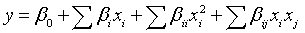
Where Y1 is the absorbance of the supernatant; Y2 is the percentage of protein precipitated; and X1 and X2 are the coded independent variables. Based on the above Equations 4 and 5, the vector b and the matrix B values are shown below:
 (7)
(7)
 (8)
(8)
The matrix B (Y1) has two real eigenvalues of opposite signs, indicating that the optimal solution to this optimization problem is a saddle point: {λi} = {-0.045, 0.131}. The matrix B (Y2) also has two real eigenvalues of opposite signs, indicating that the optimal solution is also a saddle point: {λi} = {-5.416, 5.311}.
Table 3 shows the ANOVA results to check the significance of the model parameters for both mathematical models that derived from the experimental design.
| Response | Source | DFa | SSb | MSc | F-ratiod | R2 |
|---|---|---|---|---|---|---|
| Y1 | Model | 5 | 0.253 | 0.051 | 2.125 | 60.28 |
| Error | 7 | 0.167 | 0.023 | Prob>F | ||
| Total | 12 | 0.420 | 0.177 | |||
| Y2 | Model | 5 | 818.630 | 163.730 | 16.446 | 92.15 |
| Error | 7 | 69.690 | 9.960 | Prob>F | ||
| Total | 12 | 888.32 | 0.001 |
adegree of freedom
bSum of Square
cMean sum of square
dModel MS/error MS
Table 3: Results of ANOVA analysis for the statistical model parameters for the absorbance of the supernatant (Y1), and the percentage of the protein precipitated (Y2).
Table 4 shows the lack-of-fit test to check the mathematical models adequacy. In other words, this test allows assessment if both equation models for Y1 and Y2 can adequately predict the absorbance of the supernatant and the percentage of protein precipitated, respectively.
| Response | Source | DFa | SSb | MSc | F-ratiod |
|---|---|---|---|---|---|
| Y1 | Lack-of-fit | 3 | 0.167 | 0.055 | 5857.55 |
| Pure error | 4 | 0.000 | 0.023 | Prob> F | |
| Total error | 7 | 0.167 | <0.0001 | ||
| Y2 | Lack-of-fit | 3 | 29.240 | 9.745 | 0.964 |
| Pure error | 4 | 40.450 | 10.112 | Prob>F | |
| Total error | 7 | 69.687 | 0.491 |
adegree of freedom
bSum of Square
cMean sum of square
dModel MS/error MS
Table 4: Lack-of-fit test analysis to check the model adequacy predicting the absorbance of the supernatant (Y1), and the percentage of protein precipitated (Y2).
The ANOVA results for the regression coefficients show that the regression coefficients for the model Y2 are significant considering 95% F distribution. The lack-of-fit test also shows that the model Y2 adequately fit the data and can predict the percentage of protein precipitated (Table 4). The protein absorbency recorded at 280 nm is reproducible, but the model Y1 cannot be used to adequately predict the final absorbance of the supernatant based on the lack-of-fit test [34] (Table 4). Moreover, the response surface and the contour plots, which derived from the CCD, are used to characterize the shape of the surface and can locate the optimum using computer software [33] (Figure 1).

Figure 1: Three-dimensional responses surface (A1), and contour plot (A2) showing the supernatant absorbance data (A) and those of the percentage of protein precipitated (B1 and B2) as a function of volume of protein solution and volume of TCA solution. The intersections of the two orthogonal lines, in figures (A2), and (B2) are the saddle point.
Checkpoints analysis for the prediction of model Y2
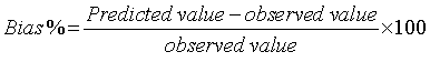
The second order statistical model (Y2) is checked in triplicate with two random points with respective (X1, X2) values of (-0.5, -0.5) and (+0.5, +0.5), in addition to the theoretically optimal point (+0.6, +1) shown in (Table 5). Bias for the fitted model (Y2) is computed using the following equation (Equation 9):
| Checkpoint # | X1 | X2 | Measured (Y2) % w/w | Predicted (Y2) % w/w | Bias |
|---|---|---|---|---|---|
| 1 | -0.5 | -0.5 | 84.62 (± 4.46) | 79.00 | -4.65 |
| 2 | +0.5 | +0.5 | 88.91 (± 2.03) | 85.59 | -4.36 |
| 3 | +0.6 | +1 | 86.92 (± 1.24) | 88.88 | +2.22 |
Table 5: Checkpoints experiments comparing measured and predicted percentage of protein precipitated.
 (9)
(9)
The results of the checkpoints analyses show that the predicted and measured values of the percentage proteins precipitated are statistically insignificant considering a 95% student’s t-distribution. The predicted percentage of protein precipitated (Y2), and the measured Y2 values, are statistically insignificant if the p-value (p) is greater than 0.05 using the student’s t-test. For checkpoint #1, the result is t = -1.78, degree of freedom (df) = 2, and p = 0.11. For checkpoint #2, t = -2.21, df = 2, and p = 0.08. For checkpoint #3, t = 2.26, df = 2, and p = 0.92. Thus, the model Y2 can accurately predict the Y2 values for a given volume of aqueous solution of BSA solution, and a given the volume of TCA solution 6.1N within the experimental design space. The Y2 values depend mainly on the volume of TCA and the interaction between both the volume of the protein solution and the volume of TCA (Figure 2). Based on the prediction profiler (Figure 3), the optimal percentage w/v of TCA in solution required to precipitate the maximum amount of protein in aqueous solution is 4.22% w/v TCA (rounded to 4% w/v). Using 4% w/v TCA, the percentage protein precipitated is 87.96 ± 1.55% w/w (n = 5).

Figure 2: Pareto chart showing the effect of the independent variables (X1, volume of protein solution; X2, volume of TCA solution), on the absorbance of the supernatant recorded at 280 nm and the percentage of protein precipitated using BCA assay. Sorted parameter estimates and their corresponding t-ratio are shown on the horizontal-axis. Bars extending beyond the vertical line indicate values reaching statistical significance (α = 0.05).

Figure 3: Prediction profiler and desirability showing the effect of the volume of protein solution and the volume of TCA solution on the absorbance of the supernatant measured at 280 nm and the percentage of protein precipitated using BCA assay.
SDS PAGE analysis
Figure 4 shows the intensity of the coomassie-stained protein band on the SDS gel for protein recovered in pellets after the precipitation phase. The intensities of the band are in agreement with the percentage of protein precipitate measured by the BCA assay. The orders of the lane are the points of the experimental design 1-9 (Table 2). Lanes 10- 11 show the band intensity for commonly used concentrations of TCA for protein precipitation; respectively 10% w/v TCA and 20% w/v TCA

Figure 4: SDS-PAGE analysis of BSA precipitation by TCA. Lane: 1, 2.00% TCA; 2, 5.78% TCA; 3, 1.35 % TCA; 4, 3.95%w/v TCA; 5, 4.35% TCA; 6, 2.50% TCA; 7, 0.95% TCA; 8, 5.30% TCA; 9, 3.18% TCA; 10, 10.00 %TCA; 11, 20.00% TCA.
Effect of TCA concentration on the percentage precipitated protein for lower concentration of proteins solution
Figure 5 shows the percentage of protein precipitate when the initial concentration of protein is within the range 0.008-0.040 mg/mL. The volume of protein solution used equals to 1 mL and the optimal concentration 4% TCA found from the experimental design is used to precipitate the proteins
Figure 6 shows the precipitate of proteins based on the final concentration of TCA solution used and the initial concentration of protein in solution. When the initial concentration of proteins is very low, between 0.024 mg/mL (total sample volume = 1mL) and 0.100 mg/ mL (total sample volume = 1 mL), there is no precipitation of protein when the final concentrations of TCA used are 10% w/v (Figure S2 shown in Supplementary file) and 20% w/v TCA (Figure S3 shown in Supplementary file), respectively. However the precipitation of protein is observed when the final concentration of TCA is 4% w/v TCA (Figure S4 shown in Supplementary file).
Application to supernatant “clarification” for tenofovir analysis
Figure 7B shows that the optimal concentration can be used to dramatically reduce the interference between BSA (20 mg/mL) and TNF while the commonly used concentration (20% TCA) (Figure 7A) is unable to reduce the interference.
The LOD and LOQ obtained using 4% w/v TCA are respectively, 0.0014 mg/mL and 0.0042 mg/mL. The LOD and LOQ also obtained using 10% w/v TCA are 0.0041 mg/mL and 0.0123 mg/mL respectively. Using 20% TCA, due to strong absorbance of the media, the absorbance of TNF cannot even be recorded as shown in Figure 7A.
Application to the precipitation of protein in human urine,
Figures 8, S5 and S6 (shown in Supplementary file) show the effectiveness of 4% w/v TCA for protein precipitation in fresh human urine containing low amount of protein. It clearly appears that 100% w/w of proteins are pelleted using 4%TCA, whereas the percentage is ∼50% w/w using 20% TCA.
Discussion
In this study, the optimal concentration of TCA required for maximal precipitation of both high (2-20 mg/mL) and low amounts of protein in aqueous solution (0.008-0.04 mg/mL) is determined using response surface methodology (RSM) with supporting biochemical, proteomic analysis, and visual data and evidence (Figure S7 shown in Supplementary file). The thorough RSM analysis clearly shows in Figure 1 that the stationary point of TCA-mediated protein precipitation is a saddle point. The precipitation of protein by TCA is specifically due to two properties, the acidity of TCA and the trichloro moiety that bear that molecule [16,17]. Molten globule’ or ‘A-state(s)’ (a “thermodynamic state, clearly different both from the native state and the denatured state of the protein), is a type of partially folded protein state [35]. The ‘A-state’ of protein is prone to stickily aggregate [16]. It is demonstrated that the precipitation of protein by TCA is governed by the formation of the sticky aggregation-like ‘A-state’ [16].
The percentage of proteins that precipitate varies from 76.29 to 92.67% w/w when the BSA concentration is in the range of 0.016-2 mg/ mL. Figure 5 and Table 2 show the percentage of protein precipitated for high and low amounts of aqueous solution of proteins, respectively. The precipitation of protein by TCA may be divided into two phases based on TCA concentration. When, the TCA concentration is less than 2% w/v, the protein precipitation is incomplete. In the second phase, as long as the concentration of TCA is greater than or equal to 2% w/v, more than 80% w/w of the protein is precipitated (Table 2). However, some traces of proteins are left in solution as shown by the absorbance recorded at 280 nm (Table 2), which is slightly above TCA absorbance alone. When the concentrations of TCA are 10% w/v and 20% w/v, respectively, 88.0% w/w and 84.9% w/w of the proteins in relatively higher concentrations (2 mg/mL) are precipitated. It is clear that the amount of protein precipitated is equally high (in comparison to that obtained with lower TCA concentration, (Figures 6, S8 and S9), but the increase of the concentration of TCA away from 4% w/v does negatively affect the outcome of the protein precipitation in many ways. For instance, the addition of TCA to relatively lower concentrations of protein solutions (less than or equal to 0.024 mg/mL and 0.1 mg/mL) in 1mL of BSA aqueous solution, so that the final concentrations of TCA are respectively 10% w/v and 20% w/v in the mixture, does not result in precipitation of protein (Figures 6, S2 and S3). In previous studies, it was reported that the addition of 20% w/v TCA to urine containing a low concentration of protein did not result in protein precipitation [18]. Moreover, it was reported that the amount of the total protein must be observed beyond 0.020-0.030 mg to observe the obvious precipitation of the protein [14]. However, this study shows that there are indeed obvious precipitations of protein when the total amount of protein is 0.016-0.020 mg for a total volume of 1 mL using 2-4% w/v TCA solution (Figures 5 and S4). It was reported in another study that when the final concentration of TCA is greater than or equal to 50- 60% w/v, most of the protein remains in solution after the addition of TCA [16,17]. The limit of detection of the BCA assay is 0.0005 mg/ mL. Thus, as low as 0.002 mg/mL, BSA is detected by the BCA assay, when the concentration of TCA used is within the range 2-4% TCA w/v, but it remained to be confirmed based on the limit of the current method based on visual inspection of pellets. There is a possibility of loss of the significant proportion of the pellets during the removing of the supernatant; especially when they are invisible to the naked eye at a relatively lower concentration of protein (<0.012 mg/mL). Thus, it is clear that higher concentration of TCA (far away from the stationary point of TCA mediated protein precipitation) is the ultimate reason why a low amount of protein in aqueous solution usually fails to precipitate in aqueous solution after the addition of TCA (Figure 6). The physicochemical explanation to the fact that higher TCA concentration fails to precipitate at a low protein concentration may be ascribed to TCA-specific physicochemical properties (density, surface tension, and polarizability). For example, the relatively high density of TCA (1630 mg/mL) may hinder relatively smaller pellet deposition at higher TCA concentrations for lower protein concentrations.

Figure 5: Percentage of protein that precipitated at initial concentration less than 40 μg/mL based on BCA assay. In this study, the final concentration of TCA is kept constant and equals to 4%w/v for a total sample volume of 1 mL.

Figure 6: Effect the concentration of TCA solution on the outcome of protein precipitation for different concentration of protein solution. The three types of concentrations of protein solution are respectively 24 μg/mL for (level 1); 100 μg /mL for (level 2) and 2000 μg/mL for (level 3).
Moreover the use of higher TCA concentrations (typically 30% w/v) is not only the waste of TCA, but also it negatively affects the quality of the sample with low recovery [16,17]. However, when the protein concentration is around the optimum point, it maintained its ‘A-state’ [16]. For example, cardiotoxin analogue III (CTX III), a protein with pI’s 9.38 with a well-known all-β-sheet protein conformation, maintained its native ‘A-state’ structure when treated with a TCA concentration below 3% w/v, while it is completely in its unfolded state when treated with 45% w/v TCA [16].
A recent study has shown that the maximum amount of precipitated protein is obtained when the concentration of TCA is between 5-40% w/v [17]. But in that study, the use of 15-45% w/v TCA was suggested for the precipitation of proteins [17]. A close comparison of the intensity of the coomassie-stained protein band on the SDS gels of this previous study clearly suggests that the maximum amount of protein precipitated is already reached with 5% w/v TCA. Thus, the result of these previous studies clearly supports our current experimental design outcome that the optimal concentration of TCA for the precipitation of protein is 4% w/v based on BCA assay. Moreover, consistent with previous studies, there is no difference among the intensities of the coomassie-stained protein band on the SDS gels (Figure 4). Using 0.008 mg/mL of BSA solution, 59.99% w/w of the protein is precipitated with 4% w/v TCA. But the percentage of precipitated protein might improve if a more advanced method, with higher resolution for the visualization of pellets, can be used to separate the pellet from the supernatant.
All the above method of precipitation of protein was done in deionized water or in simulated diluted biological fluid. We have tried to validate the effectiveness of 4% TCA for precipitation of protein in fresh human urine (within 2 hour after collection) in a sterile device with no dilution, where the concentration of protein was 20 ppm based on microalbumin assay. As shown in Figures 8 and S5, both 20% TCA and 4% TCA are effective in the precipitation of protein in fresh human urine containing as low as 20 ppm. The amount of protein precipitated was almost 100% w/w with 4% TCA whereas the amount of protein precipitated using 20% TCA was only 50% w/w based on BCA assay. The plausible explanation could be the synergic action of both TCA and the presence of acid and organic reagent such as acetone in human urine [36].

Figure 7: Interference between TNF and bovine serum albumin (BSA) and reduction of the interference by protein precipitation with the optimal concentration 4% w/v, in comparison with the 10-20% w/v concentration of TCA commonly used for protein precipitation.

Figure 8: Percentage of protein precipitated in human urine using 4% w/v TCA solution 6.1 N. 1: first collection of urine; 2: second collection of urine form the same donor. The volume of fresh human urine was kept constant and equal to1 mL.
A previous study compared the effectiveness of gold nanoparticle to TCA for the enrichment of low protein concentrations containing biological samples [18]. That study also demonstrated that TCA (20% w/v) was inefficient in enrichment for low amount of protein containing biological sample. These analyses were based on visual analysis of protein pellets. For a total protein of 8 μg, it was quite impossible to see any eventual pellet with the naked eye, whereas gold nanoparticle might have helped increase the size of the complex protein-gold nanoparticle. However based on the BCA assay in this study, 4% w/v TCA is efficient for the precipitation of low protein concentration made in deionized water (Figure 5) up to 8 ppm for a total volume of 1 ml, but it was quite impossible to see the pellet deposition because of the limitation of the naked eye. Moreover, in the screening study, this optimal concentration 4% w/v can precipitate low protein concentration containing fresh human urine for a total volume of 10 mL as shown in Figure S6. This outcome is consistent with the result of the experimental design data. As shown in Figure 2, the terms that significantly affect the precipitation of protein are the volume of TCA (X2) and the interaction between the independent variables (X1X2).
Finally, 4% w/v was efficient for the precipitation of protein up to 20 ppm (V=1 mL) both made in deionized water or in real fresh human urine. TCA(20% w/v) is unable to precipitate low protein concentration below 100 ppm made in deionized water, but amazingly it works for 20 ppm protein concentration in real fresh human urine. The discrepancy observed remained to be elucidated in future studies. Proteomics analysis have shown that human urine contains a total of 67 protein forms of 47 unique proteins were identified, including transporters, adhesion molecules, complement, chaperones, receptors, enzymes, serpins, cell signaling proteins and matrix proteins [37]. These facts suggest the potential application of this optimal condition to wide variety of protein enrichment scenarios.
This method is successfully applied for (TNF), a model microbicide, and analysis in a mixture of BSA solution (20 mg/mL). As shown in Figure 7A, it is impossible to quantify TNF (final concentration 0.01 mg/mL) when either present into BSA solution (20 mg/mL) or present in the supernatant free protein when protein is precipitated with 20% w/v TCA using the above UV spectrophotometric. However, as shown in Figure 7B, the absorbance of the supernatant free protein using 4% w/v TCA is similar to the absorbance of 4% TCA made in water which consistent with the result of the CCD further confirming complete protein removal from the media. The final absorbance of the supernatant is basically that of the residual 4% w/v TCA initially introduced for the purpose of protein precipitation. The LOD and LOQ obtained using 4% w/v TCA are significantly lower than those obtained using 10% w/v TCA. This suggests that the method for determination of TNF using 4% TCA is more sensitive than that using 10%TCA [38,39]. Moreover, the extent of tenofovir binding is not concentration-dependent and less than 1% and 7.2% bound in human plasma and serum, respectively [40]. This fact would enable better estimate of this drug or similar drug level in such biological matrixes after protein precipitation
Conclusions
This study demonstrates for the first time that response surface methodology can be used to identify the stationary point of TCAmediated protein precipitation. The optimal concentration of TCA (a saddle point) required to precipitate both low (<0.02 mg/mL) and concentrated aqueous protein solutions (2-20 mg/mL) and urine sample is a 4% w/v TCA. This finding is important because (1) low amount (2-5 times less) of TCA are required, (2) the use of optimal TCA concentration is fast and cost effective (3). This optimal concentration exhibits unprecedented and tremendous advantageous either for protein enrichment or for the analysis of xenobiotic such as tenofovir in supernatant free protein
Acknowledgements
This work is supported by award number R01 AI087304, from the National Institute of Allergic and Infectious Diseases (Bethesda, MD, USA). The content is solely the responsibility of the authors and does not necessarily represent the official view of the National Institute of Allergy and Infectious Diseases or the National Institute of Health. The first author Albert N. Ngo shows his gratitude to his advisor Dr. Bi-Botti C. Youan. The authors would like to think Mr. Scott Thompson in his assistance for the human urine collection.
References
- Kim K, Lee S, Ryu S, Han D (2014) Efficient isolation and elution of cellular proteins using aptamer-mediated protein precipitation assay. Biochem Biophys Res Commun 448: 114-119.
- Obri A, Ouararhni K, Papin C, Diebold ML, Padmanabhan K, et al. (2014) ANP32E is a histone chaperone that removes H2A.Z from chromatin. Nature 505: 648-653.
- Erde J, Loo RR, Loo JA (2014) Enhanced FASP (eFASP) to increase proteome coverage and sample recovery for quantitative proteomic experiments. J Proteome Res 13: 1885-1895.
- Manik LD, Aftab A (1999) Accurate total protein assay by mixing sample with acid, adding precipitating agent, collecting precipitate, then spectrophotometric determination of concentration; overcomes interference by common non-protein agents in solution. U.S. Patent US5900376 A.
- Wi347;niewski JR, Zougman A, Nagaraj N, Mann M (2009) Universal sample preparation method for proteome analysis. Nat Methods 6: 359-362.
- Liebler DC, Ham AJ (2009) Spin filter-based sample preparation for shotgun proteomics. Nat Methods 6: 785.
- Kim YJ, Lee HM, Wang Y, Wu J, Kim SG, et al. (2013) Depletion of abundant plant RuBisCO protein using the protamine sulfate precipitation method. Proteomics 13: 2176-2179.
- Schilcher G, Schlagenhauf A, Schneditz D, Scharnagl H, Ribitsch W, et al. (2013) Ethanol causes protein precipitation--new safety issues for catheter locking techniques. PLoS One 8: e84869.
- Wang J, Feng L, Yu W, Xu J, Yang H, et al. (2013) [Removal of high-abundance proteins in plasma of the obese by improved TCA/acetone precipitation method]. Wei Sheng Yan Jiu 42: 741-747.
- Crowell AM, Wall MJ, Doucette AA (2013) Maximizing recovery of water-soluble proteins through acetone precipitation. Anal Chim Acta 796: 48-54.
- Capriotti AL, Cavaliere C, Foglia P, Piovesana S, Samperi R, et al. (2013) Proteomic platform for the identification of proteins in olive (Olea europaea) pulp. Anal Chim Acta 800: 36-42.
- Alam A (2005) Agent for protein precipitation, a method of protein precipitation, a method of protein assay using protein precipitation agent, and a kit for protein assay. U.S. Patent US7244828 B2.
- Talei D, Valdiani A, Puad MA (2013) An effective protein extraction method for two-dimensional electrophoresis in the anticancer herb Andrographis paniculata Nees. Biotechnol Appl Biochem 60: 521-526.
- Nandakumar MP, Shen J, Raman B, Marten MR (2003) Solubilization of trichloroacetic acid (TCA) precipitated microbial proteins via naOH for two-dimensional electrophoresis. J Proteome Res 2: 89-93.
- Nandakumar MP, Cheung A, Marten MR (2006) Proteomic analysis of extracellular proteins from Escherichia coli W3110. J Proteome Res 5: 1155-1161.
- Sivaraman T, Kumar TK, Jayaraman G, Yu C (1997) The mechanism of 2,2,2-trichloroacetic acid-induced protein precipitation. J Protein Chem 16: 291-297.
- Rajalingam D, Loftis C, Xu JJ, Kumar TK (2009) Trichloroacetic acid-induced protein precipitation involves the reversible association of a stable partially structured intermediate. Protein Sci 18: 980-993.
- Wang A, Wu CJ, Chen SH (2006) Gold nanoparticle-assisted protein enrichment and electroelution for biological samples containing low protein concentration--a prelude of gel electrophoresis. J Proteome Res 5: 1488-1492.
- Asadollahzadeh M, Tavakoli H, Torab-Mostaedi M, Hosseini G, Hemmati A (2014) Response surface methodology based on central composite design as a chemometric tool for optimization of dispersive-solidification liquid-liquid microextraction for speciation of inorganic arsenic in environmental water samples. Talanta 123: 25-31.
- Chen M, Sui X, Ma X, Feng X, Han Y (2014) Application of response surface methodology to optimise microbial inactivation of shrimp and conch by supercritical carbon dioxide. J Sci Food Agric.
- Mirizadeh S, Yaghmaei S, Ghobadi Nejad Z (2014) Biodegradation of cyanide by a new isolated strain under alkaline conditions and optimization by response surface methodology (RSM). J Environ Health Sci Eng 12: 85.
- Pandiyan K, Tiwari R1, Singh S1, Nain PK2, Rana S1, et al. (2014) Optimization of Enzymatic Saccharification of Alkali Pretreated Parthenium sp. Using Response Surface Methodology. Enzyme Res 2014: 764898.
- Nandy BC, Verma V, Dey S, Mazumder B (2014) Three Levels Face Centered Central Composite Design of Colon Targeted Micro-Particulates System of Celecoxib: Screening of Formulations Variables and in Vivo Studies. Curr Drug Deliv.
- Liu Y, Chen M, Wang S, Lin J, Cai L, et al. (2014) New insight into the stereoselective interactions of quinine and quinidine, with bovine serum albumin. J Mol Recognit 27: 239-249.
- Zunszain PA, Ghuman J, Komatsu T, Tsuchida E, Curry S (2003) Crystal structural analysis of human serum albumin complexed with hemin and fatty acid. BMC Struct Biol 3: 6.
- Peng W, Ding F, Jiang YT, Sun Y, Peng YK (2014) Evaluation of the biointeraction of colorant flavazin with human serum albumin: insights from multiple spectroscopic studies, in silico docking and molecular dynamics simulation. Food Funct 5: 1203-1217.
- Meng J, Sturgis TF, Youan BB (2011) Engineering tenofovir loaded chitosan nanoparticles to maximize microbicide mucoadhesion. Eur J Pharm Sci 44: 57-67.
- Laemmli UK (1970) Cleavage of structural proteins during the assembly of the head of bacteriophage T4. Nature 227: 680-685.
- Diner S, Ozdurmu S (1977) Mathematical model for enteric film coating of tablets. J Pharm Sci 66: 1070-1073.
- Liu SB, Qiao LP, He HL, Zhang Q, Chen XL, et al. (2011) Optimization of fermentation conditions and rheological properties of exopolysaccharide produced by deep-sea bacterium Zunongwangia profunda SM-A87. PLoS One 6: e26825.
- Owen DH, Katz DF (2005) A review of the physical and chemical properties of human semen and the formulation of a semen simulant. J Androl 26: 459-469.
- Sassi AB, Isaacs CE, Moncla BJ, Gupta P, Hillier SL, et al. (2008) Effects of physiological fluids on physical-chemical characteristics and activity of topical vaginal microbicide products. J Pharm Sci 97: 3123-3139.
- Montgomery DC (2005) Design and Analysis of Experiments. 6th edition, John Wiley Sons, Inc, USA, 414-415.
- Montgomery DC (2005) Design and Analysis of Experiments. 6th edition, John Wiley Sons, Inc. USA, 390-401.
- Ohgushi M, Wada A (1983) 'Molten-globule state': a compact form of globular proteins with mobile side-chains. FEBS Lett 164: 21-24.
- Putnam DF (1971) Composition and concentrative propertives of human urine. NASA CR-1802 39-40.
- Thongboonkerd V, McLeish KR, Arthur JM, Klein JB (2002) Proteomic analysis of normal human urinary proteins isolated by acetone precipitation or ultracentrifugation. Kidney Int 62: 1461-1469.
- Leite M, Freitas A, Azul AM, Barbosa J, Costa S, et al. (2013) Development, optimization and application of an analytical methodology by ultra performance liquid chromatography-tandem mass spectrometry for determination of amanitins in urine and liver samples. Anal Chim Acta 799: 77-87.
- Lea JM, Pereira V, Pereira AC, Marques JC (2014) Rapid and sensitive methodology for determination of ethyl carbamate in fortified wines using microextraction by packed sorbent and gas chromatography with mass spectrometric detection. Anal Chim Acta 811: 29-35.
- Kearney BP, Flaherty JF, Shah J (2004) Tenofovir disoproxil fumarate: clinical pharmacology and pharmacokinetics. Clin Pharmacokinet 43: 595-612.
Relevant Topics
Recommended Journals
Article Tools
Article Usage
- Total views: 25076
- [From(publication date):
September-2014 - Jul 18, 2024] - Breakdown by view type
- HTML page views : 20128
- PDF downloads : 4948