Onset of Shoulder Muscle Activation During Internal and External Shoulder Rotation
Received: 12-Feb-2019 / Accepted Date: 19-Feb-2019 / Published Date: 27-Feb-2019 DOI: 10.4172/2165-7025.1000408
Abstract
Background: Scapulae activation and the order of muscle activation tends to be changed in patients with shoulder disorder and abnormal muscle activate pattern is the risk for shoulder disability. However, previous studies have only explored shoulder elevation in regard to the order of shoulder muscle activation. Hence, the order of muscle activation during other shoulder movements is unknown. The purpose of this study was to investigate the order of shoulder muscle activation during internal and external shoulder rotation.
Methods: Eighteen healthy subjects participated in this study. The onset of muscle activation was measured by electromyography under two conditions. Initially, the subjects started the movement of internal or external rotation at their own discretion, and then they performed isometric contraction of internal or external rotation against an external load.
Results: Following the two test conditions, results of internal rotation task showed that activation of the middle trapezius was significantly later than that of the pectoralis major and the serratus anterior. External rotation indicated that the serratus anterior was recruited after the infraspinatus. The onset of muscle activation of the middle trapezius was the same as that of the infraspinatus.
Conclusion: This result showed that the onset among shoulder muscles during internal and external rotation wasn’t the same. Especially, there was the difference of onset time among each scapular muscle. From this difference, it is thought that each scapular muscle may have different role in shoulder isometric contraction.
Keywords: Scapular muscle; Onset of muscle activation; Shoulder rotation; Shoulder disease
Introduction
The glenohumeral joint (GHJ) and the scapulothoracic joint (STJ) have important roles in the function of the shoulder complex. It is important to assess not only the GHJ but also the STJ in rehabilitation in patients with shoulder disease. There are numerous studies assessing the STJ, especially regarding scapulohumeral rhythm [1-5] and scapular muscle activation [6-8] during shoulder elevation. Some studies have identified that scapular motion and muscle activation in patients with shoulder disease differs from that in healthy subjects [9]. In particular, the upper trapezius muscle is likely to have higher activation in contrast to the lower trapezius and serratus anterior, which have lower activation during shoulder elevation [10-12]. It is therefore important to attempt to exercise these scapular muscles. Effective exercises for scapular muscle strengthening have been studied [13-15], and examinations have been undertaken to determine which exercise conditions are more likely to result in higher muscle activation [16-18].
Not only scapular motion and muscle activation, but the onset of muscle activation is also changed by shoulder disease [19]. Delay of muscle activation may result in changes in scapulohumeral rhythm, narrowing the subacromial space, changing the length-tension relationship and impairing the optimal positioning of the glenoid beneath the humeral head [20]. This will reduce the stability of GHJ, which would lead to greater mechanical advantages of deltoid and the connection between the humeral head and acromion, increasing the risk of impingement [21,22]. Therefore, therapists have to assess the onset of muscle activation.
There were various studies about shoulder muscle activation, but a number of previous studies had been performed using shoulder elevation [23-25]. Due to the limited number of studies undertaken involving other shoulder movements [26], we do not have a cohesive understanding regarding the muscle activation and the order of muscle activation during other shoulder movements. There are various directions of shoulder movement during sports situation and activity of daily living (ADL). Therapists need to understand knowledge about other than elevation.
Furthermore, the consensus has not yet been formed as how change scapular stability is. In our studies about scapular muscle activation during isometric contraction of shoulder horizontal adduction and abduction, internal and external rotation, the great difference of muscle activation was found among each scapular muscles [27,28]. Other studies also reported the difference of scapular muscle activation during shoulder motion and exercises [29-32]. It is thought that quite difference of scapular muscle activation may indicate the being of a different role in each scapular muscle. In the studies assessed to relationship about shoulder joint and trunk, these researches indicate muscle activity of the transversus abdominis occurs in advance of shoulder flexion [33,34]. The difference of the role between trunk muscles and shoulder muscles would have made another onset time. The knowledge about onset of muscle activation will be beneficial information to evaluate role of each muscle. However, due to having not enough knowledge, especially other than shoulder flexion, the change in patients with shoulder disease cannot be interpreted [35,36]. Therefore, we have to find knowledge of muscle onset during other than shoulder flexion in healthy people. The purpose of this study was to reveal the order of shoulder muscle activation during shoulder internal and external rotation in healthy subjects. The hypothesis was that the order of muscle activation is different between each scapular muscle.
Materials and Methods
Eighteen healthy men participated in this study. Subjects were excluded from this study if they had experienced upper limb and neck disease, incurred pain during the study task or engaged in hard physical exercise within the previous week. The mean anthropometric characteristics ± standard deviation (SD) were age: 25.2 ± 2.3 years, height: 170.2 ± 6.0 cm, and weight: 63.7 ± 7.5 kg. The subjects were provided with consent forms to confirm their agreement to participate in the study. The study protocol was approved by the Ethics Committee (No. 2016003).
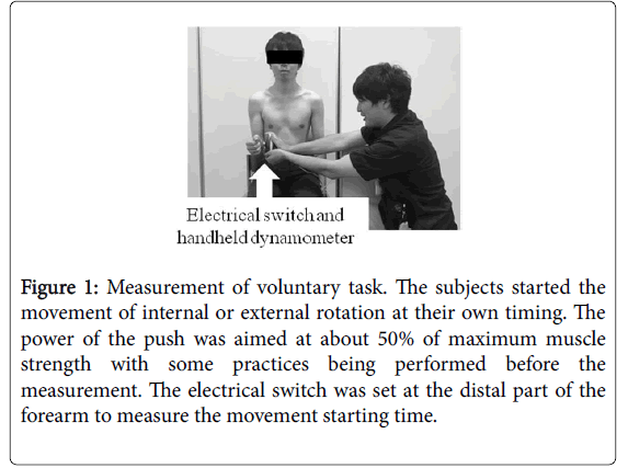
Muscle activation was assessed by electromyography during two tasks in shoulder internal and external rotation at 0 degree abduction. The tasks involved a “voluntary task,” and an “unpredictable task.” Throughout the voluntary task, subjects were measured with sitting as a starting position at 0 degree shoulder abduction, external rotation, and forearm pronation. From this starting position, they started the movement of internal or external rotation at their own timing. A handheld dynamometer was set at the distal part of the subjects’ forearm and pushed (Figure 1).
Figure 1: Measurement of voluntary task. The subjects started the movement of internal or external rotation at their own timing. The power of the push was aimed at about 50% of maximum muscle strength with some practices being performed before the measurement. The electrical switch was set at the distal part of the forearm to measure the movement starting time.
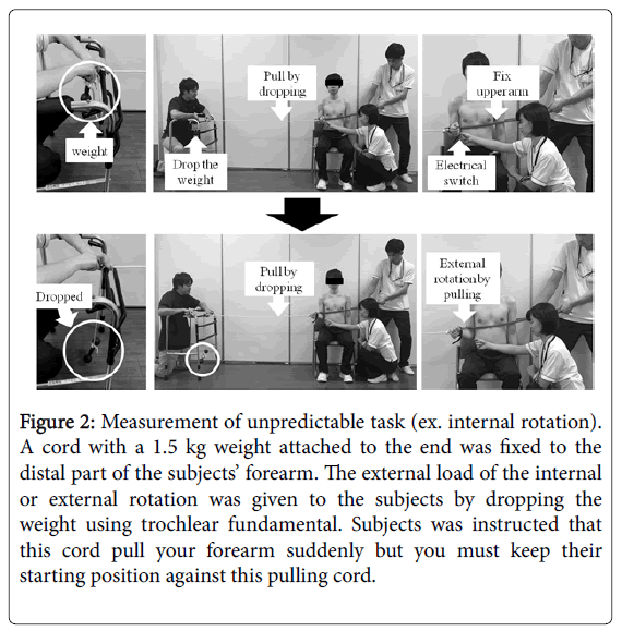
The power of the resistance by handheld dynamometer was aimed at approximately 50% of maximum muscle strength with some practices being performed before the measurement. Additionally, the electrical switch was set at the distal part of the forearm to measure the movement starting time. Similarly, the unpredictable task involved a sitting position with a 0 degree shoulder abduction, external rotation, and forearm pronation. A cord with a 1.5 kg weight attached to the end was fixed to the distal part of the subjects’ forearm. The cord was set on the level by running it on the same height adjusted device placed not in sight of subjects. The external load of the internal or external rotation was given to the subjects by dropping the weight using trochlear fundamental (Figure 2).
Figure 2: Measurement of unpredictable task (ex. internal rotation). A cord with a 1.5 kg weight attached to the end was fixed to the distal part of the subjects’ forearm. The external load of the internal or external rotation was given to the subjects by dropping the weight using trochlear fundamental. Subjects was instructed that this cord pull your forearm suddenly but you must keep their starting position against this pulling cord.
The height of the drop was set at 70 cm. The height adjusted device was fixed by the researcher’s foot throughout the task. At the beginning of the experiment, a researcher informed the subjects that “Pull your forearm by the cord suddenly but you must keep their starting position against this pulling cord.” An electrical switch was set to the subjects’ forearm by researcher’s hand to measure movement onset. Subjects were instructed to stay relaxed until receiving the external load and to keep the starting position as far as possible after receiving the external load. During the internal rotation task, the distal part of the upper arm was fixed using a resistance tube to avoid shoulder abduction movement by the external load. If the subjects incurred pain during the task, the measurement was quickly suspended. The order of the tasks was random.
Surface electromyography (EMG; MQ-8, Kissei Comtec, Japan) was used to collect raw EMG data. The EMG was recorded at a sampling rate of 1,000 Hz with electrodes (LECTRODE, Admedec, Japan) at an interelectrode distance of 20 mm. Cross-talk was minimized by careful placement of suitably sized electrodes parallel to the muscle fibers, based on standard anatomical criteria. We measured the muscle activation of agonist at GHJ and main scapular stabilizer which had been occurred high activation during isometric contraction of shoulder internal and external rotation by a previous study [28]. The target muscles were the pectoralis major, infraspinatus, serratus anterior, and middle trapezius on the dominant side. The EMG electrode position was set according to previous studies [37]. The anatomic placement of electrodes was following locations. Pectoralis major electrodes were placed the chest wall over the muscle mass (approximately 2 cm from the axillary fold), and infraspinatus electrodes were placed two fingerbreadths inferior to the center of the spina scapulae. Serratus anterior electrodes were placed obliquely over the lower fibers of the serratus anterior on the lateral thoracic cage at the level of the inferior scapula and anterior to the latissimus dorsi border just below the axilla, and middle trapezius electrodes were placed horizontally at half the distance between the spine (approximately T3-5) and vertebral border of the scapula in line with the scapular spine. Raw EMG data was filtered with a digital band-pass filter between 5 and 500 Hz.
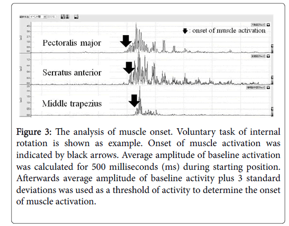
From the EMG data, the onset of muscle activation was calculated. Starting motion was judged by the electrical switch. The definition of onset of muscle activation was as follows: the average amplitude of baseline activation was calculated for 500 ms during the starting position. Subsequently, the average amplitude of baseline activity plus three standard deviations was used as a threshold of activity to determine the onset of muscle activation (Figure 3) [38,39].
Figure 3: The analysis of muscle onset. Voluntary task of internal rotation is shown as example. Onset of muscle activation was indicated by black arrows. Average amplitude of baseline activation was calculated for 500 milliseconds (ms) during starting position. Afterwards average amplitude of baseline activity plus 3 standard deviations was used as a threshold of activity to determine the onset of muscle activation.
The measured muscle was classified as the agonist at the GHJ and the STJ, and the antagonist at the STJ, on each movement direction. It has been revealed that each classified muscle indicated a different activity pattern and the agonist corresponded with high muscle activation and the antagonist with low muscle activation [28,29,40]. The classification to agonist or antagonist muscle followed the rule of scapular movement during shoulder motion. In shoulder internal rotation, the agonist muscle of the GHJ was the pectoralis major. The agonist muscle of the STJ was set at the serratus anterior because scapular internal rotation and protraction is occurred with internal shoulder rotation; scapular internal rotation and protraction is given by activation of serratus anterior. The antagonist muscle of the STJ was defined as the middle trapezius muscle because it has the opposite function of the serratus anterior. The agonist muscle of the GHJ for the external rotation was set as the infraspinatus. The middle trapezius was defined as the agonist muscles of the STJ since scapular external rotation and retraction was achieved by shoulder external rotation. Serratus anterior was defined as the antagonist muscle of the STJ for the external rotation.
In each trial, the onset of muscle activation was compared between the agonist at the GHJ, the agonist at the STJ, and the antagonist at the STJ. One-way repeated measure analysis of the variance test was used to determine if there was a difference between the three muscles: the agonist at the GHJ, at the STJ, and the antagonist at the STJ. The Tukey-Kramer test was used for post-hoc comparison with P
Results
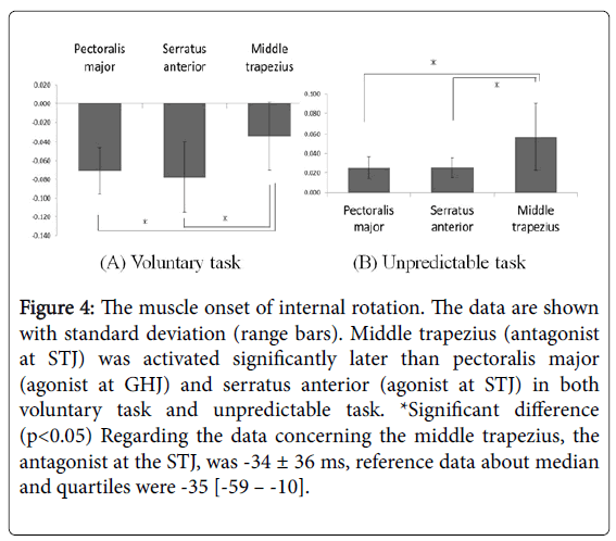
The results of internal rotation are shown in Figure 4. In the voluntary task of internal rotation, the onset of the pectoralis major, the agonist at the GHJ, was -71 ± 25 ms; the serratus anterior, the agonist at the STJ, was -78 ± 38 ms; the middle trapezius, the antagonist at the STJ, was -34 ± 36 ms. There was no significant difference between the pectoralis major and the serratus anterior. The onset of middle trapezius was significantly later than that of the pectoralis major and the serratus anterior (p<0.05). In the unpredictable task of internal rotation, the onset of the pectoralis major activation was at 25 ± 11 ms, the serratus anterior was at 25 ± 10 ms, and the middle trapezius was at 57 ± 34 ms. There was no significant difference between the pectoralis major and the serratus anterior. The middle trapezius had a significant later onset than the pectoralis major and the serratus anterior muscle (p<0.05).
Figure 4: The muscle onset of internal rotation. The data are shown with standard deviation (range bars). Middle trapezius (antagonist at STJ) was activated significantly later than pectoralis major (agonist at GHJ) and serratus anterior (agonist at STJ) in both voluntary task and unpredictable task. *Significant difference (p<0.05) Regarding the data concerning the middle trapezius, the antagonist at the STJ, was -34 ± 36 ms, reference data about median and quartiles were -35 [-59 – -10].
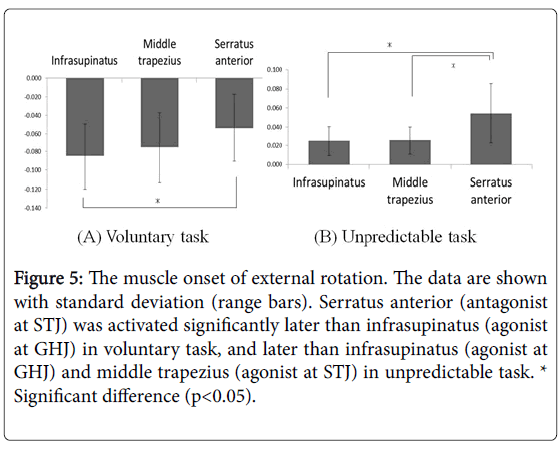
The results of external rotation are shown in Figure 5. In the voluntary task of external rotation, the onset of infraspinatus activation, the agonist at the GHJ, was -85 ± 36 ms; at the middle trapezius, the agonist at the STJ, was -75 ± 38 ms, and at the serratus anterior, the antagonist at the STJ, was -54 ± 36 ms. The serratus anterior had a significant later onset than the infraspinatus (p<0.05). Under unpredictable tasks, the onset of the infraspinatus activation was 25 ± 15 ms, the middle trapezius was 26 ± 15 ms, and the serratus anterior was 54 ± 31 ms. There was no difference between the infraspinatus and the middle trapezius muscles, but serratus anterior was recruited significantly later than the infraspinatus and the middle trapezius (p<0.05). Overall, the onset of the agonist activation at the GHJ and the agonist at the STJ was the same; however, the antagonist at the STJ was delayed to these two muscles.
Figure 5: The muscle onset of external rotation. The data are shown with standard deviation (range bars). Serratus anterior (antagonist at STJ) was activated significantly later than infrasupinatus (agonist at GHJ) in voluntary task, and later than infrasupinatus (agonist at GHJ) and middle trapezius (agonist at STJ) in unpredictable task. * Significant difference (p<0.05).
Discussion
It was revealed that there is the difference of muscle onset between the agonist at STJ and the antagonist at STJ. This study identified that the antagonist at STJ started activation after the agonist at the GHJ and the STJ. There are many studies on agonists and antagonists affecting other joints. It had been measured the onset of forearm muscles during movement of the wrist [41,42]. There are also some studies regarding antagonist muscles and the effect on knee and elbow joints [43-45]. From these studies, it was revealed that the antagonist had minimal muscle activation during isometric, isotonic, and isokinetic contraction, and that the function of the antagonist was to control the movement caused from the activation of the agonist and to aid stability by co-contraction with the agonist [46-49]. Additionally, there were some studies about agonist and antagonist at STJ. In the study on scapular muscle activation during isometric contraction of shoulder horizontal adduction and abduction, the agonist at STJ had high activation and the antagonist at STJ had minimal activation [27,31]. Compared to these studies, the results of this study have similar. The characteristic of scapular muscles resembles muscles on other joints, and therefore, the concept as agonist or antagonist might be able to be applied to scapular muscles. Therefore, each scapular muscle have to be assessed individually in clinical situation.
In this study, the experiment was performed under two tasks; voluntary task and unpredictable task. In the voluntary task, the subjects could move their arms at their own timing and onset of muscle activation is faster than the starting time of joint movement, indicating that the onset of muscle activation had a negative value. In the unpredictable task, the muscle activation trigger occurred when subjects were pulled their arms by the external load. The arms were firstly moved by the external load and then the muscle was recruited, consequently, the onset of muscle activation indicated a positive value. We had considered that the activation order of these two conditions might be different, but the results showed that the order of muscle activation was similar. This finding indicates that the activation pattern will not be changed by either the instruction for the subjects to start movement at their own timing and at the reaction of the external load in shoulder joint. And the scapular muscle activation does not commence before the onset at the GHJ muscle. This result indicates that order of exercise for GHJ and STJ was not need to be sticked.
Reaction time differences are influenced by the characteristics of test movement, arthrology, the distance that the neural signal must travel, and motor unit characteristics of the tested joint and muscle. In shoulder joint, muscle latent muscle reaction timing of scapular muscles and rotator cuff muscles was 40-70 ms in response to sudden movement [26,50]. The latency of wrist joint muscle was 20-25 ms [51]. About elbow joint muscle, sudden elbow joint perturbations produced latency of 25-60 ms for biceps brachii and brachialis [52]. Compared to these studies, onset values of the scapular muscles that we report are similar, however a little earlier. In this study, an electrical switch was set to the subject’s forearm by researcher’s hand to measure movement onset because an electrical switch didn’t react in case of setting by mechanical device. This mechanical device couldn’t give enough pressure to subject’s forearm for making electrical switch react. This manual setting of an electrical switch by researchers might have influenced to whole reaction time value, however this didn’t influence to the order of muscle activations.
There were some limitations in this study. First, the subjects were young healthy adults, and hence, the results may not apply to patients with shoulder disease. It had been reported that EMG showed delayed onset of the serratus anterior and earlier onset of the upper trapezius activity in patients with impingement [19,35], resulting in inconclusive data between subjects and patients. This result is useful to compare healthy people; however, we need to conduct a study to understand the characteristics of patients. Furthermore, these results might be changed by age. Teunis reported that overall prevalence of rotator cuff abnormalities increased with age [53], and rotator cuff tear may change muscle activation order. In retrospect, a larger sample size needed to be studied as it would provide more reliable data. Finally, the force of the external load set involved only one pattern in this study; consequently, it would be necessary to confirm the results by incorporating a heavy or light force external load. In daily living activity and sports activities, various levels of muscle activation are observed, and therefore, an examination with several load variations will be useful.
References
- Kibler WB (1998) The role of the scapula in athletic shoulder function. Am J Sports Med 26: 325-337.
- Kibler WB, Uhl TL, Maddux JW, Brooks PV, Zeller B, et al. (2002) Qualitative clinical evaluation of scapular dysfunction: a reliability study. J Shoulder Elbow Surg 11: 550-556.
- Lefèvre-Colau MM, Nguyen C, Palazzo C, Srour F, Paris G, et al. (2018) Kinematic patterns in normal and degenerative shoulders. Part II: Review of 3-D scapular kinematic patterns in patients with shoulder pain, and clinical implications. Ann Phys Rehabil Med 61: 46-53.
- McQuade KJ, Smidt GL (1998) Dynamic scapulohumeral rhythm: the effects of external resistance during elevation of the arm in the scapular plane. J Orthop Sports Phys Ther 27: 125-133.
- Chen SK, Simonian PT, Wickiewicz TL, Otis JC, Warren RF (1999) Radiographic evaluation of glenohumeral kinematics: a muscle fatigue model. J Shoulder Elbow Surg 8: 49-52.
- Leong HT, Ng GY, Chan SC, Fu SN (2017) Rotator cuff tendinopathy alters the muscle activity onset and kinematics of scapula. J Electromyogr Kinesiol 35: 40-46.
- Magarey ME, Jones MA (2003) Dynamic evaluation and early management of altered motor control around the shoulder complex. Man Ther 8: 195-206.
- Malmström EM, Olsson J, Baldetorp J, Fransson PA (2015) A slouched body posture decreases arm mobility and changes muscle recruitment in the neck and shoulder region. Eur J Appl Physiol 115: 2491-2503.
- Voight ML, Thomson BC (2000) The role of the scapula in the rehabilitation of shoulder injuries. J Athl Train 35: 364-372.
- Ludewig PM, Reynolds JF (2009) The association of scapular kinematics and glenohumeral joint pathologies. J Orthop Sports Phys Ther 39: 90-104.
- Michener LA, Sharma S, Cools AM, Timmons MK (2016) Relative scapular muscle activity ratios are altered in subacromial pain syndrome. J Shoulder Elbow Surg 25: 1861-1867.
- Cools AM, Witvrouw EE, Declercq GA, Vanderstraeten GG, Cambier DC (2004) Evaluation of isokinetic force production and associated muscle activity in the scapular rotators during a protraction-retraction movement in overhead athletes with impingement symptoms. Br J Sports Med 38: 64-68.
- Decker MJ, Hintermeister RA, Faber KJ, Hawkins RJ (1999) Serratus anterior muscle activity during selected rehabilitation exercises. Am J Sports Med 27: 784-791.
- Ekstrom RA, Soderberg GL, Donatelli RA (2005) Normalization procedures using maximum voluntary isometric contractions for the serratus anterior and trapezius muscles during surface EMG analysis. J Electromyogr Kinesiol 15: 418-428.
- Moseley JB Jr, Jobe FW, Pink M, Perry J, Tibone J (1992) EMG analysis of the scapular muscles during a shoulder rehabilitation program. Am J Sports Med 20: 128-134.
- Batbayar Y, Uga D, Nakazawa R, Sakamoto M (2015) Effect of various hand position widths on scapular stabilizing muscles during the push-up plus exercise in healthy people. J Phys Ther Sci 27: 2573-2576.
- Gioftsos G, Arvanitidis M, Tsimouriset D, Kanellopoulos A, Paras G, et al. (2016) EMG activity of the serratus anterior and trapezius muscles during the different phases of the push-up plus exercise on different support surfaces and different hand positions. J Phys Ther Sci 28: 2114-2118.
- Larsen CM, Juul-Kristensen B, Olsen HB, Holtermann A, Søgaard K (2014) Selective activation of intra-muscular compartments within the trapezius muscle in subjects with Subacromial Impingement Syndrome. A case-control study. J Electromyogr Kinesiol 24: 58-64.
- Worsley P, Warner M, Mottram S, Gadola S, Veeger HE, et al. (2013) Motor control retraining exercises for shoulder impingement: effects on function, muscle activation and biomechanics in young adults. J Shoulder Elbow Surg 22: 11-19.
- Ayatollahi K, Okhovatian F, Kalantari KK, Baghban AA (2017) A comparison of scapulothoracic muscle electromyographic activity in subjects with and without subacromial impingement syndrome during a functional task. J Bodyw Mov Ther 21: 719-724.
- Kibler WB (1991) Role of the scapula in the overhead throwing motion. Contemp Orthop 22: 525-532.
- Sahrmann Sh (2002) Diagnosis and Treatment of Movement Impairment Syndromes. Mosby, Missouri, USA.
- Rebelled GM, Rojas VG, Valdez EM, Valdes EM, Xie HB (2016) The recruitment order of scapular muscles depends on the characteristics of the postural task. J Electromyogr Kinesiol 31: 40-47.
- Moraes GF, Faria CD, Teixeira-Salmela LF (2008) Scapular muscle recruitment patterns and isokinetic strength ratios of the shoulder rotator muscles in individuals with and without impingement syndrome. J Shoulder Elbow Surg 17: 48-53.
- Larsen CM, Søgaard K, Chreiteh SS, Holtermann A, Juul-Kristensen B (2013) Neuromuscular control of scapula muscles during a voluntary task in subjects with subacromial impingement syndrome. A case-control study. J Electromyogr Kinesiol 23: 1158-1165.
- Brindle TJ, Nyland JA, Shapiro R (2007) Scapulothoracic latent muscle reaction timing comparison between trained overhead throwers and untrained control subjects. Scan J Med Sci Sports 17: 252-259.
- Ijiri T, Takagi R, Suzuki T (2017) Isometric contraction of scapular muscles activities during horizontal abduction and adduction of the shoulder. J Nov Physiother 7: 1-5.
- Suzuki T (2017) Clinical Physical Therapy. In: The function of scapular muscle: 1-25, InTech, Rijeka, Croatia.
- Uga D, Nakazawa R, Sakamoto M (2016) Strength and muscle activity of shoulder external rotation of subjects with and without scapular dyskinesis. J Phys Ther Sci 28: 1100-1105.
- Jung SH, Hwang UJ, Kim JH, Gwak GT, Kwon OY (2017) Effects of horizontal shoulder abduction and adduction on the activity and strength of the scapular protractors. J Electromyogr Kinesiol 37: 155-159.
- Miyasaka J, Arai R, Ito T, Shingu N, Hasegawa S, et al. (2017) Isometric muscle activation of the serratus anterior and trapezius muscles varies by arm position: a pilot study with healthy volunteers with implications for rehabilitation. J Shoulder Elbow Surg 26: 1166-1174.
- Alizadehkhaiyat O, Hawkes DH, Kemp GJ, Frostick SP (2015) Electromyographic analysis of shoulder girdle muscles during common internal rotation exercises. Int J Sports Phys Ther 10: 645-654.
- Allison GT, Morris SL, Lay B (2008) Feedforward responses of transversus abdominis are directionally specific and act asymmetrically: implications for core stability theories. J Orthop Sports Phys Ther 38: 228-233.
- Hodges PW, Richardson CA (1997) Feedforward contraction of transversus abdominis is not influenced by the direction of arm movement. Exp Brain Res 114: 362-370.
- Phadke V, Ludewig PM (2013) Study of the scapular muscle latency and deactivation time in people with and without shoulder impingement. J Electromyogr Kinesiol 23: 469-475.
- Baets LD, Jaspers E, Janssens L, Deun SV (2014) Characteristics of neuromuscular control of the scapula after stroke: A first exploration. Front Hum Neurosci 8: 1-8.
- Moeller CR, Bliven KC, Valier AR (2014) Scapular muscle-activation rations in patients with shoulder injuries during functional shoulder exercises. J Athl Train 49: 345-355.
- Silva L, Marta S, Vaz J, Fernandes O, Castro MA, et al. (2013) Trunk muscle activation during golf swing: Baseline and threshold. J Electromyogr Kinesiol 23: 1174-1182.
- Yassin M, Talebian S, Takamjani IE, Maroufi N, Ahmadi A, et al. (2015) The effects of arm movement on reaction time in patients with latent and active upper trapezius myofascial trigger point. Med J Islam Repub Iran 29: 1-9.
- Shin AR, Lee JH, Kim DE, Cynn HS (2018) Thera-Band application changes muscle activity and kyphosis and scapular winging during knee push-up plus in subjects with scapular winging: The cross-sectional study. Medicine 97: e0348.
- Irlbacher K, Voss M, Meyer BU, Rothwell JC (2006) Influence of ipsilateral transcranial magnetic stimulation on the triphasic EMG pattern accompanying fast ballistic movements in humans. J Physiol 574: 917-928.
- Liang N, Yamashita T, Ni Z, Takahashi M, Murakami T, et al. (2008) Temporal modulations of agonist and antagonist muscle activities accompanying improved performance of ballistic movements. Hum Mov Sci 27: 12-28.
- Bazzucchi I, Sbriccoli P, Marzattinocci G, Felici F (2006) Coactivation of the elbow antagonist muscles is not affected by the speed of movement in isokinetic exercise. Muscle Nerve 33: 191-199.
- Kronberg M, Németh G, Broström LA (1990) Muscle activity and coordination in the normal shoulder. An electromyographic study. Clin Orthop Relat Res 257: 76-85.
- Remaud A, Cornu C, Guével A (2009) Agonist muscle activity and antagonist muscle co-activity levels during standardized isotonic and isokinetic knee extensions. J Electromyogr Kinesiol 19: 449-458.
- Mari S, Serrao M, Casali C, Conte C, Martino G, et al. (2014) Lower limb antagonist muscle co-activation and its relationship with gait parameters in cerebellar ataxia. Cerebellum 13: 226-236.
- Aagaard P, Simonsen EB, Andersen JL, Magnusson SP, Bojsen-Møller F, et al. (2000) Antagonist muscle coactivation during isokinetic knee extension. Scand J Med Sci Sports 10: 58-67.
- Bazzucchi I, Riccio ME, Felici F (1983) Tennis players show a lower coactivation of the elbow antagonist muscles during isokinetic exercises. J Electromyogr Kinesiol 18: 752-759.
- Humphrey DR, Reed DJ (1983) Separate cortical systems for control of joint movement and joint stiffness: reciprocal activation and coactivation of antagonist muscles. Adv Neurol 39: 347-372.
- Brindle TJ, Nyland J, Shapiro R, Caborn DN, Stine R (1999) Shoulder proprioception: latent muscle reaction times. Med Sci Sports Exerc 31:1394-1398.
- Goodin DS, Aminoff MJ (1992) The basis and functional role of the late EMG activity in human forearm muscles following wrist displacement. Brain Res 589: 39-47.
- Bennett DJ (1993) Electromyographic responses to constant position errors imposed during voluntary elbow joint movement in human. Exp Brain Res 95: 499-508.
- Teunis T, Lubberts B, Reilly BT, Ring D (2014) A systematic review and pooled analysis of the prevalence of rotator cuff disease with increasing age. J Shoulder Elbow Surg 23: 1913-1921.
Citation: Ijiri T, Urabe Y, Maeda N, Sasadai J, Suzuki T (2019) Onset of Shoulder Muscle Activation During Internal and External Shoulder Rotation. J Nov Physiother 9: 408. DOI: 10.4172/2165-7025.1000408
Copyright: © 2019 Ijiri T, et al. This is an open-access article distributed under the terms of the Creative Commons Attribution License, which permits unrestricted use, distribution, and reproduction in any medium, provided the original author and source are credited.
Select your language of interest to view the total content in your interested language
Share This Article
Recommended Journals
Open Access Journals
Article Tools
Article Usage
- Total views: 7659
- [From(publication date): 0-2019 - Nov 17, 2025]
- Breakdown by view type
- HTML page views: 6699
- PDF downloads: 960

