Observations on Veterinary Toxicology in Short
Received: 05-Jan-2023 / Editor assigned: 07-Jan-2023 / PreQC No. jvmh-23-86908 / Reviewed: 20-Jan-2023 / QC No. jvmh-23-86908 / Revised: 23-Jan-2023 / Manuscript No. jvmh-23-86908 / Accepted Date: 29-Jan-2023 / Published Date: 30-Jan-2023 QI No. / jvmh-23-86908
Abstract
Given that it deals with such a broad range of poisons, veterinary toxicology is a very complicated field. The study of toxicology covers a wide range of topics, such as the physical and chemical characteristics of many poisons (including medications, food and feed additives, common poisons, consumer goods, and specific synthetics), their distribution in the body, and their effects on the environment. The majority of synthetics that are currently used often in animal poisonings are manmade substances. A common occurrence in crisis veterinary medicine is severe harm. Additionally, veterinary toxicology is concerned with how to address disease issues brought on by damages. Currently, it’s necessary to clearly incorporate veterinary toxicity into ecological medication and coordinate veterinary medication.
Keywords
Veterinary; Toxicology; Medication
Introduction
The concept of poisonous reactions depends on the poison as well as the degree, duration, and force of the openness as well as the characteristics of the exposed person (i.e., species, orientation, age, previous infection states, wholesome status, and earlier openness to the specialist or related compounds). The numerous variations in how domestic, marine, wild, and zoo species react to toxins greatly complicate the field of veterinary toxicology. Of course, there are a wide range of distinct factors that can affect how dangerous a synthetic is overall. The biggest challenge facing modern veterinary toxicologists understands the full profile of each poison, including its harmfulness components. In veterinary toxicology, toxicoses are evaluated, toxins are identified, described, and their fate in the body is determined, and toxicoses are treated. The current state of animal health and food safety clearly shows the relevance of veterinary toxicology, as evidenced by the recent global melamine contamination in pet and swine feed, the disease and death caused by pet jerky treats, and worries over the usage of beta-agonists in food animals. Due of the rarity of cases seen in a clinical environment, veterinary toxicology can be difficult. A toxicant or poison is another name for a toxic agent. The redundant term “biotoxin” is occasionally used to refer to a poison created by a biologic source (such as venoms or plant poisons). Generally speaking, a toxicant is a poisonous substance that is either the primary product or a result of human action (eg, pesticides manufactured for commercial use, dioxins produced as a byproduct of industrial processes). The disease brought on by a hazardous agent is referred to by the terms toxicosis, poisoning, and intoxication. The term “toxicity” (sometimes wrongly used in place of “poisoning”) describes the quantity of a toxic chemical [1-4] required to have a negative effect. Effects occurring within the first 24 hours are referred to as acute toxicosis. Chronic toxicosis refers to the side effects brought on by extended exposure (>3 months). To describe the considerable time between acute and chronic, terms like subacute and subchronic are utilised. Toxic consequences vary depending on dose. Effects from a dose can be unnoticeable, therapeutic, poisonous, or fatal. Toxicant concentration is indicated as parts per million or parts per billion, and a dose is the amount of a substance per unit of body weight. In addition to being employed for tissue levels, these quantitative expressions are also used for feedstuffs, water, and air.
Materials and Method
Animals’ absorption of toxic agents
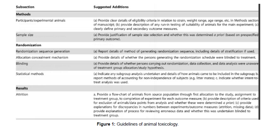
It is possible for substances to be absorbed by the GI tract, skin,lungs, eyes, mammary glands, uterus, as well as injection sites. Although localised harmful effects are possible, the cell must be partially or completely destroyed and absorbed in order to be affected. The main component influencing absorption is solubility. While lipidsoluble chemicals are typically readily absorbed, even though intact skin, insoluble salts and ionised compounds are not. For instance, barium is poisonous, yet barium sulfate’s limited absorption makes it suitable for use in intestinal contrast radiography. A harmful substance is translocated or distributed to reactive locations, including storage depots, through the bloodstream. The liver is the organ most frequently involved in intoxication because it receives the portal circulation (and detoxification). Receptor sites are necessary (Figure 1) for the selective deposit of exogenous substances in different tissues. Chemical solubility in water is a major factor in how easily it may be distributed. Polar or aqueous-soluble substances typically pass through the kidneys for excretion, whereas lipid-soluble substances are more likely to pass through the bile and build up in fat depots. The organ or tissue where a poison has the greatest impact on an animal may not always have the highest concentration of that toxin (the target organ).
Animal toxic agents’ distribution
The largest quantities of lead may be detected in the bone, neither a tissue that is neither a target for toxic effects nor a valid tissue for toxicologic interpretation. For effective [4-7] organ selection for analysis, knowledge of the translocation properties of hazardous substances is required.
Animals’ metabolism of toxic agents
Toxic substances are metabolised or biotransformed by the body as part of a “effort to detoxify.” In some cases, xenobiotic agents that have been metabolised are more harmful than the initial substance. Lethal synthesis is the term for this. Many organophosphorous insecticides undergo metabolism, producing metabolites that are more hazardous than the original (or parent) chemicals (eg, parathion to paroxan). The metabolism comprises two stages. Phase I comprises mechanisms for oxidation, reduction, and hydrolysis. These hepatic enzyme-catalyzed processes often transform foreign substances into derivatives for Phase II reactions. However, products from Phase I may be excreted in their original form if polar solubility allows for translocation. Phase II primarily entails reactions that involve synthesis or conjugation. Glucuronides, acetylation byproducts, and combinations with glycine are examples of typical conjugates. Rarely does xenobiotic agent metabolism take place via a single route. The majority is usually retained or eliminated as metabolites, with only a small portion being excreted unmodified. Between species, there are significant variances in metabolic systems. For instance, cats are unable to conjugate substances like morphine and phenols because they lack types of glucuronyl transferase. In some cases, an earlier exposure to a harmful chemical triggers an enzyme response that increases tolerance to subsequent exposures.
Results and Discussion
Animal excretion of toxic agents
Most hazardous substances and their metabolites are excreted by the kidneys. In females, the mammary gland and the GI tract are both used for some excretion. In the bile, several polar and highmolecular- weight substances are expelled. These substances undergo an enterohepatic cycle when they are eliminated by the liver through bile, reabsorbed by the intestine, and then brought back to the liver. For some harmful substances, milk serves as a conduit for elimination. The excretion rate may be the main cause for worry because some toxic substances can leave harmful residues in animals used for food production. Excretion rates can be significantly influenced by a number of variables, including dose, method of administration, and animal condition. Through glomerular filtration, passive tubular diffusion, and active tubular secretion, toxic substances are eliminated in the kidney. The anatomic site where xenobiotic excretion takes place is unique in terms of the damage it causes to the kidney. The proximal tubules, glomeruli, medulla, papilla, and loop of Henle are excretion sites. The most frequent location of damage brought on by toxicants is the proximal convoluted tubule. Prostaglandin synthase, prostaglandin reductase, and cytochrome P450 are the significant Phase I enzymes [5-11] found in the kidney. The concentration of the Phase I enzyme cytochrome P450 in the kidney is 10% lower than that in the liver. UDPglucuronosyltransferases (UGT), sulfotransferases, and glutathione- S-transferase are significant Phase II enzymes that are located in the kidneys. Phenylbutazone, a medication, targets the medulla and papilla, numerous plant toxins target the tubules, fluoride targets the loop of Henle, and immune complexes target the glomeruli. Half-life (t12), which is the amount of time needed for the disappearance of half of the compound, is used to express the elimination or disappearance (by metabolic change) of a chemical from an organ or the body. The concentration of the substance typically affects the rate of elimination. First-order kinetics is defined as a constant fraction (for example, 12) removed per unit of time. The rate of elimination might be governed by a metabolic response. Zero-order kinetics is the elimination of a fixed amount per unit of time. There may probably be variations in elimination rates between various bodily compartments. Elimination that starts out quickly (from the plasma or central component, for example) and then slows down from the periphery component is referred to as a two-compartment system (eg, liver, kidney, or fat).
Exposure-related variables in veterinary toxicology
The main issue is dose, because it’s rarely clear how much a dangerous chemical is consumed. The length and frequency of exposure are crucial. Absorption, translocation, and maybe metabolic processes are impacted by the exposure route. The timing of hazardous chemical exposure in relation to stressful events or dietary consumption could also play a role. When some poisonous substances are consumed, emesis may happen if the stomach is empty, but if it’s partially full, the material may be retained and toxicosis may happen. Environmental variables including temperature, humidity, and barometric pressure have an [11] impact on how much is consumed and even when some harmful substances appear. Seasonal or climatic variations are associated with a number of mycotoxins and dangerous plants.
Clinical Factors Influencing Toxic Agent Activity in Animals
A harmful agent’s solubility is influenced by its chemical makeup, which in turn affects absorption. In general, lipid-soluble or nonpolar compounds are absorbed more quickly than polar or ionised ones. The hazardous compound’s accessibility for absorption is also influenced by the vehicle or carrier. The toxicity of isomers, especially optical isomers, varies. Hexachlorocyclohexane (lindane), for instance, has a higher toxicity in its gamma isomer than in its other isomers.
Conclusion
Relevant information and samples should be delivered to a diagnostic lab. To create a plan for a laboratory inquiry, a thorough history is required, and it can be helpful in a legal situation. Details should be provided. For instance, noting CNS signals is insufficient because most animals show some degree of CNS indicators prior to passing away. Actions and symptoms should be described in detail.
Acknowledgement
The authors appreciate VetScan for providing the preoperative CT pictures.
Conflict of Interest
There are no conflicts of interest, according to the authors.
Ethics Statement
This study did not need to be submitted to the local ethics and welfare council since all diagnostic studies and begun therapies were a regular component of clinical procedures.
References
- Alsan M (2015) The effect of the tsetse fly on African development. Am Econ Rev 105: 382–410.
- Swallow BM (2000) Impact of trypanosomiasis on African agriculture. PAAT Technical and Scientific Series.
- Shaw APM, Wintd B GC, GRW, Mattiolie RC, Robinson TP, et al. (2014) Mapping the economic benefits to livestock keepers from intervening against bovine trypanosomosis in Eastern Africa. Prev Vet Med 113:197–210.
- NTTICC (2004) National Tsetse and Trypanosomosis Investigation and Control Center.
- Franco JR, Cecchi G, Priotto G, Paone M, Diarra A, et al. (2018) Monitoring the elimination of human African trypanosomiasis: Update to 2016. PLoS Negl Trop Dis 12: 1–16.
- Shaw A, Wint W, Cecchi G, Torr S, Waiswa C, et al. (2017) Intervening against bovine trypanosomosis in eastern Africa: mapping the costs and benefits. Food and Agriculture Organization of the United Nations PAAT Technical and Scientific Series.
- Meyer A, Holt HR, Oumarou F, Chilongo K, Gilbert W, et al. (2018) Integrated cost-benefit analysis of tsetse control and herd productivity to inform control programs for animal African trypanosomiasis. Parasites and Vectors 11:1–14.
- Tekle T, Terefe G, Cherenet T, Ashenafi H, Akoda KG, et al. (2018) Aberrant use and poor quality of trypanocides: a risk for drug resistance in south western Ethiopia. BMC Vet Res 14: 4.
- Mulandane FC, Fafetine J, Abbeele J Van Den, Clausen P-H, Hoppenheit, A, et al. (2017) Resistance to trypanocidal drugs in cattle populations of Zambezia Province, Mozambique. Parasitol Res 117: 429–436.
- Vreysen MJB, Seck MT, Sall B, Bouyer J (2013) Tsetse flies: Their biology and control using area-wide integrated pest management approaches. J Invertebr Pathol 112.
- Scoones I (2014) The politics of trypanosomiasis control in Africa. STEPS Working Paper 57 Brighton STEPS Centre.
Indexed at, Google Scholar, CrossRef
Indexed at, Google Scholar, CrossRef
Indexed at, Google Scholar, CrossRef
Indexed at, Google Scholar, CrossRef
Indexed at, Google Scholar, CrossRef
Citation: Alberto G (2023) Observations on Veterinary Toxicology in Short. J VetMed Health 7: 168.
Copyright: © 2023 Alberto G. This is an open-access article distributed under theterms of the Creative Commons Attribution License, which permits unrestricteduse, distribution, and reproduction in any medium, provided the original author andsource are credited.
Select your language of interest to view the total content in your interested language
Share This Article
Recommended Journals
Open Access Journals
Article Usage
- Total views: 1277
- [From(publication date): 0-2023 - Sep 23, 2025]
- Breakdown by view type
- HTML page views: 882
- PDF downloads: 395

