Case Report Open Access
Novel Diagnostic Tool CISH to Demonstrate CMV DNA by Light Microscopy-A Case Report of CMV Colitis
Barodawala SM1, Majethia NK2* and Chadha K3
1Department of Histopathology, Grant Medical College and Sir JJ Group of Hospitals, Maharashtra, India
2Department of Pathology, Grant Medical College and Sir JJ Group of Hospitals, Maharashtra, India
3Department of Oncopathology, Grant Medical College and Sir JJ Group of Hospitals, Maharashtra, India
- *Corresponding Author:
- Majethia NK
Grant Medical College and Sir JJ Group of Hospitals
Maharashtra, India
Tel: 02223735555
Email: nikhilmajethia@gmail.com
Received date: August 03, 2016; Accepted date: September 19, 2016; Published date: September 27, 2016
Citation: Barodawala SM, Majethia NK, Chadha K (2016) Novel Diagnostic Tool CISH to Demonstrate CMV DNA by Light Microscopy-A Case Report of CMV Colitis. J Gastrointest Dig Syst 6:472. doi: 10.4172/2161-069X.1000472
Copyright: © 2016 Majethia NK, et al. This is an open-access article distributed under the terms of the Creative Commons Attribution License; which permits unrestricted use; distribution; and reproduction in any medium; provided the original author and source are credited.
Visit for more related articles at Journal of Gastrointestinal & Digestive System
Abstract
Cytomegalovirus (CMV) infections are generally thought to be opportunistic especially in the setting of an immunosuppressed host. We present an unusual case of CMV colitis with liver abscess observed in an immunocompetent patient. The right diagnosis was made after implementation of Chromogenic In Situ Hybridization (CISH) on colonic biopsy tissue. Our purpose is therefore to familiarize fellow colleagues and clinicians with possible role of CISH in the diagnosis of this entity.
Keywords
Chromogenic in situ hybridization; Cytomegalovirus; Colitis
Introduction
The prevalence of CMV infection is high, ranging from 30%-100%, depending on age and race of population under evaluation. Most cases of cytomegalovirus (CMV) colitis occur in immunocompromised patients, including those with congenital or acquired immunodeficiency diseases, those receiving immunosuppressive drugs and transplant recepients. Only a small number of immunocompetent patients with CMV colitis have been reported worldwide [1-3]. CMV has a propensitiy to involve rapidly growing tissue such as granulation tissue in an ulcer bed [4]. We present an unusual case of CMV colitis with liver abscess observed in a immunocompetent patient. The right diagnosis was made after implementation of Chromogenic in situ Hybridization (CISH) on colonic biopsy tissue. Our purpose is therefore to familiarize fellow colleagues and clinicians with possible role of CISH in the diagnosis of this entity.
Case Report
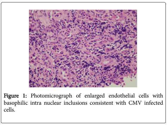
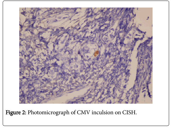
A 69 year old male presented with diarrhea, gastrointestinal bleed, abdominal pain and fever. Ultrasonography of the abdomen examination revealed a solitary liver abscess and caecal thickening. Endoscopic evaluation showed swollen and thickened walls of colon with multiple erythematous patches and ulcerative changes in caecum and right colon. The patient also had antral gastritis. The clinical diagnosis was amoebic colitis. A caecal biopsy was submitted for histopathologic examination. It revealed moderate colitis with focal ulceration and submucosal granulation tissue. The latter showed proliferating capillaries and fibroblasts along with dense neutrophilic and moderate lymphoplasmacytic infiltrate. Periodic acid-Schiff (PAS) stain did not reveal trophozoites of Entamoeba histolytica ruling out the clinical diagnosis. The noteworthy feature was the presence of enlarged endothelial cells with basophilic intra nuclear inclusions (owl eye) consistent with CMV infected cells (Figure 1 and 2).
The tissue was subjected to Chromogenic in situ hybridization (CISH) for CMV using Zytofast Digoxigenin-labeled oligonucleotide probe for the detection of Cytomegalovirus (CMV) DNA. The CMV DNA in the cells or tissue links to the secondarily polymerized enzyme-conjugated antibody to this DNA. The chromogenic substrate is then linked to this antibody which leads to the formation of a color precipitate that is visualized by light microscopy. Following the report of CMV infection, the patient received antiviral therapy and he is doing well.
Discussion
Case reports for CMV infection in colon have been well documented in immunocompetent patients, the first case reported in 1992 [5,6], sigmoid colon being the most common site [1]. In immunocompetent patients, diabetes mellitus, renal failure, severe trauma, sepsis, shock, burns, cirrhosis, are predisposing like in our case. Our patient presented with multiple ulcers in caecum along with a liver abscess. imitating other more common conditions, like amoebic colitis. Other differential diagnosis include ischemic colitis, pseudomembranous colitis and Inflammatory bowel disease [7]. However , these were ruled out on histopathological examination and histochemical stains i.e. PAS stain. Due to presence of large cells with basophilic intranuclear inculsions surrounded by a clear halo (owl eye) in endothelial cells a diagnosis of cytomegalovirus infection was suspected and confirmed by CISH for CMV. Until recently, the diagnosis of CMV infection had been limited to serological conversion, culture and detection of the virus by cytopathic effects on body fluids or tissue or the identification of typical CMV inclusion bodies in biopsy specimens. The application of mRNA or DNA probes specific for CMV has been shown to be sensitive and specific in adjunct to histological criteria. Similarly, detection of CMV antigens like pp65 with IgM monoclonal antibodies using immunohistochemistry can be carried out [8], CISH is a type of Bright field in situ hybridization (BRISH) which can be carried out on the slide prepared from the paraffin block. The BRISH includes CISH and SISH where in a chromogen (e.g.DAB) and silver are used to highlight the DNA /RNA of infectious agents like HPV DNA, EBER RNA of EBV (Epstein Barr virus in cases of lymphoma) [9] and CMV DNA. The latter is what we report in our study. It is to be noted that the specificity of demonstrating CMV DNA by CISH is 100%. The use of in situ hybridization is not new as it is more popularly used as Fluorescence in situ hybridization (FISH) for demonstrating HER-2/neu in breast carcinoma [10] cases and prognosticating hemato-lymphoid malignancies. It offers a real advantage by providing a cytomorphologic link where in slides can be analyzed by an anatomical pathologist using a light microscope [11].
Conclusion
The purpose of documenting this case is to highlight the fact that although in situ hybridization technique is already being utilized in oncology practice but its role in confirming infectious etiologies on tissues is less explored. Our 3 years experience in recognizing HPV, EBV, CMV has optimized many patients management. This is one such case where CMV illustration by CISH could help treat the patient. CISH is a novel the tool for diagnosing such cases where other methods cannot be used and can be an integral part of armamentarium available to the surgical pathologist.
References
- Lin MW, Liang JT, Chang KJ, Lee PH, Lin BR (2008) Cytomegalovirus Colitis in anImmunocompetentPatient: Report of a Case and Review of theLiterature. J Soc Colon Rectal Surgeon 19: 27-32.
- Karakozis S, Gongora E, Caceres M, Brun E, Cook JW, et al. (2001) Life-threateningcytomegalovirus colitis in theimmunocompetentpatient: report of a case and review of theliterature. Dis Colon Rectum 44: 1716-1720.
- Lee CS, LowAH, Ender PT, Bodenheimer HC (2004) Cytomegalovirus colitis in animmunocompetentpatientwith amebiasis: case report and review of theliterature. Mt Sinai J Med71: 347-350.
- Christidou A, Zambeli E, Mantzaris G(2007) Cytomegalovirus and InflammatoryBowelDisease: pathogenicity, diagnosis and treatment. Ann Gastroenterol 20: 110-115.
- Blair SD, Forbes A, Parkins RA (1992) CMV colitis in animmunocompetentadult. J R SocMed 85: 238-239.
- Paparoupa M, Schmidt V, Weckauf H, Ho H, SchuppertF (2016) CMV Colitis in ImmunocompetentPatients: 2 Cases of a DiagnosticChallenge. Case RepGastrointestMed
- Subbalaxmi MVS, Kumar VG, Chandra N, Raju YSN (2014) Cytomegalovirus colitis in a human immunodeficiency virus seropositive individual withmoderatelysevereimmunodeficiency. TheJ ClinSci Res 3: 42-45.
- Weiss LM , Chen YY (2013) EBER in situ hybridizationfor Epstein-Barr virus. Methods in Molecular Biology 999: 223-230.
- Garrido E, Carrera E, Manzano R, Lopez-Sanroman A (2013) Clinicalsignificance of cytomegalovirusinfection in patientswithinflammatoryboweldisease. WorldJournal of Gastroenterology 19: 17-25
- Gupta D, Middleton LP, Whitaker MJ,AbramsJ (2003) Comparison of fluorescence and chromogenic in situ hybridizationfordetection of HER-2/neuoncogene in breastcancer. American journal of clinicalpathology119 : 381–387.
- Nitha H, Grogan TM (2013) Bright Field in situ hybridizationmethodstodiscover gene amplifications and rearrangements in clinicalsamples. Methods in Molecular Biology 986: 341-352.
Relevant Topics
- Constipation
- Digestive Enzymes
- Endoscopy
- Epigastric Pain
- Gall Bladder
- Gastric Cancer
- Gastrointestinal Bleeding
- Gastrointestinal Hormones
- Gastrointestinal Infections
- Gastrointestinal Inflammation
- Gastrointestinal Pathology
- Gastrointestinal Pharmacology
- Gastrointestinal Radiology
- Gastrointestinal Surgery
- Gastrointestinal Tuberculosis
- GIST Sarcoma
- Intestinal Blockage
- Pancreas
- Salivary Glands
- Stomach Bloating
- Stomach Cramps
- Stomach Disorders
- Stomach Ulcer
Recommended Journals
Article Tools
Article Usage
- Total views: 11337
- [From(publication date):
October-2016 - Apr 05, 2025] - Breakdown by view type
- HTML page views : 10498
- PDF downloads : 839


