Editorial Open Access
Non-invasive Diagnosis of Fetal Lung Prematurity with GLHW Ultrasound Tissue Characterization
1Department of Obstetrics and Gynaecology, Tottori University Medical School, Yonago, Japan
2Department of Obstetrics and Gynaecology, Hamamatsu Medical Centre, Hamamatsu, Japan
- *Corresponding Author:
- Maeda K
Department of Obstetrics and Gynaecology
Tottori University Medical School
Yonago, Japan
Tel: 81859226856
E-mail: maedak@mocha.ocn.ne.jp
Received date: December 25, 2016; Accepted date: December 27, 2016; Published date: December 31, 2016
Citation: Maeda K, Serizawa M (2016) Non-invasive Diagnosis of Fetal Lung Prematurity with GLHW Ultrasound Tissue Characterization. J Preg Child Health 3:e136. doi:10.4172/2376-127X.1000e136
Copyright: © 2016 Maeda K, et al. This is an open-access article distributed under the terms of the Creative Commons Attribution License, which permits unrestricted use, distribution and reproduction in any medium, provided the original author and source are credited.
Visit for more related articles at Journal of Pregnancy and Child Health
Abstract
Non-invasive diagnosis of fetal lung immaturity. Ultrasound gray level histogram width (GLHW) tissue characterization was studied in fetal lung. GLHW ratio of fetal lung and liver was multiplied by gestational weeks, and fetal lung immaturity was diagnosed, if the product was less than 29. Ninety six per cent of neonatal respiratory distress syndrome (RDS) was predicted noninvasively in immature fetal lung diagnosed by GLHW tissue characterization. Fetal lung immaturity diagnosed by GLHW ultrasound tissue characterization predicted neonatal RDS noninvasively.
Keywords
Fetus; Lung; Liver; Lung immaturity; Neonatal RDS; Non-invasive diagnosis; Ultrasound; Gray level histogram; GLHW; Tissue characterization.
Introduction
The maturity of fetal lung was diagnosed by the lamellar body count, L/S ratio or PG of amniotic fluid obtained by invasive amniocentesis in the past as there was risk in the amniocentesis with needle, non-invasive method was desired. Although ultrasound tissue characterization was studied [1], its clinical study was unable in the past, because special computer and particular software were desired. As Maeda wished to study non-invasive ultrasound tissue characterization in clinical medicine particularly. In obstetrics and gynaecology, using the gray level histogram prepared in common ultrasonic B-mode devices, where the B-mode gray level was objectively using the gray level histogram presented in common ultrasound B-mode devices enabling to diagnose tissue character in daily practice [2,3].
Calculation of GLHW value and its standardization
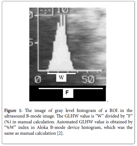
The gray level histogram was detected by 1 cm2 region of interest (ROI) on the ultrasonic B-mode image to display the gray level histogram and its analysis data, where the histogram base width length was divided by the full 64 bits length to obtain the gray level histogram width (GLHW) value (%), and analysed histogram data (Figure 1). The GLHW of the ultrasound phantom RMI412A ‘(Radiation Measurement Inc. Middleton, WI, USA) was measured to study clinical nature of GLHW. The phantom GLHW value was stable among B-mode image devices of Aloka, Toshiba, etc. (Tokyo, Japan) and a 3D ultrasound device B-mode (GE Helthware, USA). The GLHW value was stable if the image gain was changed, while the value increased when the image contrast was large, therefore, the GLHW was measured at the lowest image contrast [2].
Figure 1: The image of gray level histogram of a ROI in the ultrasound B-mode image. The GLHW value is �W� divided by �F� (%) in manual calculation. Automated GLHW value is obtained by �%W� index in Aloka B-mode device histogram, which was the same as manual calculation [2].
GLHW values of fetal lung and liver
GLHW of fetal lung was low in the lung of young fetuses, while the GLHW of fetal liver was almost constant; therefore, fetal lung GLHW was divided by the GLHW of fetal liver to standardize the fetal lung GLHW [4,5].
The maturity of fetal lung was detected when the ratio of fetal lung GLHW and fetal liver GLHW multiplied by gestational weeks was 29 or more, and the 3 parameter indexes was less than 29 in immature fetal lung, where the sensitivity was 96% and specificity was 72% (Figure 2). Since immature fetal lung causes neonatal Respiratory Distress Syndrome (RDS), the sensitivity and specificity of the GLHW index to predict neonatal RDS was 96% and specificity was 72%, which were the largest among various indices to predictt fetal lung immaturity and neonatal RDS, namely, fetal lung immaturity study arrived at the goal to detect almost all immature lung and neonatal RDS using GLHW (Figure 2).
As the technique to detect fetal lung immaturity was totally noninvasive, GLHW is able to be studied repeatedly in preterm pregnancy, and also estimate the effect of steroid to promote fetal lung maturity repeatedly. It will be possible to discuss whether artificial surfactant would be administered or not observing the results of GLHW analysis of fetal lung in prenatal stage.
The GLHW tissue characterization enabled the diagnoses of gynaecologic tumour malignancy, fetal brain periventricular echo density, meconium stained amniotic fluid predicting fetal asphyxia and amniotic aspiration syndrome, and possible diagnosis of adult liver diseases.
General application of ultrasonic tissue characterization is possible using the histogram of common ultrasound B-mode imaging devices in GLHW technique. Non-invasive technique of GLHW tissue characterization in the diagnosis of fetal lung immaturity will further promote the perinatal medicine. Further application of GLHW in tissue characterization will promote various pathologic diagnosis including adult studies in the future.
References
- Akaiwa A (1989) Ultrasonic attenuation character estimated from backscattered radiofrequeny signals in obstetrics and gynecology. Yonago Acta Medica 32: 1-10.
- Maeda K, Akaiwa A, Kihaile PE (1993) Ultrasound tissue characterization. Ultrasound in obstetrics and gynecology. Little Braun, Boston pp: 55-59.
- Maeda K, Utsu M, Yamamoto N, Ito T (2002) Clinical tissue characterization with gray level histogram width in obstetrics and gynecology. Ultrasound Rev Obstet Gynecol 2: 124-128.
- Maeda, Serizawa M (1999) Echogenicity of fetal lung and liver quantified by the gray-level histogram width. Ultrasound Med Biol 25: 201-206.
- Maeda K, Utsu M, Yammoto N, Serizawa M (1999) Echogenicity of fetal lung and liver quantified by the grey-level histogram width. Ultrasoun in Med & Biol 25: 201-208.
- Seizawa M, Maeda K (2010) Noninvasive fetal lung maturity prediction based on ultrasonic grey level histogram width. Ultrasound in Med & Biol 36: 1998-2010.
- Maeda K, Utsu M, Yamamoto N, Ito T (2002) Clinical tissue characterization with gray level histogram width in obstetrics and gynecology. Ultrasound Rev Obstet Gynecol 2: 124-128.
Relevant Topics
Recommended Journals
Article Tools
Article Usage
- Total views: 3219
- [From(publication date):
December-2016 - Dec 18, 2024] - Breakdown by view type
- HTML page views : 2452
- PDF downloads : 767


