Nocardial Infection Following Elective Knee Video Arthroscopy in an Immunocompetent Adult
Received: 26-Jul-2022 / Manuscript No. JIDT-22-70436 / Editor assigned: 27-Jul-2022 / PreQC No. JIDT-22-70436 (PQ) / Reviewed: 16-Aug-2022 / QC No. JIDT-22-70436 / Revised: 23-Aug-2022 / Manuscript No. JIDT-22-70436 (R) / Published Date: 30-Aug-2022 DOI: 10.4172/2332-0877.1000511
Abstract
Background: Nocardia spp are Gram-positive aerobic microorganisms that belong to the Nocardiaceae family, that most frequently cause infections in immunocompromised individuals and that are uncommonly associated to surgical site infections, especially following orthopedic surgery. The authors present a case of nocardial infection following an elective knee video arthroscopy in an immunocompetent adult and empathize the importance of the etiological diagnosis and treatment of this infection.
Case presentation: A 37-year-old immunocompetent female presented with painful edema, increased temperature and redness in the right knee, with the formation of nodules and abscesses one year after being submitted to a video arthroscopic procedure. Laboratory findings showed normochromic and normocytic moderate anemia and an elevated erythrocyte sedimentation rate, and MRI showed dense fluid collections in the medial portion of the knee. Nocardia spp was retrieved from tissue samples, and the patient was successfully treated with meropenem and trimethoprim-sulfamethoxazole for twelve months.
Conclusion: This is a case report of nocardial infection following elective orthopedic surgery in an immunocompetent patient. Nocardia spp is not a frequent pathogen in such SSI’s, but should be considered whenever chronic or recurrent nodular skin lesions are present, not only in immunocompromised hosts.
Keywords: Nocardial infections; Surgical site infections; Orthopedic surgery; Sulfamethaoxazole
Abbreviations
PCR-RLFP: Restriction Fragment Length Polymorphism Polymerase Chain Reaction; MALDI-TOF MS: Matrix-Associated Laser Desorption Time Of Flight Mass Spectrometry; SSI: Surgical Site Infection; CDC: Centers for Disease Control
Introduction
Preterm Nocardial infections are caused by different species of the genus Nocardia, which are intracellular Gram-positive aerobic bacteria that belong to the Actinomycetales order and to the Nocardiaceae family. They are saprophytes organisms found in soil, sand, water, plants, dust, feces and decayed matter, especially in tropical and subtropical areas [1-4]. These are partially acid-fast, filamentous ramified organisms that infect human beings through inhalation, ingestion, direct contact with contaminated environment or through direct inoculation to the skin following trauma or surgery [1,5,6]. There are more than one hundred species of Nocardia spp, with different distribution throughout the world and different patterns of antimicrobial resistance, and N. asteroides, N. farcinica, N. nova, N. transvalensis, N. brasiliensis, N. abscessus and N. cyriacigeorgica are the most common identified pathogens out of the thirty pathogenic species [5]. Infections can be observed in all ages and are more frequent in the elderly and in immunocompromised individuals, and although there has been a hospital outbreak of person-to-person transmission of Nocardia described in the United Stated, this mode of transmission is extremely rare [3]. The incidence of nocardial infection is 500 to 1000 cases per year in the United States and 150 to 200 cases per year in France. There is a lack of epidemiological information about Nocardia spp. in Brazil, with some case series published by Lacaz, Chedid and Castro [6]. Person-to-person transmission is rare, although it has been reported as a cause of an outbreak in the United States [3,7]. Nocardial infections can be local (pulmonary) or systemic, and mortality of this disease can be as high as 50% if there is brain abscess in immunocompromised hosts [1-4,6].
Cutaneous nocardiosis can occur either by systemic dissemination or by direct inoculation of the pathogen into the skin following trauma or surgery [1,3,4]. It is more frequently observed in the lower extremities and has a chronic course, presenting with edema at the site of inoculation, erythematous plaques, nodules and abscesses with granulous discharge, mimmetizing sporotrichosis [1,8].
Species identification can be achieved by traditional biochemical tests such as casein decomposition and resistance to lisozim, although these tests cannot identify some species. Modern molecular testing through Restriction Fragment Length Polymorphism Polymerase Chain Reaction (PCR-RLFP) has allowed identification of new species, and sequencing of the 16S rRNA is considered the gold-standard for Nocardia species identification, presenting as a fast, inexpensive and trustful technique [6]. Matrix-Associated Laser Desorption Time of Flight Mass Spectrometry (MALDI-TOF MSTM) can be useful for identifying slow-growing organisms such as Nocardia spp, even though its data bank is still limited [9].
Nocardia species differ in clinical susceptibility to antibiotics, thus the importance of identifying the species for the correct treatment [2,5,7]. Treatment of nocardial infections can vary from three to twelve months, and can be four to six months for cutaneous disease without muscle involvement [7]. Trimethoprim-sulfamethoxazole is classically the drug of choice for the treatment of nocardial infections overall, but the resistance of this pathogen to this drug can be as high as 42%. Carbapenems, third-generation cephalosporins, amikacin, linezolid, daptomycin and minocycline are key options for treatment of Nocardia [5,10]. The burden of Surgical Site Infections (SSI) is both human and financial, and these infections represent 29% of the healthcare-acquired infections worldwide [11]. SSI are the third most frequent nosocomial infection in Brazil, corresponding to 14% to 16% of all healthcare-acquired infections, with an incidence of 11% [12-15].
Overall, Staphylococcus spp are the most frequent bacteria associated to SSI, especially in orthopedic SSI’s, followed by Gram-negative rods such as E. coli, K. pneumoniae and P. aeruginosa. Orthopedic SSI’s determine increase of two weeks in length of hospital stay, double the risk of re-admissions, increase costs by 300% and decrease patients’ quality of life [16,10]. Open fractures and dislocations of the lower limbs are mostly secondary to high energy trauma, such as transit accidents, and the risk of infection is proportional to the severity of the lesions, such as the Gustillo-Anderson classification for open fractures [16].
Primary orthopedic nocardial infections are rare [10,11,13], and orthopedic SSI’s due to this pathogen are uncommon, with only seventeen cases described [2]. According to Yong, et al., fourteen cases of septic arthritis following orthopedic surgery were reported to date, and they report a case of nocardial infection following reconstruction of anterior cruciate ligament repair with tendon allograph [2]. The authors report a case of nocardial surgical site infection following an elective knee video arthroscopy in a young immunocompetent adult and empathize the singularity of this pathogen in SSI, as well as the importance of correct etiological diagnosis and management of this infection [17-22].
Case Study
A 37-year-old female sought medical attention at an Infectious Diseases private office in January 2017 reporting a transit accident in 2010, in Rio de Janeiro, Brazil, when she had an open-right knee dislocation treated with Steinmann pins. These pins were removed thirty days after the injury, she was discharged to her home town in Southern Brazil and remained free of pain and inflammatory signs in the knee for the following twelve months, although reporting symptoms suggestive of arthrofibrosis.
The patient consulted an orthopedic surgeon for those complaints in her home town and was submitted to an elective knee video arthroscopy for synovectomy in 2011. She reported no symptoms for another twelve months following the procedure, when she noticed painful edema, increased temperature and redness in the right knee, with the formation of nodules and abscesses, which were drained and treated with cephalexin for seven days. No pathogen was identified at that time. This situation recurred several times from 2012 to 2017 and was treated by the same physician with first generation cephalosporins or fluoroquinolones associated with drainage, with no etiological identification.
The patient then became pregnant and decided to seek for an Infectious Diseases specialist opinion. She denied fever or other systemic symptoms, and also denied additional trauma do the knee other than the one in 2010 and the video arthroscopy in 2011.
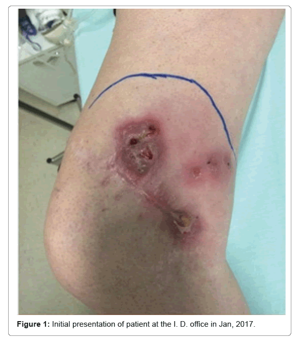
She presented with a large painful area of redness on the medial distal face of her right thigh with six abscessed nodules (Figure 1). No intra-articular effusion could be detected through physical examination, and the knee presented with important range limitation due to pain and possible chronic fibrosis of the joint.
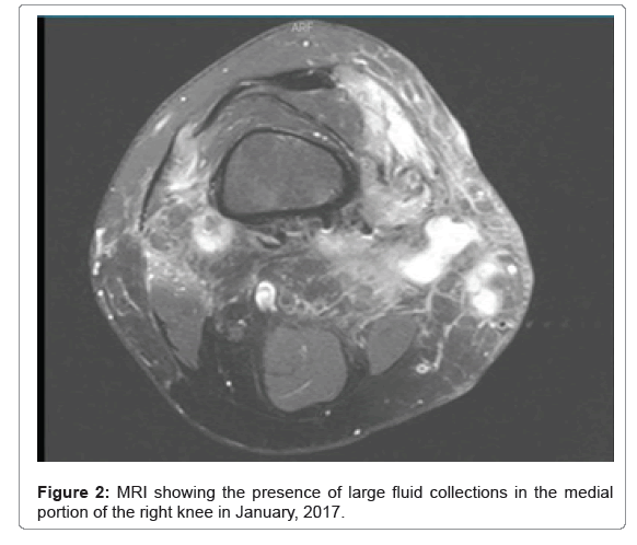
Laboratory findings showed normochromic and normocytic moderate anemia and an elevated erythrocyte sedimentation rate. The magnetic resonance images showed the presence of dense fluid collections in the medial portion of the right knee that extended to the posterior and lateral compartments through the deep adipose plans, eventually fistulizing through the skin (Figure 2).
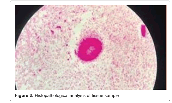
The patient was submitted to open drainage of the abscesses, and sporiform fungal-like structures were identified in the histopathological analysis of the samples, within a chronic diffuse inflammatory neutrophilic infiltrate of the skin, subcutaneous tissue and muscle. Nodular formations were observed, as well as muscle necrosis (Figure 3).
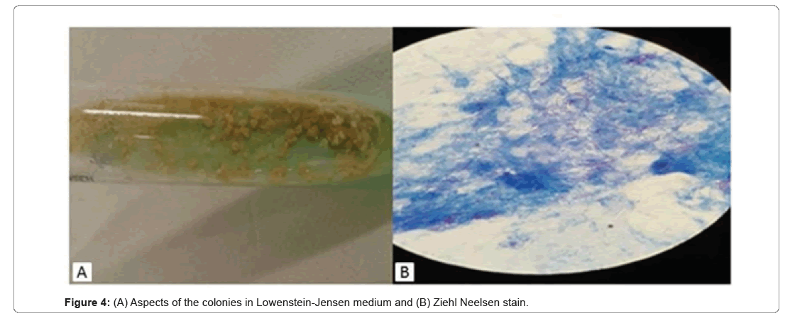
A partially acid-fast Gram-positive long ramified rod was retrieved from wrinkled and dry colonies that grew in Lowenstein-Jensen medium (Figures 4A and 4B). A Polymerase Chain Reaction (heat-shock protein 70 PCR-RFLP) was negative for Mycobacteria spp.
Nocardia sp was identified through mass spectrometry Matrix-Assisted Laser Desorption/Ionization Time of Flight (Maldi-TOFTM) using the Myla and SARAMIS data bases. The protocol for this procedure was consistent with the one described by Salleb, et al., in which the inactivation and breaking of the cell wall release proteins, favoring a safe manipulation of the microorganism.
Although the sample was analyzed through polymerase chain reaction of the 16S rRNA and through mass spectrometry (Maldi-TOFTM) in a reference laboratory, the species could not be fully identified.
As the patient was 12-weeks pregnant and trimethoprim-sulfamethoxazol is a class D drug for pregnancy, treatment with meropenem was started and maintained until child delivery, when the sulfonamide was then started and maintained for twelve months – even though the species was not identified and susceptibility tests were not provided.
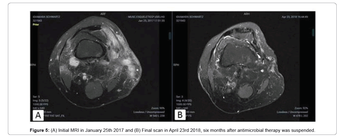
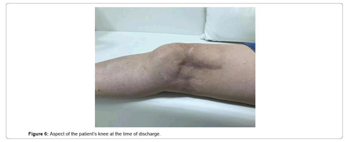
The patient needed another three surgical debridements during the treatment, and was successfully discharged from the Infectious Diseases follow up six months after the completion of the treatment, with complete resolution of the infection (Figures 5A, 5B and 6).
Results and Discussion
Even though Gram-positive cocci are the most frequent pathogens associated to orthopedic SSI’s, chronic, atypical or recurrent infection, symptoms should point to differential etiological diagnosis. Adequate diagnosis and treatment of any infection provides better outcomes and must always be pursued. Nocardial infections pose as a challenge to physicians, especially in low- resources scenarios, where pathogen identification cannot always be achieved. As Nocardia sp. is an acid-fast rod that grows in Löwenstein-Jensen medium, it can easily be mistaken by some rapid-growing Mycobacterium species, and the correct identification of the pathogen is crucial for therapeutic success.
The identification of Nocardial species is difficult and is often only achieved through sequencing or molecular techniques, such as PCR-RFLP [14], or Maldi- TOF™. These Technologies are not available country-wide in Brazil, especially in low-resource areas, far away from capital cities, which was what happened in this case. Even though Nocardia spp was retrieved from the tissue samples, the pathogen was only identified to the genus and not the species. Some issues may have contributed to that, such as the age of the colony, the need for sending the sample to a reference laboratory facility and the limited size of the Maldi-TOFTM data bank [7,9]. Gene sequencing not always can identify the species, as there are some interspecies polymorphisms within the 16SrRNA gene, making it very difficult to distinguish between related Nocardia species [22]. Identification of pathogens is crucial for treatment of any infection, and the adequate treatment of nocardial infections include surgical treatment and debridement as well as directed antimicrobial therapy. Although there are no optimal antimicrobial regimens established, sulfonamides are the cornerstone agents for treatment of nocardial infections [7,20,21]. As some Nocardia spp show high levels of sulfonamide resistance, empirical treatment of severe nocardial infections may involve combinations regiments, such as sulfonamides and carbapenems or third-generations cephalosporins, for example [21-25]. Current recommendation from the Centers for Disease Control (CDC) is that sulfonamides are appropriate for treating infections during the first trimester of pregnancy as long as no other suitable alternative drug is available, being assigned as class C agents during pregnancy [22,23]. As treatment should preferably be individually based [24], the authors chose a carbapenem-based initial treatment regimen, transitioning it to a sulfonamide-based regimen once pregnancy ended.
Although it is impossible to establish with a good amount of certainty, the fact that the patient did not report any infection signs or symptoms until twelve months after the elective surgical procedure points to this being the inoculation moment. Nocardia spp is not a frequent pathogen in such SSI’s but should be considered whenever chronic or recurrent nodular skin lesions are present, not only in immunocompromised hosts.
Nocardia spp has been isolated from hospital water by Hassan and Ali [15], good practices to cleansing and disinfection of surgical equipment, as well as strict local hospital policies towards complying with federal legislation for not reprocessing single use arthroscopic material are crucial for preventing SSI’s of any kind.
Nocardia farcinica had been reported to cause surgical site infections in two outbreaks, although neither of these reports identified a definite source and mode of transmission [17,18]. Also, post-operative mediastinitis caused by Nocardia was well documented in literature in several publications [26,27].
Wenger et al. described a surgical site infection caused by N. farcinica in open heart surgeries in Newark in 1991 and 1992, and the time between surgery and symptoms ranged from 21 to 45 days. The only common variable between the four described patients was the exposure to one specific anesthesiologist, in which the pathogen was isolated from his hands. The pathogen was phenotypically similar to the ones isolated from the patients.
Conclusion
In all these reports, the signs and symptoms of nocardial infection developed within a few weeks after the surgical procedure, and what is remarkable in the present case is that the patient was free of symptoms for twelve months after the second surgical procedure and denied any additional trauma. Also, the patient presented in this case report was treated in two different facilities, by two different orthopedic surgeons and different anesthesiology teams, and no investigation concerning staff colonization could be performed. This is a case report of nocardial infection following elective orthopedic surgery in an immunocompetent patient. Nocardia spp is not a frequent pathogen in such SSI’s but should be considered whenever chronic or recurrent nodular skin lesions are present, not only in immunocompromised hosts.
Acknowledgment
We are thankful to Instituto Hermes Pardini, Instituto de Patologia do Oeste, Cíntia Dias e Jerso Menegazzi for their contribution.
Authors Contribution
Study concept and design: CCP and MDL Acquisition of data: CCP and MDL. Drafting of the manuscript: CCP, MDL, and NBA. Critical revision of the manuscript for important intellectual content: CCP, MDL, NBA and MAM. All authors read and approved the final manuscript.
Consent for Publication
Written informed consent was obtained from the patient publication of this case report. We thank her for providing consent to publish this case report.
Competing Interests
The authors declare that they have no competing interests.
References
- Fatahi-Bafghi M (2018) Nocardiosis from 1888 to 2017. Microb Pathog 114:369-384.
[Crossref] [Google Scholar] [PubMed]
- Yong EX, Cheong EY, Boutlis CS, Chen DB, Liu EYT, et al. (2015) Nocardia septic arthritis complicating an anterior cruciate ligament repair. J Clin Microbiol 53:2760-2762.
- Sharma NL, Mahajan VK, Agarwal S, Katoch VM, Das R, et al. (2008) Nocardial mycetoma: Diverse clinical presentations. Indian J Dermatol Venereol Leprol 74:635-640.
[Crossref] [Google Scholar] [PubMed]
- Baldi BG, Santana ANC, Takagaki TY (2006) Pulmonary and cutaneous nocardiosis in a patient treated with corticosteroids. J Bras Pneumol 32:592-605.
[Crossref] [Google Scholar] [PubMed]
- Baio PVP, Ramos JN, Santos LSd, Soriano MF, Ladeira EM, et al. (2013) Molecular identification of nocardia isolates from clinical samples and an overview of human nocardiosis in Brazil. PLoS Negl Trop Dis 7:e2573.
[Crossref] [Google Scholar] [PubMed]
- Schlaberg R, Fisher MA, Hanson KE (2014) Susceptibility profiles of nocardia isolates based on current taxonomy. Antimicrob Agents Chemother 55:795-800.
- Smit LHM, Leemans R, Overbeek BP (2003) Nocardia farcinica as the causative agent in a primary psoas abscess in a previously healthy cattle inspector. Clin Microbiol Infect 9:445-448.
- Jenks PJ, Laurent M, McQuarry S, Watkins R (2014) Clinical and economic burden of Surgical Site Infection (SSI) and predicted financial consequences of elimination of SSI from an English hospital. J Hospital Infect 86:24-33.
[Crossref] [Google Scholar] [PubMed]
- Khot PD, Bird BA, Durrant RJ, Fisher MA (2015) Identification of Nocardia species by Matrix-Assisted Laser Desorption Ionization–Time of Flight mass spectrometry. J Clin Microbiol 53:3366-3369.
[Crossref] [Google Scholar] [PubMed]
- Ercole FF, Chianca TCM. Surgical Site Infection in patients submited to hip arthroplasty. Latin Americ J Nurs 10:157-165.
- Allegranzi B, Nejad SB, Combescure C, Graafmans W, Attar H, et al. (2011) Burden of endemic health-care-associated infection in developing countries: Systematic review and meta-analysis. Lancet 377:228-241.
[Crossref] [Google Scholar] [PubMed]
- Ercole FF, Chianca TCM, Duarte D, Starling CEF, Carneiro M (2011) Surgical Site Infection in patients submitted to orthopedic surgery: The nnis risk index and risk prediction. Rev Latino-Am Enfermagem 19:269-276.
[Crossref] [Google Scholar] [PubMed]
- Mu Y, Edwards JR, Horan TC, Berrios-Torres SI, Fridkin SK (2011) Improving risk-adjusted measures of Surgical Site Infection for the national healthcare safety network. Infect Control Hosp Epidemiol 32:970-986.
[Crossref] [Google Scholar] [PubMed]
- Brown-Elliott BA, Brown JM, Conville PS, Wallace RJ (2006) Clinical and laboratory features of the Nocardia spp. based on current molecular taxonomy. Clin Microbiol Rev 19:259-282.
[Crossref] [Google Scholar] [PubMed]
- Shojaei H, Rahdar AH (2016) First report of isolation of Nocardia otitidiscaviarum from hospital water. J Coastal Life Med. 4.
[Crossref] [Google Scholar] [PubMed]
- Lima ALLM, Zumiotti AV, Uip DE, Silva JS (2004) Fatores preditivos de infecção em pacientes com fraturas expostas nos membros inferiores. Acta Ortop Bras 12.
- Exmelin L, Malbruny B, Vergnaud M, Provost F, Boiron P, et al. (1996) Molecular study of nosocomial nocardiosis outbreak involving heart transplant recipients. J Clin Microbiol 34:1014-1016.
[Crossref] [Google Scholar] [PubMed]
- Blumel J, Blumel E, Yassin AF, Schmidt-Rotte H, Schaal KP (1998) Typing of Nocardia farcinica by pulsed-field gel electrophoresis reveals an endemic strain as source of hospital infections. J Clin Microbiol 36:118-122.
[Crossref] [Google Scholar] [PubMed]
- Wenger PN, Brown JM, McNeil MM, Jarvis R (1998) Nocardia farcinica sternotomy site infections in patients following open heart surgery. J Infect Dis. 178:1539-1543.
[Crossref] [Google Scholar] [PubMed]
- Ukai Y, Fujimoto N, Fujii N, Shirai M, Wakabayashi M, et al. (2012) Case of muscle abscess due to disseminated nocardiosis in a patient with autoimmune hemolytic anemia, and review of the published work. J Dermatol 39:466-469.
[Crossref] [Google Scholar] [PubMed]
- Schlaberg R, Fisher MA, Hanson KE (2014) Susceptibility profiles of nocardia isolates based on current taxonomy. Antimicrob Agents Chemother 58:795-800.
[Crossref] [Google Scholar] [PubMed]
- Mazzaferri F, Cordioli M, Segato E, Adami I, Maccacaro L, et al. (2018) Nocardia infection over 5 years (2011-2015) in an Italian tertiary care hospital. New Microbiol. 41:136-140.
[Google Scholar] [PubMed]
- Committee Opinion No. 717. Sulfonamides, nitrofurantoin, and risk of birth defects. American College of Obstetricians and Gynecologists. Obstet Gynecol 130:e150-e152.
- You Y, Chen W, Zhong B, Song Z, Yang X (2018) Disseminated nocardiosis caused by Nocardia elegans: A case report and review of the literature. 46:705-710.
[Crossref] [Google Scholar] [PubMed]
- Fatahi-Bafghi M (2018) Nocardiosis from 1888 to 2017. Microb Pathogen 114:369-384.
[Crossref] [Google Scholar] [PubMed]
- Verghese S, Kurian VM, Maria CF, Padmaja P, Elizabeth SJ, et al. (2003) Nocardia asteroides Mediastinitis Complicating Coronary Artery Bypass Surgery. J Assoc Physicians India 51:1009-1010.
[Google Scholar] [PubMed]
- Thaler F, Gotainer B, Teodori G, Dubois C, Loirat P (1992) Mediastinitis due to Nocardia asteroids after cardiac transplantation. Int Care Med 182:127-128.
[Crossref] [Google Scholar] [PubMed]
Citation: Ponzi CC, Martins MD, Alves NB, Catapan K (2022) Nocardial Infection Following Elective Knee Video Arthroscopy in an Immunocompetent Adult. J Infect Dis Ther 10:511. DOI: 10.4172/2332-0877.1000511
Copyright: © 2022 Ponzi CC, et al. This is an open-access article distributed under the terms of the Creative Commons Attribution License, which permits unrestricted use, distribution, and reproduction in any medium, provided the original author and source are credited.
Select your language of interest to view the total content in your interested language
Share This Article
Recommended Journals
Open Access Journals
Article Tools
Article Usage
- Total views: 2983
- [From(publication date): 0-2022 - Nov 18, 2025]
- Breakdown by view type
- HTML page views: 2473
- PDF downloads: 510

