Case Report Open Access
Nocardia farcinica Multiple Brain Abscesses in an Immunocompetent Patient
| Luisa Vinciguerra1*, Carmen Murr2 and Julian Böse1,2 | |
| 1Department GF Ingrassia, Section of Neurosciences, University of Catania, Via Santa Sofia, 78 - 95123 Catania, Italy | |
| 2Department of Neurology, Heidelberg University, INF 400, 69120 Heidelberg, Germany | |
| Corresponding Author : | Luisa Vinciguerra Department GF Ingrassia, Section of eurosciences University of Catania. Via Santa Sofia 78 - 95123 Catania, Italy Tel: +393488551601 E-mail: luisavinciguerra.doc@gmail.com |
| Received: July 11, 2015 Accepted: July 13, 2015 Published: July 16, 2015 | |
| Citation: Vinciguerra L, Murr C, Böse J (2015) Nocardia farcinica Multiple Brain Abscesses in an Immunocompetent Patient J Neuroinfect Dis 6:182. doi:10.4172/2314-7326.1000182 | |
| Copyright: © 2015 Vinciguerra L, et al. This is an open-access article distributed under the terms of the Creative Commons Attribution License, which permits unrestricted use, distribution, and reproduction in any medium, provided the original author and source are credited. | |
| Related article at Pubmed, Scholar Google | |
Visit for more related articles at Journal of Neuroinfectious Diseases
Abstract
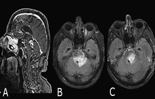
Background: Nocardiosis is a rare infection caused by Nocardia species, that usually causes a solitary abscess as the most common manifestation in the central nervous system. Nocardia farcinica appears more virulent than the other subspecies, and the infection generally occurs in immunocompromised patients. We describe a case of primary multiple brain abscesses from N. farcinica in an immunocompetent host. Case report: A 66-year-old immunocompetent man was admitted with a 6-week-history of night sweats and progressive fatigability followed by intermittent diplopia, left facial nerve palsy, aphasia, dysmetria, loss of balance, disorientation and drowsiness. He had no history of fever, headache, vomiting or seizures. Cerebral magnetic resonance imaging (cMRI) displayed multiple supratentotial lesions and a wide confluent ponto-mesencephalic hyperintensity on fluid-attenuated inversion recovery (FLAIR) images. Most of the lesions showed ring gadolinium enhancement and partially diffusion restriction. Mild pachymeningeal thickening and enhancement were also detected. The brain biopsy and the microbiological evaluation revealed branching gram-positive bacilli, confirmed as Nocardia farcinica. Because of hydrocephalus occlusus caused by the space-occupying brain stem lesion the patient required temporary external ventricular drainage. Treatment with intravenous imipenem and cotrimoxazole was administered for one month and consecutively oral cotrimoxazole was maintained, with substantial improvement of neurological conditions. Conclusions: CNS norcardial infections may manifest as multiple brain or spinal cord lesions, diffuse cerebral inflammation, and meningitis mimicking other neurological disorders, especially tumors, and possibly delaying diagnosis and treatment. The mortality rates estimated for multiple abscesses due to Nocardia farcinica are significant. This infection might progress despite a specific therapy and tend to relapse. Early identification, appropriate and prolonged treatment are crucial for a favorable outcome.
| Keywords |
| Nocardiosis; Cerebral abscess; Antibiotic therapy. |
| Introduction |
| Nocardiosis is a rare infection caused by the aerobic Actinomyces species of Nocardia, which is ubiquitous in the environment [1,2]. The infection is generally acquired by inhalation, but is also transmitted through cutaneous disease, insect bite, trauma, surgery, and vascular catheters [3-5]. Nocardia shows to have a special tropism for the neural tissue. Solitary abscess represents the most common manifestation in the central nervous system (CNS), accounting for 1% to 2% of all cerebral abscesses [2]. Nocardia farcinica appears more virulent than the other subspecies, since the infection may cause disseminated lesions, tend to relapse and have a higher antibiotic resistance (especially to thirdgeneration cephalosporins) [6,7]. Furthermore, Nocardiosis usually affects immune compromised patients [1,4,8]. |
| Case Report |
| A 66-year-old immune competent man with medical history of hypoththyroidism and diabetes mellitus was referred for a 6-weekhistory of night sweats and progressive fatigability followed in the last week by intermittent diplopia, memory disturbance and loss of balance. Fever, headache, vomiting and seizures were not reported. On admission the neurological examination of the afebrile patient revealed disorientation, drowsiness, left facial nerve palsy, dysconjugate eye movements, motor aphasia, dysmetria at the finger-to-nose and heel-to-shin maneuvers bilaterally, dysdiadochokinesia and severe gait ataxia. cMRI displayed multiple supratentotial lesions (in the right preand post-central gyrus, left frontal and temporal lobe bilaterally) and a wide nodular heterogeneous hyperintensity on FLAIR images in the left midbrain and pons, with the involvement of cerebellar hemisphere and the basal ganglia of the left side and an adjacent mass effect. Most of the lesions showed partially diffusion restriction and ring gadolinium enhancement (Figure 1A and 1B). Mild pachymeningeal thickening and enhancement were also detected. An initial diagnosis of tumor was made and the patient started on dexamethasone. Apart from the diabetes, history did not indicate immune deficiency, and travel-, workand exposition history were unremarkable. Extensive laboratory data only revealed a slightly decreased white blood cell count, but the absolute values were unremarkable, serum immunoglobulin levels were within normal range and human immunodeficiency virus serologic test was negative. Cerebrospinal fluid (CSF) analysis showed mild pleocytosis (16/mcL), hyperproteinorrachia (165 mg/dl), hypoglicorrachia (103 mg/dl), high albumin CSF/serum concentration quotient (20.5), and intrathecal IgM and IgA synthesis. Biopsy of the left temporal lobe lesion revealed the presence of an abscess containing methylamine silver staining filamentous structures, resembling Nocardia spp. The following microbiological evaluations on brain tissue and blood were positive for Nocardia farcinica. The antibiotic susceptibility revealed a cefotaxime-resistance. Because of hydrocephalus occlusus caused by the space-occupying brain stem lesion the patient required temporary external ventricular drainage. He had a focal seizure, so he was started on levetiracetam. Furthermore, treatment with intravenous imipenem (1 g qid) and cotrimoxazole (960/4800 mg daily) was administered for one month with substantial improvement of neurological conditions. After four weeks of treatment the patient was conscious but disorientated, had recovered from diplopia and facial nerve palsy, while aphasia, dysmetria and gait had substantially improved. Consecutively, oral cotrimoxazole was maintained, as part of the strategy to continue the therapy for one year at the dose of 480 mg four times a day. The onemonth follow-up cMRI showed a regression of supratentorial and brain stem lesions (Figure 1C). |
| Discussion |
| In this report we present a case of primary multiple brain abscesses due to N. farcinica in an immune competent patient. CNS norcardial infections may manifest as single abscess, multiple brain or spinal cord lesions, diffuse cerebral inflammation, and meningitis mimicking neoplasms, vasculitis, and stroke [1,7,9]. The mortality rates estimated for Nocardia brain abscess are 55% and 20% in immune compromised and immune competent patients, respectively; however, these rates rise for multiple abscesses and if the infection is due to the subspecies N. farcinica [1,3,10,11]. Nocardial brain abscesses are not common and Nocardiosis in immune competent individuals is even less frequent, being the infection usually observed in immune compromised hosts [1,3,4,8]. Symptomatology could be insidious, variable and without fever or other clear infective symptoms, delaying diagnosis and treatment. No therapeutic guidelines are available, however antibiotics should probably be maintained for up to 12 months in order to prevent relapses. In conclusion, despite the low incidence no cardial infection should be evaluated in the differential diagnosis of cerebral lesions in both immune deficient and immune competent patients, because early identification and appropriate treatment are crucial for a favorable outcome of this potentially life-threatening infection. |
References
- Kennedy KJ, Chung KH, Bowden FJ, Mews PJ, Pik JH, et al. (2007) A cluster of nocardial brain abscesses. Surg Neurol 68: 43-9.
- Lin YJ, Yang KY, Ho JT, Lee TC, Wang HC, et al. (2010) Nocardial brain abscess. J Clin Neurosci 17: 250-253.
- Alijani N, Mahmoudzadeh S, Hedayat Yaghoobi M, Geramishoar M, Jafari S (2013) Multiple brain abscesses due to nocardia in an immunocompetent patient. Arch Iran Med 16: 192-4.
- Lederman ER, Crum NF (2004) A case series and focused review of nocardiosis: clinical and microbiologic aspects. Medicine (Baltimore) 83: 300.
- Beaman BL, Beaman L (1994) Nocardia species: host-parasite relationships. Clin Microbiol Rev 7: 213.
- Uhde KB, Pathak S, McCullum I Jr, Jannat-Khah DP, Shadomy SV (2010) Antimicrobial-resistant nocardia isolates, United States, 1995-2004. Clin Infect Dis 51: 1445
- Kumar VA, Augustine D, Panikar D, Nandakumar A, Dinesh KR, et al. (2014) Nocardia farcinica brain abscess: epidemiology, pathophysiology, and literature review. Surg Infect (Larchmt) 15: 640-6.
- Lerner PI (1996) Nocardiosis. Clin Infect Dis 22: 891-903.
- Nandhagopal R, Al-Muharrmi Z, Balkhair A (2014) Nocardia brain abscess. QJM 107: 1041-2.
- Mamelak AN, Obana WG, Flaherty JF, Rosenblum ML (1994) Nocardial brain abscess: treatment strategies and factors influencing outcome. Neurosurgery 35: 622-31.
- Al Tawfiq JA, Mayman T, Memish ZA (2013) Nocardia abscessus brain abscess in an immunocompetent host. J Infect Public Health 6: 158-61.
Figures at a glance
 |
| Figure 1 |
Relevant Topics
- Bacteria Induced Neuropathies
- Blood-brain barrier
- Brain Infection
- Cerebral Spinal Fluid
- Encephalitis
- Fungal Infection
- Infectious Disease in Children
- Neuro-HIV and Bacterial Infection
- Neuro-Infections Induced Autoimmune Disorders
- Neurocystercercosis
- Neurocysticercosis
- Neuroepidemiology
- Neuroinfectious Agents
- Neuroinflammation
- Neurosyphilis
- Neurotropic viruses
- Neurovirology
- Rare Infectious Disease
- Toxoplasmosis
- Viral Infection
Recommended Journals
Article Tools
Article Usage
- Total views: 14587
- [From(publication date):
August-2015 - Apr 05, 2025] - Breakdown by view type
- HTML page views : 10061
- PDF downloads : 4526
