Nocardia Brain Abscess Masquerading as Tuberculous Brain Abscess
Received: 12-Apr-2023 / Manuscript No. JNID-23-93908 / Editor assigned: 14-Apr-2023 / PreQC No. JNID-23-93908(PQ) / Reviewed: 28-Apr-2023 / QC No. JNID-23-93908 / Revised: 04-May-2023 / Manuscript No. JNID-23-93908 (R) / Accepted Date: 10-May-2023 / Published Date: 11-May-2023 DOI: 10.4172/2314-7326.1000446
Abstract
Nocardia brain abscess is a rare clinical entity [1]. It accounts for 1%-2% of all cerebral abscesses [2]. It typically occurs in immunocompromised patients with impaired host defences usually cell mediated immunity but cases in immunocompetant hosts have also been reported [3].Cerebral nocardiosis is associated with highest mortality and morbidity among all brain abscess caused by microorganisms [4]. Nocardiosis is liable to be misdiagnosed owing to the non- specific clinical manifestations, laboratory and imaging findings. The most common variant affecting the brain is Nocardia farcinica. We report a rare case of brain abscess caused by Nocardia farcinica in a 36-year-old male.
Keywords
Infectious disease; Nocardiosis; Brain abscess; Nocardia farcinica; MALDITOF-MS; Mortality
Introduction
Nocardia is an aerobic filamentous gram positive bacterium which is found in the soil. Nocardia is acquired by direct inhalation or via skin contact. Immunocompromised people and those with pre-existing lung disease are highly susceptible .Nocardiosis is associated with Pulmonary alveolar proteinosis, tuberculosis, chronic granulomatous disease, IL- 12 deficiency, auto antibodies to GM-CSF. Lung infection is the most common presentation of Nocardiosis. Isolated CNS infection develops in approximately 9% individuals. Nocardia farcinica has a higher risk of dissemination to CNS. Clinical forms of Nocardia farcinica infection include pulmonary or pleural infections, brain abscesses and skin or soft tissue infections [5]. Focal neurologic deficits, altered consciousness, seizures, visual disturbances and ataxia are common presentations in CNS nocardiosis. However, the mortality rate of nocardial brain abscesses is as high as 30%, which is much higher than the 10% caused by other bacteria [6].
The diagnosis of N. farcinica using conventional methods is difficult, and the misdiagnosis may lead to inappropriate treatments and delay the therapy, resulting in the high mortality of the disease. Routine diagnostic methods are blood, cerebrospinal fluid, and brain aspirate culture, and craniotomy is generally performed to remove a lesion and obtain a specimen.
However, in recent years matrix assisted laser desorption ionizationtime of flight mass spectrometry (MALDI-TOF MS) has emerged as an important tool for microbial identification and diagnosis. During the MALDI-TOF MS process, microbes are identified using either intact cells or cell extracts. The process is rapid, sensitive, and economical in terms of both labor and costs involved. The introduction of this technique would contribute to early and accurate diagnosis of the disease, avoid surgery, and reduce mortality.
Case presentation
A 36 year, old male presented with complaints of headache, vomiting for a duration of two weeks and altered sensorium, seizures for a day. No history of fever in the past 3 months. Past history is significant for recurrent episodes of extra-pulmonary tuberculosis. No history of any surgery or any steroid abuse in the past. On Examination, at admission he was drowsy, arousable to verbal stimulus, had papilledema and neck stiffness however he didn’t have any weakness or cranial nerve deficits.
He had clear lung sound without rales or wheezing. The heartbeat was regular without any murmur. There was no tenderness or rebound tenderness in the abdomen.
Routine blood tests were within normal limits except for erythrocyte sedimentation rate, which was 86 mm/h. Human immunodeficiency virus (HIV) antibody, hepatitis B surface antigen and hepatitis C antibody were all negative. Chest and abdomen radiographs were normal.
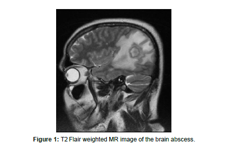
CT Head was done immediately which revealed relatively well defined heterodense lesion with peripheral hyperdense area involving Right parieto-occipital lobe with adjacent edema, mass effect and midline shift suggestive of a granulomatous etiology. MRI Brain suggested features of tubercular abscess rupture with ventriculitis [Figure 1].
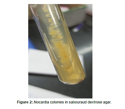
The presumptive diagnosis was CNS tuberculosis, all though all tests were negative for mycobacterium. However, the patient did not benefit from antituberculous treatment. CSF study revealed increased WBC counts with neutrophilic predominance and increased protein and very low sugars. CSF CBNAAT was inconclusive. Meanwhile CSF culture results indicated the growth of Nocardia. Modified Ziehl Neelsen staining of CSF Culture using 1% sulfuric acid revealed; thin, filamentous, branching partially acid- fast bacteria in a background of many polymorphonuclear leukocytes, suggestive of Nocardia [Figure 2].The growth of CSF culture on Sabouraud dextrose agar showed buff colored dry, cerebriform colonies [Figure 3]. The growth was confirmed by Matrix Assisted Laser Desorption Ionization Time of Flight Mass Spectrometry (MALDITOF-MS) as Nocardia farcinica. The Anti-TB treatment was stopped and he was prescribed Cotrimoxazole with imepenem but succumbed to his illness weeks later.
Discussion
Nocardia species are Gram- positive, aerobic, branching filamentous bacteria belonging to Actinomycetales, which are usually found in the soil. Three main species cause humans infection, including N asteroides, N brasiliensis, and N caviae, N asteroides is the most commonly isolated. Here, we are reporting a brain abscess caused by the rarer species, i.e., N farcicina. Nocardia infection commonly arise in the immunocompromised states including organ transplants, leukemia, HIV, steroid abuse.
Our patient did not have any of these predisposing factors. Usually nocardial pathogen spread to the CNS hematogenously. The involvement of the CNS was found in nearly half of all disseminated nocardial infections in the literature. In this report, we present a case of primary brain abscess due to Nocardia farcinica in an immune competent patient. CNS norcardial infections may manifest as single abscess, multiple brain or spinal cord lesions, diffuse cerebral inflammation, and meningitis mimicking neoplasms, vasculitis, and stroke. The mortality rates estimated for Nocardia brain abscess are 55% and 20% in immune compromised and immune-competent patients, respectively; however, these rates rise for multiple abscesses and if the infection is due to the subspecies N. farcinica. Nocardial brain abscesses are not common and Nocardiosis in immune competent individuals is even less frequent.
To treat a nocardial brain abscess, a 12- month course of therapy is recommended. TMP- SMX is currently accepted as the first- line treatment for nocardiosis.
Conclusion
Diagnosis of primary and systemic nocardiosis is difficult due to paucity of clinical and laboratory signs of infection. A comprehensive list of differential diagnosis and meticulous diagnostic evaluation of the patients is of paramount importance to prevent morbidity and mortality associated with delayed or missed diagnosis.
Acknowledgement
Not applicable.
Conflict of Interest
Author declares no conflict of interest.
References
- Massimiliano B, Beretta S, Farina C, Ferrarimi M, Vittorio C (2005) Medical treatment for nocardial abscesses. Case report J Neurol 252: 1120-1121.
- Grond SE, Schaller A, Kalinowski A, Tyler KA, Jha P (2020) Nocardia farcinica brain abscess in an immunocompetent host with pulmonary alveolar proteinosis: a case report and review of the literature. Cureus 12: 11494.
- Beaman BL, Burnside J, Edwards B, Causey W (1976) Nocardial infections in the United States, 1972-1974. J Infect Dis 134:286-289.
- Kennedy KJ, Chung KHC, Bowden FJ, Mews PJ, Pik J, H et al. (2007) A cluster of nocardial brain abscesses. Surg Neurol 68:43-49
- Corti ME, Villafañe-Fioti MF (2003) Nocardiosis: a review. Int J Infectious Dis 7:243-250.
- Cassir N, Million M, Noudel R, Drancourt M, Brouqui P (2013) Sulfonamide resistance in a disseminated infection caused by Nocardia wallacei: a case report. J Med Case Rep7:103.
Indexed at, Google Scholar, Crossref
Indexed at, Google Scholar, Crossref
Indexed at, Google Scholar, Crossref
Indexed at, Google Scholar, Crossref
Indexed at, Google Scholar, Crossref
Citation: Goutham Krishna TC, John JK, Aswathi Raveendran UV (2023) Nocardia Brain Abscess Masquerading as Tuberculous Brain Abscess. J Neuroinfect Dis 14: 446. DOI: 10.4172/2314-7326.1000446
Copyright: © 2023 Goutham Krishna TC, et al. This is an open-access article distributed under the terms of the Creative Commons Attribution License, which permits unrestricted use, distribution, and reproduction in any medium, provided the original author and source are credited.
Share This Article
Recommended Journals
Open Access Journals
Article Tools
Article Usage
- Total views: 1829
- [From(publication date): 0-2023 - Apr 17, 2025]
- Breakdown by view type
- HTML page views: 1582
- PDF downloads: 247


