Research Article Open Access
New Aspects of Therapy of Hepatocellular Carcinoma Egyptian Patients
| Ragaa Hosny Mohamad1*, Mohamad Gamil Abdelmoneim El-Said2, Zekry Khalid Zekry3, Amal Mohamad Al-Bastawesy4, Rashwan Mohamad Farag5, Hussien Abdel-RahmanAl-Mehdar6, Abdulbaset Abdelsalam Elfighia7, Yahia Mahmoud Ismail8, Elsayed Ibrahim Elshayeb 9, Abdel Fatah Mohsen Badawy10, Sabry Mohamad Sharawy1 and Mahmoud Mohamad Elmarzabani11 | |
| 1Clinical Biochemistry, Cancer Biology, National Cancer Institute, Cairo University, Egypt | |
| 2Surgical Oncology National Cancer Institute, Cairo University, Egypt | |
| 3Medical Oncology, National Cancer Institute, Cairo University, Egypt | |
| 4Food Technology Research Centre, Cairo University, Egypt | |
| 5Medical Biochemistry, Ain Shams University, Egypt | |
| 6Faculty of science, Biology Department, Immunology Unit, King Abdulaziz University, Jeddah, Saudi Arabia | |
| 7Medical Biochemistry, Tripoli University, Libya | |
| 8Medical Oncology, National Cancer Institute, Cairo University, Egypt | |
| 9Internal Medicine, Faculty of Medicine, Menofyia University, Egypt | |
| 10Organic Chemistry, Petroleum Research Centre, Egypt | |
| 11Drug Research and Experimental Pharmacology, National Cancer Institute, Cairo University, Egypt | |
| *Corresponding Author : | Ragaa Hosny Mohamad Mohamadeen Al-Tahtawy Cancer Biology Department National Cancer Institute Cairo University, Fom Al-Khalig Street Post No:11234, Cairo, Egypt Tel: 0020100185686 Fax: +0223644720 E-mail: ragaatahtawy@gmail.com/td> |
| Received May 14, 2014; Accepted December 30, 2014; Published January 06, 2015 | |
| Citation: Mohamad RH, El-Said MGA, Zekry ZK, Al-Bastawesy AM, Farag RM, et al. (2015) New Aspects of Therapy of Hepatocellular Carcinoma Egyptian Patients. Biochem Physiol 4:150. doi:10.4172/2168-9652.1000150 | |
| Copyright: © 2015 Mohamad RH, et al. This is an open-access article distributed under the terms of the Creative Commons Attribution License, which permits unrestricted use, distribution, and reproduction in any medium, provided the original author and source are credited. | |
Visit for more related articles at Biochemistry & Physiology: Open Access
Abstract
Background: The purpose of this study was design to investigate the potential impact of chemo and hormonal therapy drugs either with natural oils or other with vitamin E on HCC Egyptian patients.
Methods: A retrospective analysis of medical records was performed on 100 unresectable HCC Egyptian patients in stage A or B who were identified through the National Cancer Institute Cairo University and Menoufia University, from 2010 to 2013. All of patients had been received systemic chemo and hormonal and therapy (5-fluorouracil, Oxaloplatin and tamoxifen) either with five type of natural oils or other with vitamin E. Clinical picture , hematological test, metabolic parameters, telomerase activity, VEGF, MAD, GSH, zinc, ER ,PR, KRAS, BRAF and NRAS has been assessed. Survival curves were also analyzed at pre-and post-treatment. All patients were applied to do regular exercise, control their diet before receiving treatment. The survival rate was determined.
Results: the present data indicate that oral NS.O and CUR.O showed greatly kept hematological finding, serum levels of telomerase activity, VEGF, GSH, MAD ALT, AST, GGT, bilirubin, albumin and zinc to the normal levels (p < 0.001), the toxicity has been reduced without affecting response to chemo and hormonal therapy and survival , the quality of life, clinical picture and survival seemed to be better.Fortunately the depletion in the serum levels of the VEGF and the telomerase activity correlates with good of clinical picture compared with other groups under investigation.
Conclusion: Using Chemo and hormonal therapy, in combination with oral NS.O and CUR.O was identified as the most suitable treatment for HCC patients resulted in improving survival, maximizing clinical benefit, and minimizing risk of chemo and hormonal therapy toxicity. The telomerase activity and VEGF levels can be used as markers to predict survival and response to treatment in HCC patients in the future.
| Keywords |
| Evening Primrose oils; Fish oil; Nigella Sativa L.oil; Curcumin oil; HCC; Chemotherapy; Telomerase activity; MDA; GSH; VEGF; Survival |
| Abbreviations |
| PUFAs-3 Polyunsaturated fatty acids; EPAEicosapentaenoic acid; DPA- Docosapentaenoic acid; DHADocosahexaenoic acid; HCC- Hepatocellular carcinoma; NSO-Nigella Sativa L.oils oil; FO-fish oil; EPO-Evening Primrose oil; VEGF-Vascular endothelial growth factor; MAD-Malondialdehyde(lipid peroxide); GSH-Glutathione reduced; ALT-Alanine aminotransferase; ASTAspartate aminotransferase; GGT-Gamma glutamyl transferase; T.B-Total bilirubin; Alb-Albumin; PCR-Polymerase Chain Reaction; TBARS-Thiobarbituric acid reactive substances; GLC-Gas liquid chromatography; TRAP-Telomeric Repeat Amplification Protocol; TMX-Tamoxifen; 5-Fu-5-Flurouracil; CK-Creatine kinase; LDHLactate dehydrogenase |
| Introduction |
| Hepatocellular carcinoma (HCC) represents is the fifth most common human cancer worldwide accounting for an estimated 500,000 fatal outcomes per year [1]. The medical challenges presented by HCC lie not just in the high incidence of the disorder, but also in its generally unfavorable clinical course [2]. Current treatment options, including surgery, radiotherapy, adjuvant chemotherapy and hormone therapy, may not be adequate to significantly reduce the current morbidity and mortality of Liver cancer and the most cases it is associated with the presence of liver cirrhosis and has a poor prognosis with an overall median survival of 8 months in Austria [3]. |
| Wide arrays of flavonoids and phenolic substances some vitamins and their derivatives, especially those present in dietary and medicinal plants may be used as adjunctive nutritional therapy, have been reported to possess substantial antioxidant, anti-inflammatory, anticarcinogenic, pathways regulating cell cycle, apoptosis, gene expression,and antimutagenic effects [4]. Cancer can be considered a chronic disease of the genome that may be influenced at many stages in its natural history by adjunctive nutritional therapy are potentially willing to alteration to not only to slow the progression and reduce the risk of developing HCC [5] or attenuate the pro-oncogenic effects of different (viral or chemical) carcinogenic agents on the induction, progression of liver cell transformation [6]. GLA may increase the effects of anti cancer treatments and it may also achieve sensitisation effects of anti cancer treatments [7], such as doxorubicin, cisplatin, carboplatin, idarubicin, mitoxantrone, tamoxifen, vincristine, and vinblastine. Another study found that women with breast cancer who took -linolenic acid (GLA( had a better response to tamoxifen (a drug used to treat estrogen sensitive breast cancer) than those who took only tamoxifen [8]. |
| The Evening Primrose oil, contain -linolenic acid (GLA), exerts selective cytotoxic effects on cancer cells without affecting normal cells by inducing membrane destabilization for tumor cells in the cell cycle [9]. Moreover, it so sensitize cells to the effects of other anticancer therapies including antimitotic drugs such as 5-flurouracil, paclitaxel, docetaxel, and vinorelbine, and antiestrogen such as tamoxafin [10]. Also the gamma-linolenic acid (GLA) has a synergistic effect with cytotoxic drugs as gemcitabine at concentrations that correspond to in vivo therapeutic doses and against pancreatic adenocarcinoma cell lines in vitro [11]. |
| Consumption of fish oil reduces the risk of HCC, because it is a rich source of n-3 polyunsaturated fatty acids (PUFAs), such as eicosapentaenoic acid (EPA), docosapentaenoic acid (DPA), and docosahexaenoic acid (DHA) [12]. Thakkar et al. [13] showed that fish oil (F.O) prevents gentamicin and cyclosporine-A-induced nephrotoxicity. Rani et al. [14] proved that the of colorectal cancer model treatment with the 5-Fluorouracil (5-FU) only has low therapeutic response rate and severe side effects but when they using FO which was rich in n-3 polyunsaturated fatty acids in combination with 5-Fluorouracil they proved that the supplementation of FO is potentially a promising option for increasing the therapeutic potential and mitigating the side effects and toxicity profile of 5-FU. |
| Nigella Sativa L.oils are very nutritious as it includes mineral, 35% fixed oils, 1.5% essential (volatile) oil, 21% proteins, alkaloids a pentacyclic triterpene saponins [15] and alpha (α)-hederin [16]. The fixed oil is composed mainly of fatty acids as linoleic (C18:2), oleic (C18:1), palmitic (C16:0) and stearic (C18:0) acids [17]. Thymoquinone (TQ) is the most pharmacologically active ingredient found abundantly with its derivatives such as dithymoquinone, thymohydroquinone (TQ), and thymol [18]. TQ is considered as potent anti-oxidant [19], anti-carcinogenic and anti-mutagenic agent [20]. Moreover, a low dose of thymoquinone induced breast cancer cell death [21]. Forunatly, thymoquinone and/or its analogues may have clinical potential as an anticancer agent alone or in combination with chemotherapeutic drugs such as cisplatin in breast cancer cell [22]. |
| Alpha (α)-hederin, a pentacyclic triterpene saponin was also reported to have potent in vivo antitumor activity [16] inhibits cell proliferation and cellular viability [23]. The fatty acids like Linoleic Acid (LA) and Gamma-Linolenic Acid (GLA) help strengthen and maintain cell integrity, heal skin conditions like acne, eczema, psoriasis, reduce wrinkles, and heal wounds [24]. N. sativa seed oil induce no harmful effects on the liver, it posses strong antioxidant property which control lipid peroxidation activity, reduced the genetic instability that can lead to carcinogenesis [25] and disregulation in cell cycle and apoptosis [26]. |
| Curcumin is a yellow, naturally occurring polyphenolic phytochemical derived from the powdered rhizome of the herb Curcuma longa Linn [27]. Several studies on animal model studies have shown that Curcumin suppresses carcinogenesis in, stomach [28], colon [29], breast [30], and liver [31]. Chemopreventive activities of Curcumin are thought to be involving up-regulation of carcinogen-detoxifying enzymes [32] and antioxidants [33], suppression cyclooxygense-2 expression [34], and inhibition nuclear factor-κB release [35]. |
| HCC has no effective systemic therapy currently exists. Recombinant [alpha]-interferon (INF) has been suggested to have some antitumor efficacy in this illness, and synergism with 5-fluorouracil (5-FU) has been reported in several gastrointestinal malignancies but there was no significant benefit from treatment with 5-FU and IFN in patients with HCC [3]. Their supplementation with vitamin E could prevent free radical induced damage to the tissues and could prevent lipid per-oxidation by scavenging hydroxyl radical and enhances immune responses for the elderly people [36]. |
| Angiogenesis are considered to be important for HCC progression [37]. Vascular endothelial growth factor (VEGF) is a well known, potent angiogenic factor which enhances vascular permeability, promoting the extravasation of protein to form a stromal matrix and tumor invasion [28]. |
| Increased telomerase activity strongly correlates with increased malignant potential and stage; in addition, genomic instability associated with loss of telomere sequences correlates with a late stage development of colonic carcinomas [38] Telomerase activity was also highly expressed in malignancy [39]. On the other hand, in humans, over 85% of malignant tumors express telomerase activity whereas most somatic tissues do not present. Until recently, there was no standard medical treatment for unresectable Hepatocellular carcinoma (HCC). |
| The main goal of the present designed pilot trial to focus on using chemo and hormonal therapy either with Vitamin E or with five type natural oils (the negilla sativa, Curcumin, Evening Primrose, and fish oils). Study the value of natural products and dietary phytochemicals, including omega-three polyphenole and sesquiterpenoids on prolonged survival maximize clinical benefit clinical picture, minimize toxicity of chemo and hormal therapy in unresectable HCC patients. |
| Materials |
| • 5- Fluorouracil obtained from Roche - Company (USA) |
| • Oxaloplatin obtained from Roche - Company (USA) |
| • Tamoxifen (TAMX) obtained from Novartis Company (USA) |
| • Leucovorin; Lederle Company ( Hong Kong) |
| • α- tocopherol (vitamin E) was obtained from Pharco Pharmaceuticals, Alexandria-Egypt. (Each capsule contains 100 mg vitamin E) |
| • Nigella sativa L. oils obtained in capsular form (each capsule contains 1000 mg) from Al-Kahira Pharmaceuticals and Chemical Industries Company Cairo-Egypt |
| • Curcuminoids were provided in a capsule form (each capsule contains 1000 mg) as oil obtained from Alleppey finger turmeric in a standardized, Good Manufacturing Practice formulation. (C3 Complex, Sabinsa Corp. India) |
| • Evening primrose seed oil (Oenothera Biennis L. family Oenotheraceae): Commercial name: Primaleve. Each EPO capsule contains 500 mg evening primrose oil obtained from Efamol Ltd. |
| • Fish oil: Commercial name; EPA. Each capsule contains 129 mg fish oil (Incromega Croda Chemicals Europe Ltd, UK) |
| • All chemicals used in this study were purchased from Sigma Chemical Co.USA with the highest grade and purity |
| Methods |
| The ingredient analysis of Negilla Sativa, Curcumin, Evening Primrose and Fish oils |
| Separation technique: Separation of fatty acid composition in various oils was carried out by a combination of thin layer chromatography (TLC) [40] and gas liquid chromatography (GLC) techniques [41]. The oils were saponified by refluxing with 10% alcoholic potassium hydroxide. After dilution with distilled water, the unsaponifiable fraction was extracted with ether. Both aqueous (saponifiable) and non-aqueous (unsaponifiable) portions were separated by separating funnel. The ether was evaporated, and the extract was weighed and kept for further investigation. |
| Etherification: The separated lipid fraction by TLC was esterified with methanolic sulphuric acid (85:15, v/v) according to according to Nelson and Kornberg [42]. |
| Qualitative determination: The unsaponifiable fractions of the natural oils (NS,Curcuim oil ,FO and EPO) were also analyzed by GLC with a CR-6A Chromatopac integrator using a 2.1 m x 3 mm (i.d.) glass column packed with GP 10% SP 2330 on a 100/120 Chromosorb (R) WAW (Suppelco, USA) 190 oC as isothermal temperature. |
| Identification and/or detection of hydrocarbons and sterols content of the unsaponifiable matter was performed using silica gel G-60, 230- 400 (Merck, Germany) with petroleum ether:diethyl ether:acetic acid (80:20:1, 85:15:1, v/v) according to Khan et al. [43]. |
| The retention times and co-injection with the available authentic reference standard compounds [cholesterol, campesterol, β-sitosterol, β-amyrine (Merck, Germany)] were estimated .The chromatogram was developed with iodine vapors according to Sims and Larose [44] or Rhodamin 6 G (Basic Red 1) and detected under UV. The concentrations of the fatty acids were determined by integration of the areas of their peaks by help of the laboratory data system. The relative amounts were expressed as percentage of the sum. |
| Analysis of volatile compounds by GC-MS:Volatile oil of turmeric rhizomes extract was obtained by steam distillation. The oil was diluted to 1:100 in methanol before being injected into a gas chromatographymass spectrometer (GC-MS). The injection volume was 1 μl. analyses were carried out with an Agilent Technologies model 6890 N) mass spectrometer coupled to a Quadrupole mass selective detector (model 5973 inert). The ionization voltage was 70eV. HP-Innowax capillary column (30m x 0.25 mm i.d., 0.25mm film thickness) was used for the separation. The oven temperature was programmed as 50-240° C at 4 °C/min. |
| Experimental and clinical studies |
| I-Experimental study: The present experiment was designed to prove the Nigella Sativa L.oils can be use as a novel delivery system to improve the bioavailability of Curcumin.Anand et al. [45] reported that the Curcumin, extremely low serum levels, limited tissue distribution, apparent rapid metabolism and short circulation half-life are the underlying causes of its low oral bioavailabilityand /or permeability of the bioactive in the GIT. |
| Acorrding to study performed by Deepak et al. [46] who proved that natural products which possess delivery systems have been used to enhance the effectiveness of drug and food materials and to decrease the dosage required, natural products are more voluntarily absorbed than synthetic drugs, hence, for enhancing the bioavailability of the drugs, there should be novel drug response to peripheral stimuli i.e. nutritional alters, infection, inflammation and other chronic diseases. Natural products NPs are characteristically derived secondary metabolites; isolated from organisms (such as bacteria, fungi, protozoans, animals as well as plant material which possess leaves, fruit, seed, bark, root, stem, or other parts of the plants entirely that contain as active ingredients such as juices, gums, fatty oils, essential oils and many other substances of these plants for example Nigella Sativa L. [42] and Black Pepper oils [47] which contain phytochemical compound is thymoquinone had been proven in enhancing the circulating bioavailability of several drugs and nutrients and it is quiet challenging to obtain a satisfactory bioavailability [48]. So, the following experiments had been design to prove the Nigella Sativa L.oils use as a novel delivery system to improve the bioavailability of Curcumin compared with black pepper in animal model and volanuteer human. The procedures followed are given below: |
| A-Animal model test |
| Male Albino rats (n = 15ΓΆΒ?Β?) at 12 weeks of age were used in the study were divided into three identical groups five animal for each. The animals were purchased from Animal house of National Cancer Institute (Cairo, Egypt). For acclimatization, no more than three animals were housed in each Polypropylene cage, under good laboratory conditions (temperature 25 ± 3 °C; 55 ± 5% relative humidity) with 12 h dark and light cycle for minimum of 7 days. During this period they had free access to normal rat chow (Specialty Feeds, Memphis, TN, USA) and tap water ad libitum. At the day of dosing, all rats were weighed and examined in detail for physical abnormalities. The study protocol was approved by the Animal Care and Use Committee (Approval No. 2011/14), National Cancer Institute –CairoUniversity-Egypt. |
| All test groups of animals contain five animal (N=5) for each administratered via intrapreitoneal (i.p) and systemic routes, Animals were fasted 12 h prior to dosing and the food was redistributed two hours after the administration. The pharmacokinetic profile was determined analysed by HPLC for both three groups of animals. The protocol of the animal’s treatment was presented as follows: |
| (A) Curcumin alone (0.4 gm/Kg of body weight of animal) was administratered via oral routes using a stainless steel feeding needle to albino male rate (N=5), sacrificed 1 hour later and concentration of Curcumin in plasma analysed by HPLC |
| (B) Curcumin (0.4 gm/Kg of body weight of animal) was administratered via oral routes using a stainless steel feeding needle then injected Nigella Sativa L.oils (0.1gm/Kg) in 0.1ml of 10% ethanol saline via tail vein intrapreitoneally (i.p) to albino male rate(N=5),sacrificed at the indecatted Curcumin and Nigella Sativa L.oils times (2.30, 60 and 120 minutes), the concentration of Curcumin and Nigella Sativa L.oils in plasma analyzed by HPLC |
| (C) Curcumin oil (0.4 gm/Kg of body weight of animal) was administratered via oral routes using a stainless steel feeding needle then injected Black pepper oils (0.1gm/Kg) in 0.1ml of 10% ethanol saline via tail vein intrapreitoneally (i.p) to albino male rate(N=5). Sacrificed at the indecatted Curcumin and Black pepper oils times (2.30, 60and 120 minutes, the concentration of Curcumin and Black pepper oils in plasma analysed by HPLC |
| B-In volunteers human |
| Two dietary supplement clinical trials were conducted with eighteen healthy volunteers male, investigating the co-administration of a highly refined.The trial involved 12 volunteers devided in three groups contain 6 human for each for comparing the absorption concentration of Curcumin in Case if using a liquid formulation of the Curcumin (2000mg) + Nigella Sativa L.oils (500 mg/kg), Curcumin (2000mg) + Black piprene (500 mg/kg) and Curcumin alone (2000mg) with a randomized crossover period of two weeks. The pharmacokinetic profile was analysed in serum by HPLC. |
| Clinical study: The present study was designed to Show the therapeutic approach of synergistic effects of natural oil or vitamen E when use with chemo-hormonal therapy in treatment of unrespectable inoperable HCC patients. |
| Patients |
| A retrospective analysis of medical records was performed on 100 male unrespectable inoperable HCC patients in stage A or B who were identified through the Medical Oncology department National Cancer Institute Cairo University and Gastroenterology and Hepatology departments Menoufia University between 2013 and 2010 mean age of 63.0 ± 14.1 years, to study the synergistic effects of natural oil when use it after with treatment chemo-hormonal therapy of unrespectable inoperable HCC patients. Finally, 20 healthy control subjects were selected from the staff working in NCI Cairo University with the same matched with other groups of patients under investigated in age and sex. |
| To be eligible to participate in the study, patients must meet the following criteria: (a) All study participants provided written informed consent prior to therapy. (b) C.T guided biopsy for proven of the hepatocellular carcinoma (HCC) histologically based on the guidelines of the National Cancer Institute-Cairo-Egypt. (c) Tumor staging was performed according to UICC classification and the severity of liver diseases was classified according to the Child-Paugh classification as described by Sherlock and Docley [49]. (d) Eastern Cooperative Oncology Group performance status was 0 or 1; (e) Total bilirubin concentration was ≤1.0 mg/dL; serum creatinine concentration was ≤1.5 mg/dL; (f) Prothrombin time was <13 seconds; activated partial thromboplastin time was <30 seconds; (g) Patients suffering from autoimmune disease or metabolic disorders affecting the liver or previously received any therapy was excluded form this study and (h) complete blood count was normal. (i) All of the patients before treatment did not use any antitumor drugs. (k) age >28 years; (l). Tumor cannot be resected (m). Another 20 volunteers subjects also they were selected from the staff working in NCI Cairo University were enrolled to use Nigella Sativa oil for enhancing the bioavalability of Curcumin oil compared with black pipper oil. |
| The clinical protocol was reviewed and approved by the Institutional Review Board (IRB) No. (5) at 9/8/2012 NCI. Eg of the National Cancer Institute, Cairo University, Egypt. The study was conducted and carried out in accordance with the Helsinki Declaration. |
| Workup and Evaluation |
| The patients were divided into five equal groups. All the patients controlled their diet and do regular exercise. KRAS, BRAF, NRAS in PBMCs and ER liver tissues were done for all patients under study. Clinical examination and laboratory tests were done including complete blood picture, liver and kidney functions, and alpha-fetoprotein (AFP). Abdominal ultrasound and abdomen and pelvis computerized tomography (CT) were essentially done as baseline for all patients to determine the extent of involved parenchyma, and the presence or absence of macro vascular compression, displacement or invasion. |
| The radiographic appearance of the tumor at diagnosis “PRETEXT” was used to assign the pretreatment extent of the tumor and “POSTTEXT” for patients received preoperative chemotherapy [50]. All patients had also baseline CT chest and bone scan as part of their disease staging. Evaluation of disease response was reviewed using Response Evaluation Criteria in Solid Tumors (RECIST) [51].Tumor marker level and imaging studies were carried out at the main checkpoints of therapy; post treatment and at end of treatment protocol and at regular intervals during follow up or as elsewhere required for evaluating a suspected event (progression/recurrence). Histopathology and survival data of the patients under study were systematically reviewed and verified full history review. |
| Therapeutic approach of the protocol |
| The present results revealed that the KRAS mutation detected at codons 12, 13 and 61 of KRAS, (G12A and G13D), BRAF mutation detected at codons V600E of BRAF (V600E mutation), NRAS mutation detected at codons 12, 13 and 61 of NRAS (G12A, Q61K and A146T). Mutations were detected in KRAS, BRAF and NRAS in the tumor tissues of HCC may not respond to target therapy and not recommended to use it. Moreover the therapeutic approach included chemotherapy for inoperable tumors. Combined chemo and hormonal therapy in form of 20 mg/m2 Leucovorin (about 5 mg) i.v. dissolved in 200 mL sterile water, over at least 3 minutes, followed with 5-fluorouracil (5-FU) 500 mg/m2 i.v., on day 1 and oxaliplatin 100 mg/m2 on Day 2, was given on as neoadjuvant basis for stage III patients in accordance to RECIST [51] guidelines, at the same time all five groups which contain 20 patient for each. Al of them they were received tamoxifen 20mg/day for 14 days each month, after 48 hour of systematic therapy had been given the groups received vitamin E or foure type of natural oils individually described as the following protocol: |
| Group 1: 20mg/m2 Leucovorin+5-FU 500 mg/m2 on day 1 and oxaliplatin 100 mg/m2 on Day 2 as well as tamoxifen 20mg/day were given for 14 days each month + at the third day patients received100 mg vitamin E capsule two times daily. |
| Group 2: Patients received 20mg/m2 leucovorin+5-FU 500 mg/ m2 on day 1 and oxaliplatin 100 mg/m2 on Day 2 as well as tamoxifen 20mg/day were given for 14 days each month + at the third day patients received 1000 mg Nigella Sativa L.oils capsule two times daily. |
| Group 3: Patients received 20mg/m2 Leucovorin+5-FU 500 mg/ m2 on day 1 and oxaliplatin 100 mg/m2 on Day 2 as well as tamoxifen 20mg/day were given for 14 days each month + at the third day patients received 1000 mg Nigella Sativa L.oils capsule two times daily and 4000 mg Curcumin oil capsule two times daily. |
| Group 4: Patients received 20mg/m2 Leucovorin+5-FU 500 mg/ m2 on day 1 and oxaliplatin 100 mg/m2 on Day 2 as well as tamoxifen 20mg/day were given for 14 days each month + at the third day patients received 129 mg of fish oil capsule two times daily. |
| Group 5: Patients received 20mg/m2 Leucovorin+5-FU 500 mg/ m2 on day 1 and oxaliplatin 100 mg/m2 on Day 2 as well as tamoxifen 20mg/day were given for 14 days each month + at the third day patients received 2 mg of primrose oil capsule two times daily. |
| Group 6: Healthy control subjects were selected from the staff members working in NCI Cairo University. |
| Collection of blood samples |
| Three blood samples were collected; one part in heparinzed tubes for separation of PBMCs by a Ficoll Hypaque density gradient and they were stored at -80 °C until use, the second part of blood sample was collected in test tubes containing EDTA for determination of complete blood picture, percentage of prothrombin concentration (PC%) and platelet count. The third part was left to clot at room temperature and centrifuged at 3000 rpm for 10 min and the clean non-haemolyzed supernatant serum was removed with a Pasteur pipette into the dry clean sterile appendorf and stored after labeling at -80°C until use for biochemical analysis. All samples were obtained at the time of the initial evaluation from the normal controls and from the treated HCC patients before chemotherapy and/or natural oil. For all patients physical examination, serum hepatitis B surface antigen (HBsAg) and HCV antibody were determined by enzyme immunoassay using commercially available kits (EIAII , Abott Laboratories , USA). |
| Parameters under investigation: |
| 1. Serum Zinc levels were determined by atomic absorption against standard references [52] |
| 2. Serum transaminases (ALT, AST) [53], total bilirubin [54], serum albumin [55], Gamma glutamyl transferase (GGT) [56] |
| 3. Serum malondialdehyde (MDA) level was measured as thiobarbituric acid reactive substances (TBARS) with a specific EIA kit (Cayman Chemical, Ann Arbor, MI, USA) at 530–540 nm [49]. |
| 4. Serum GSH was determined according to the modified method performed by Jollow et al. [50] |
| 5. Samples are tested using Cobase®KRAS, BRAF, NRAS mutation kit which is Ce marked under the European IVD direction 98/79/EC.This assay uses aproprietary method for extracting cell-free DNA from HCC tumor in praffine block and PCR assay to amplify small DNA fragment, enrich for mutant alleles and detect them by massively parallel sequencing. HGVS nomenclature according to Gebank reference: NM_004985.3. It was traditionally used, the Kras, BRAFand NRAS for all patients were estimated and the results mutation in all genes under study, so the targeted therapy is not recommended at that cases. |
| 6. ER and PR is assess in the serial biopsies were taken from HCC patients before and after treatment with tamoxifen ,the ER and PR reports were performed by using the H&E-stained slides and paraffin-embedded tissue blocks that corresponded to the whole sections on which the initial ER and PR immunohistochemical assays were performed. ER and PR staining was scored from 0 to 3+ based on the percentage of tumor nuclei staining and the staining intensity as follows: 0, 3% or fewer of tumor nuclei stained; 1+, more than 3% of nuclei stained weakly; 2+, more than 3% of tumor nuclei stained moderately intensely; and 3+, more than 75% of nuclei stained strongly. |
| 7. Telomerase activity was measured in the peripheral blood mononuclear cells (PBMCs) through the use of the TRAPEZE TM Telomerase Detection Kit (Oncor Co., Gaithersburg, MD, USA), which is a modification of the TRAP assay bymethed described by Kim et al. [51] |
| Telomeric Repeat Amplification Protocol (TRAP) |
| The used TS primer (5’-AATCCGTCGAGCAGAGTT-3’) for the TRAP reaction was labeled with g-32P-ATP (3,000 Ci/mmol, 10 mCi/ mL) and T4 polynucleotide kinase. The total amount of the TRAP reaction solution was 25 mL: 2.5 mL of the 10×TRAP buffer (200 mM Tris-HCl, pH 8.3, 15 mM MgCl2, 630 mM KCl, 0.5% Tween 20, 10 mM EGTA, 0.1% BSA), 0.5 mL of the 50× dNTPs mix (25 mM each of dATP, dTTP, dGTP, and dCTP), 1 mL of the 32P-TS primer, 0.5 mL of the TRAP primer mix (RP primer, K1 primer, TSK1 template), 0.2 mL of Taq polymerase (5 units/mL; Takara Co., Otsu, Japan), 18.3 mL of distilled water and 2 mL of the specimen containing telomerase. The solution was allowed to react at 30 °C for 30 min in a thermal cycler (Mastercycler 5330, Eppendorf Co., and Germany). Next, the reaction was terminated by heating for 30 sec at 94 oC. Amplification of the reaction product was repeated 30 times; one unit of amplification consisted of 30 sec at 94 oC and 30 sec at 60 oC. Loading dye (0.25% bromophenol blue, 0.25% xylene cyanol, 50% glycerol, 50 mM EDTA, pH 8.0) was put into each reaction tube, and electrophoresis was conducted on 12.5% polyacrylamide gel and 0.25× TBE buffer. The results were analyzed by using a phosphorimager (Molecular Dynamics Co., USA). The samples having only band of 36 bp were considered negative. The samples with accompanying TRAP products that were longer by 6 bp units (for examples, 50 bp and 56 bp, 62 bp and 68 bp, in addition to 36 bp) were considered positive. The Human Embryonic Kidney (HEK) 293 cells line was derived from human embryonic kidney, and was demonstrated to produce adenovirus. It served as a positive control and distilled water was used for the negative control. |
| Statistical analysis |
| Statistical analysis was performed with SPSS version12. Quantitative variables were summarized using mean and SD values. Qualitative data were summarized using frequencies and percentage. Comparison between different measured values before and after treatment within each group was analyzed using paired-t test. Quantitative data were expressed as means ± standard deviation (x±sd ). Differences were considered significant when P < 0.05 and highly significant when P <0.01 [57]. Kaplan-meier method was employed to delineate survival curve and log rank test for comparisons of survival time. |
| Results |
| The results of the separation of the three tested oils (F.O, EP.O, and CUR.O & NS.O) by TLC & GLC chromatography with an internal standard (19:0) used for calibration are shown in Table 1. |
| The major seven fatty acids were 14:0, 16:0, and 16:1n7, 18:1n9, 20:1n9, 20:5n3, 22:1n11 and 22:6n3. The table (2(shows that linoleic acid; 18:2n6 (73.212%) and (3.49%) is the major fatty acid in EPO and FO, respectively. The fatty acids of the NSO detected significant amounts of linoleic acid is 20.09%, γ-linolenic (GLA) (7.26%) and (50.46%) and oleic (23.80%) and (16.01%) acids in addition to minor amounts of Palmitic acid (12.02%) and (6.53%) and stearic acid )2.66%) and (2.64%) and others were found. The present data of GLC analysis (Table 2) also proved that palmitic, oleic, myristic, stearic acid γ-linolenic and linolenic are found in all the three oils used in the present study. The major eight fatty acids were 14:0, 16:0, and 16:1n7, 18:1n9, 20:1n9, 20:5n3, 22:1n11 and 22:6n3. |
| The Table 2 shows that linoleic acid; 18:2n6 (73.212%) and (20. 90%) is the major fatty acid in FO and EPO, respectively. Significant amounts of linoleic acid and (50.46%) which considered the major fatty acid were detected in the Nigella Sativa L.oils. The Table 2 shows that the γ-linolenic (GLA); 18:2n6 (20.451, 12.61, and 10.39)% in NSO, FO and EPO respectively, Oleic acid 18:1ω-9 (15.21, 8.56, and2.470)% respectively. Moreover Table 2 shows also that (α-linolenic (ALA) C18:3 ω-6) (5.44, 0.93 and 0.172)% in NSO, FO and EPO respectively. In addition to minor amounts of myristic acid (C14:0), palmitic acid C16:0) and stearic acid acid (C18:0) were found in the NS.O, FO and EPO as appeared in Table 2. The primrose oil has the highest concentration of linolenic acid (73.49%) followed by Nigella Sativa L.oils (50.460) and fish oil (20.09%). Nigella Sativa L.oils has the highest concentration of (GLA) γ-Linolenic acid (18:3ω-6 (20.451). Alfa-linolenic acid is present in excess in NS (6.20%) versus, 0.93% in FO and completely absent in primrose oil (0.172%) while the oleic acid (56.24-58.88%) was the major fatty acid found in the Curcumin oil sample and the saturated fatty acids mainly myristic acid (15.210%) and palmitic acid (7.026%) were found in the same sample. Other unsaturated fatty acids were linoleic acid (10.90%), γ-linolenic acid (5.15%), Alfa linolenic acid 4.46% and Eicosapentaenoic acid (3.25%). |
| Compounds identified through mass spectra library matching for the peaks were listed at the table 2. |
| Fractionated for Volatile Oils of Curcumin |
| The volatile oil of Curcumin contained a complex mixture consisting of a high proportion of ar-turmerone, alpha-turmerone, beta-turmerone, beta- Sesquiterpenoid hydrocarbone zingiberenol, γ-curcumene, sesquiterpenes, and zingiberene. The major constituents are being sesquiterpenes and polyphenole whereas the fraction of monoterpenes was small were listed in Table 3. Compounds identified through mass sectra library matching for the peaks were listed at the table. *Classification of compound obtained from references [58,59]; **Pharmacological activity information obtained from references [22,60]. |
| Fractionated for Volatile Oils of Negilla sativa |
| The steam-distillation of Negilla sativa L. (N.S) gave a yellowish volatile oil; its chemical composition is listed in Table 4 which revealed twenty five compounds constituting 86.7%. Its main contents are thymoquinone (14.43%) and hydroquinone (20.22%), the major constituents in NSO are being a large content from sesquiterpenoids, phenyl propanoids, sesquiterpenes with high levels of trans-anethole and p-cymene.). Compounds identified through mass spectra library matching for the peaks were listed at the table. *Classification of compound obtained from references [58,59]; **Pharmacological activity information obtained from references [22,60]. |
| Exprimental obvservation |
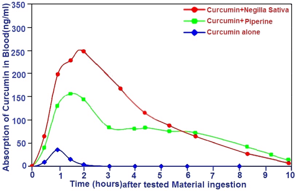
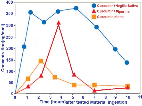
| The results for using Nigella sativa L.oils as a novel delivery system to pmprove the bioavailability of Curcumin compared with Black pepper. Figure 1 and 2 in the present study demonstrate that the Nigella Sativa L.oils and black pepper oils enhanced the oral bioavailability of Curcumin in both rats and humans at doses that were devoid of adverse side effects However but the Nigella sativa L. oils enhanced the oral bioavailability of Curcumin more than and black pepper oils, Nigella Sativa L.also enhanced the oral bioavailability of Curcumin in both rats and humans certain differences between rats and humans with respect to Curcumin were evident. |
| Figure 1 in the present diagram in the present study revealed that the plasma distribution of Curcumin administrate along with Nigella Sativa L.oils or black pepper oil via systemic routes and via tail vein intrapreitoneally (i.p) in albino rat.The present data demonstrate that Nigella sativa L.oils and black pepper oils enhanced the oral bioavailability of Curcumin (A) in both rats and humans at doses that were devoid of adverse side effects. However, Nigella sativa L. oils (B) enhanced the oral bioavailability of Curcumin more than and black pepper oils (C) at the indecatted times (2.30, 60 and 120 mintues). |
| Figure 2 in the current study demonstrate that the plasma distribution of Curcumin administrate along with using a liquid formulation of the Nigella sativa L.oils and black pepper in volunteer enhanced the oral bioavailability of Curcumin in humans at doses that were devoid of adverse side effects. However, Nigella sativa L. oils enhanced the oral bioavailability of Curcumin more than and black pepper oils in volunteer humans, the study shows that Nigella Sativa L.oils enhances the serum concentration and bioavailability of Curcumin in both rats and humans. this effect is probably may be due to increased absorption of Curcumin in the presence of Nigella sativa L.oils after using black pepper oil.As a result, Nigella sativa L.oils is five-to sixe times more bioavailable than ordinary 95% extract. Just 500 mg dose extract is equivalent to taking 262.1 ng, but “95%” Curcumin extract of this new bioavailability (Nigella sativa L.oils) enhanced Curcumin 160.8 ng of plant-bound Curcumin extract with piperine. In human trial, Nigella sativa L.oils delivered 5.22 times more Curcumin to the bloodstream than the ordinary stand alone Curcumin product and 4.04 times more Curcumin to the bloodstream than the plant-bound Curcumin extract with piperine Figure 2. |
| Clinical observation |
| On the other hand, in this work, the biological effects of these oils showed that the individual clinical data in HCC patients before and after treatment is listed in Table 5 and 6 and indicated that there is a good improvement of the clinical picture of the patients such as ascites, edema, jaundice, etc. were better in groups who receiving NSO+CUR. Oil added to chemo-hormonal therapy. Also the hematological findings (Hb, WBCs, PC% and PLT count) in patients who received these oils were kept within the normal range.Further NSO in added to Curcumin oil (CUR.O) more effect than other groups under investigation as shown in Table 7. |
| Table 8 cleared that the serum levels of ALT, AST, GGT, total bilirubin and albumin in patients received chemotherapy with NSO in added to CUR.Owere ameliorated and significantly improved and the levels of non-enzymatic antioxidant parameter, such as Zn and the levels of enzymatic antioxidant significantly elevated in serum levels of GSH and significantly decreased MDA serum levels in groups of patients who received chemo and hormonal therapy in addition to NSO and CUR.O compared with chemo and hormonal therapy in addition to vitamin E P<0.05. Furthermore, there was negative significant correlation between the levels of lipid per-oxidation product MDA and serum Zinc and the correlation coefficient (r) equals -0.720. Moreover Zinc has positive significant correlation to MDA. Correlation coefficient (r) reaches -0.775. |
| There is a positive correlation between telomerase activity and serum Zinc, MAD and VEGF. Table 9 revealed that the VEGF levels mean were significantly correlated with the expression of telomerase activity in the PBMCs (P=0.002). Moreover the depletion in the serum levels of the VEGF and the telomerase activity in PBMCs correlates with increasing the survival time Tables 9 and Figure 3 in patient who received 5-Fu+TMX + NSO+CUR.O (G3) compared with other group under study especially group of patients who received 5-Fu+ oxaliplatin +TMX +vitamine E(G1). The present data in Table 10 proved that the oestrogen receptor (ER) has been appositive effect in all the serial biopsies were taken form HCC patients to assess changes in ER expression during treatment. So, tamoxifen (anti-estrogen drug) is recommended to use it. The tamoxifen might be effective in patients with better prognosis and confirm that there is no survival advantage for treating HCC patients with tamoxifen in addition to supportive therapy without use of GLA. Negilla sativa (GLA) and Primrose oil (GLA) GLA was well tolerated with no major side effects. 5-FU+oxaloplatin +TAM +GLA or beta- Sesquiterpenoid hydrocarbone and zingiberene (objective response) cases achieved a significantly faster clinical response vs. group received 5-FU+TAM+VitE which was served as control, evident at the week- 12 biopsy on treatment (P=0.010) and at the week- 24 biopsy on treatment (P=0.010). There was significant reduction in ER expression in both two groups of treatment one treated with 5-FU+oxaloplatin +TAM +GLA in Negilla sativa oil (GLA)+beta- Sesquiterpenoid hydrocarbone in Curcumin oil and other gruops treated with 5-FU+oxaloplatin +TAM +GLA in Negilla sativa (GLA), than those groups like5-FU+TAM Fish oil (GLA), TAM+ Primrose oil (GLA) and FU+TAM+VitE, objective responders sustaining greater ER fall than tamoxifen counterparts (at the week- 12 biopsy P=0.002, 0.021. 0.030; 0.034 and at the week- 24 biopsy P= 0.001 P=0.026; P=0.019,0.034). |
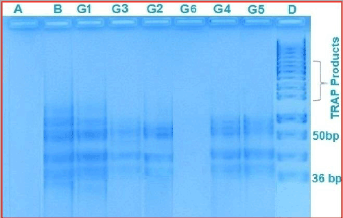
| Figure 3 in the present work revealed that the Representative results of TRAP-Silver Staining Assay on Agarose gel electrophoresis of PCR products in the PBMCs of HCC patients shows: Negative controls lanes positive controls at different concentrations,(A) that is means the telomerase were negative in the groups under investigation like cases of healthy control cases group (G6) lane, Nigella sativa L.oil + curcumin oil group(G3) lane, Nigella sativa L. oils group (G2) lane, , Fish oil group (G4) lane. Positive controls lane (B) were seen in the cases pre treatment (B) lane and in the cases who received 5-Fu+ oxaliplatin +TMX +Primrose oil group G5 lane, and other HCC patient group who received standard treatment of chemotherapeutic agents as 5-Fu+ oxaliplatin +TMX combined with vitamin E G1 lane. Lane (D) standard of telomerase activity. |
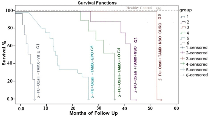
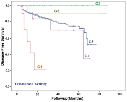
| In Figure 4 the Kaplan–Meier survival curve showed significant difference in survival time among five groups (p<0.001). In addition, the survival time in control group was dramatically different from that in five treatment groups (p<0.01). These findings suggest different treatments can prolong the survival time, which was the most evident in the Group3 in which the patients underwent 5-Fu+ oxaliplatin + Nigella Sativa L.oils +Curcumin oil (G3) therapy, had the best median survival rate of 54 months, followed by patients receiving G2=5-Fu+ oxaliplatin +TMX +Nigella sativa L.oils (44 months), G4=5-Fu+ oxaliplatin +TMX + Fish oil (37 months). G5= 5-Fu+ oxaliplatin +TMX +Primrose oil (26 months) and 5-Fu+ oxaliplatin +TMX +vitamin E G1 (7 months). |
| Figure 5 revealed that the disease-free survival rate in HCC patients with high telomerase expression (green line) was significantly lower than that in patients with low telomerase expression (blue line) gave significant results in improving survival rate (P<0.001) as presented in Figure 4. |
| In the present study considered a new designs for trials to demonstratethe herbal agents activity in human subjects and try to discuss the possible targets of bioactive herbal agents at the hepatocarcinogenesis and highlight their role at each individual stage on the most common herbal prescriptions and extracts thought to be useful in less toxicity and prevention or the combination of differentiation-inducing agents with chemotherapy might be a potential way of sensitization for chemotherapy to treat HCC patients. Moreover, the patients who regularly exercise and received natural oils after chemotherapy treatment have a much lower risk of cancer recurrence, no disturbances of the gastrointestinal tract, improved the performance status, the clinico-pathological data in HCC patient groups showed an improvement in their clinical picture , disappeared all the side effects symptoms can destroy cancer by blocking blood vessel growth, compared to people who are physically inactive and received vitamin E. it seems interesting that they showed a good improvement in performance status and the antitumor effect more than the other groups under investigations in the HCC patient’s had been received NSO than others oils. In this study, were almost, kept within the normal range in patients treated with natural oils. |
| Discussion |
| Malignant transformation of hepatocytes may occur through repetitive liver injury in a context of inflammation, regeneration and cirrhosis and oxidative DNA damage Levrero [61]. Thus it is difficulty to give sufficient dose of chemotherapy for HCC patient because of the poor liver function [62]. So, chemotherapy adds little to overall survival of HCC patients because of the low dose of drug and the low sensitivity of HCC cells [63]. A spectrum of natural sesquiterpenoids from herb oil has displayed anti-oxidant, anti-inflammatory and anti-carcinogenic properties [64]. |
| Phytochemical compounds are emerging as a new generation of anticancer agents with limited toxicity in cancer patients [22]. Curcuma oil, consisting of multiple sesquiterpenoids, is steam-distilled from the rhizome of Curcuma longa [65]. Although Curcumin has been found as an important bioactive compound in Curcuma longa, poor systemic absorption of Curcumin was also reported [66]. Another study showed that curcuma sesquiterpenoids from the volatile oil fraction have been found with similar bioactivities to Curcumin [67]. Some sesquiterpenoids with anticancer activity have been identified such as turmerone and β-elemene [60]. Recently, Li et al. [64] proved that the treatment with curcuma oil can decrease the incidence of HCC. Curcuma oil inhibits cell growth and induces cell death in Hepa1-6 cells. Curcuma protected mice with hepatic injury from inflammatory and oxidative stress. Curcuma oil can inhibit hepatoma cell growth in vivo and in vitro. Evening Primrose oil (EPO), comes from the seeds of the Evening Primrose plant. Like black seed oil and borage oil, EPO contains gamma linolenic acid (GLA), a fatty acid that the body converts to a hormone-like substance called prostaglandin E1 (PGE1) [68]. |
| Lientjies [69] showed that the Prostaglandin (E1) helps to stop platelets sticking together to form clots, lower blood pressure, inhibits help lower cholesterol levels, inhibits development of osteoarthritis reduce pain and inflammatory reactions and promotes excretion of excess sodium via the kidneys. Also they reported that the body manufactures E3 from eicosapentaenoic acid(EPA), the last step but one of the omega-3 chain, both E3 and E1 prevent production and activity of arachidonic acid from cell membranes but E3 more strongly than E1, therefore, prostaglandinE1and E3 could be beneficial in less severe inflammation, reducing joint tenderness and swelling, safe and without side-effects [70] .The presence of highest concentration of γ-linolenic acid (GLA) with other essential fatty acids & derived thymoquinone in this study may improve the clinical picture of HCC patients and could inhibit eicosanoid generation in leukocytes and membrane lipid peroxidation [71]. |
| A novel molecular explanation concerning the anticancer actions of GLA may relate to its ability to regulate oncoproteins [72], the ω-6 polyunsaturated fatty acid, GLA, is found in several plant oils, and used as a herbal medicine having antitumor activity which significantly diminished proteolytic cleavage of the extracellular domain of the Her-2/neu–coded p185Her-2/neu tyrosine kinase onco-protein and consequently, its activation. Also, GLA reduced Her-2/neu protein levels in the Her-2/neu over expressing in ovarian cancer, breast cancer and gastrointestinal tumor derived in vitro [73]. Gamma linolenic acid may have activity against endocrine-sensitivity with low systemic toxicity in breast cancer patients treated with primary therapy [74]. |
| Lui et al. [75] reported that the ω-3PUFA together with chemotherapy drug can enhance chemotherapy sensitivity as well as it may has anti-tumor effect, this could be attributed to an induced effect of the polyunsaturated fatty acids and in particular γ-linolenic acid and eicosapentaenoic acid in this oil. The present data proved that the consumption of n-3 fatty acids in fish oil reduces the risk of HCC showed good antioxidant activities but less than the group of patients who received NSOand Curcumin oils and other groups of patients who received NSO only in addition anticancer drugs only despite the fact that the major constituents of these oils are eicosapentaenoic acid (EPA), docosapentaenoic acid (DPA), and docosahexaenoic acid (DHA) could be protect against the development of some types of cancers but little is known about its association with hepatocellular carcinoma (HCC) [76]. |
| The present sudy shwed that the angiogenic factors such as VEGF in HCC patients had been regulated and reduced when they received Nigella sativa L.oils+Curcumin oils as natural product inaddition to anticancer drugs as 5-florouracil and oxaloplatin as well as tamoxifen than other groups who received anticancer drugs such as 5-florouracil and oxaloplatin as well as tamoxifen and /or Nigella sativa L.oils or Evening Primrose oil or fish volatile oil as natural product. these data are agree with data documented in Kenny et al. [77] which they proved that the serum levels of the angiogenic factors such as transforming growth factor β and VEGF had been regulated and reduced, this due to the Fixed oil of Nigella sativa and derived thymoquinone have the ability to inhibit eicosanoid generation in leukocytes and membrane lipid peroxidation [78]. |
| The present data shows a high telomerase expression and increasing VEGF in serum were shown to be independent factors associated with poor prognosis these data are agree with data documented by Meyerson [37] they proved that the increasing in the telomerase activity strongly correlates with increased malignant potential and stage. In addition to that, genomic instability associated with loss of telomere sequences correlates with a late stage development of colonic carcinomas and over 85% of malignant tumors express telomerase activity whereas most somatic tissues do not in humans [38]. |
| The present study proved that telomerase was expressed as a high percent in the malignant HCC which was similar to the results in a search previously performed by Zarghami et al. [78] they proved that in the malignant bladder with a reverse relationship between serum zinc deficiency levels and increasing telomerase activity in this cancer. This also agrees with the present data which proved a reverse relationship between serum zinc deficiency levels and the increasing telomerase activity. Furthermore, the present data proved that the high serum levels in VEGF were related with a high expression of telomerase activity in the PBMCs. In the current study the presence of telomerase activity in the PBMCs of HCC patients had been confirmed but after treatment with anticancer drug in added to natural oil specically Nigella Sativa oil as well as Curcumin oil the telomerase expressionnearly had been absent, and survival could be better than survival in its absence. These data had been similar to that was documented by Hong et al. [79]. This could be related with a limited number of patients categorized in different clinicopathological parameters. Thus, it may be useful to demonstrate these results in a larger number of patients’ pool. These results may indicate that telomerase could be a promosing marker for hepatocellar carcinomas in the future. |
| Skender et al. [80] stated that the use of n-3 polyunsaturated fatty acids could be a relatively non-toxic form of supportive therapy for improving colon cancer treatment and slowing down or preventing its recurrence. However, it is necessary to use them with caution, based on solid scientific evidence of their mechanisms of action from the molecular to the cellular and organism levels. The significant improvement of liver enzymes level and other liver function parameters in HCC patients treated with the selected natural oils can also be explained by their role in scavenging the free radicals and pro-oxidants and reduce their damaging effect. These data are in agree with El- Dakhakhny et al. [81] who proved that the NSO caused a significant reduction of AST, ALT and also agree with Leung [82] who reached also to the same conclusion. Moreover, Madan et al. [83] proved that the combination regimen containing chemotherapy in addition to vitamin E appears to be effective in normalizing amino transferases among nonalcoholic fatty liver disease suffering patients. The nutritional non-enzymatic antioxidants were increased in their serum levels in HCC patients as regards Nigella sativa oil and fish oil supplementation when compared with control subjects this agree with data performed by Khanzode et al. [84] they reported that the Zinc which is not a proper antioxidant but is an important cofactor incorporated into an intracellular antioxidant enzyme (superoxide dismutase) can ameliorate the dysfunction state occurring in HCC suffering individuals. |
| Tomlinson and DeFrain [85], they reported that, also, it plays in maintaining structural integrity and health of the hoof and udder. Moreover, Also, Wright et al. [86] they proved that trace elements as zinc showed restoration towards normal level after natural oils supplementation due to improved disease state since the cause affecting their changes were diminished. Supplementation of Zn, during times of oxidative stress may reduce oxidative damage to white blood cells and increase disease resistance. |
| The present data revealed that the GSH is an important defense mechanism against cellular damage by free radical formation and their propagation. Also the significant decrease in MDA after oils administration in HCC patients help acceptance of ameliorating effect of these selected natural medical herbs to decrease the risk of this disease. These results go with study performed by Abdalla [87] they stated that high levels of glutathione (GSH) are needed for cellular functions, signal transduction and protection against certain carcinogens. However, low GSH levels render cells more vulnerable to ROS attacks. Increased ROS activates intracellular oncogenic pathways or mutate a tumor suppressor gene pathway, which will activate a tumorigenesis process. These antioxidants help restoration towards normal level of liver function after treatment with natural oils supplementation, and this can led to improve the diseased state [88]. |
| In the current study the KRAS, BRAF and NRAS and ER in all HCC patients were done and its results proved that the KRAS, BRAF and NRAS was mutated so the targeted therapy is not recommended for that HCC Cases. The previous study revealed that tamoxifen might be effective in HCC patients confirm that there is no survival advantage for treating HCC patients with tamoxifen in addition to supportive therapy, since estrogen receptors (ERs) are present in approximately one-third of HCCs, these tumors could potentially benefit from ER blockade with tamoxifen [89]. Nevertheless, several prospective randomized trials and a systematic review of tamoxifen in patients with advanced HCC have failed to show a survival benefit or improved functional status, the lack of efficacy may be due to presence of variant ERs in ΗCC [90]. Chemotherapy has not demonstrated any benefit for the addition of tamoxifen [91]. Another study reported by Zeeneldin and Saadeldin [91] confirmed that, there is no survival advantage for treating HCC patients with tamoxifen in addition to supportive therapy. |
| From fortunately, the current data presented in the Table 9 proved that the serial biopsies were taken form HCC patients to assess changes in oestrogen receptor (ER) expression during treatment and the ER is highly positive in all of in HCC tissue Cases in groups of HCCs patients treated with tamoxifen (anti-estrogen drug) induced ER downregulation in groups received natural oils which contained GLA and eicosapentaenoic acid than other received vitamin E this result had been documented in the study preformed by Gaudio et al. [92] which they proved that the treatment of HCC patient have positive ER liver tissue with anti-estrogens like drug (tamoxifen) in addition of the GLA it reduced levels of ERs and to inhibit hepatocyte proliferation and regulating, differentiation, angiogenesis and survival. |
| The apparent enhancement of tamoxifen-induced ER downregulation by the GLA and eicosapentaenoic acid as a useful adjunct to 5-FU and tamoxifen in endocrine-sensitive HCC cancer. Furthermore the GLA as a useful adjunct to 5-FU and tamoxifen had been which achieved a significantly reduced levels of ERs, inhibited angiogenic factors VEGF and telomerase Activity , faster and maximize clinical benefit in response; minimize the toxicity, improving patient survival, moreover the GLA and eicosapentaenoic acid were well tolerated with no major side effects in both treatment arms with 5-FU+TAM+ Nigella Sativa(GLA), than the other groups treatment with 5-Fu+TAM+Fish oil (GLA), 5-Fu+TAM+ Primrose oil (GLA) and 5-FU+TAM+ Vitamin E, respectively. |
| In conclusions the present study concluded that the chemo and hormonal therapy either with natural oils capsules especially nigella sativa and Curcumin oils or other with vitamin E was identified as the most suitable treatment for unresectable HCC patients resulted in improving survival, maximizing clinical benefit, and minimizing risk of chemo and hormonal therapy toxicity, kept greatly all serum parameters levels within the normal range.The current also study showed that the telomerase activity and VEGF levels can be used as predict survival and response to treatment markers in HCC patients in the future. |
| Depletion in the serum levels of the VEGF as an angiogenic factor and in the telomerase activity were correlated with good prognosis and decrease malignant potential, enhance the quality of life and the survival seemed to be better in the absence of telomerase expression in HCC Egyptian patients. Further research must be performing with more patients to explore how the chemo and hormonal therapy either with natural oils capsules especially nigella sativa and Curcumin oils or other with vitamin E prolonging the survival of patients. Also it must be required to perform further investigation to prove the enhancement of tamoxifen-induced ER down-regulation by GLA. |
| Acknowledgements |
| Most of gratitude goes to our patients whom we are learning from. Authors would also like to thank all physicians and nursing staff at the Medical Oncology and Cancer Biology Departments, NCI, Cairo University who participated in care giving to our patients and to their families. |
References
|
Tables and Figures at a glance
| Table 1 | Table 2 | Table 3 | Table 4 | Table 5 |
| Table 6 | Table 7 | Table 8 | Table 9 | Table 10 |
Figures at a glance
 |
 |
 |
 |
 |
| Figure 1 | Figure 2 | Figure 3 | Figure 4 | Figure 5 |
Relevant Topics
- Analytical Biochemistry
- Applied Biochemistry
- Carbohydrate Biochemistry
- Cellular Biochemistry
- Clinical_Biochemistry
- Comparative Biochemistry
- Environmental Biochemistry
- Forensic Biochemistry
- Lipid Biochemistry
- Medical_Biochemistry
- Metabolomics
- Nutritional Biochemistry
- Pesticide Biochemistry
- Process Biochemistry
- Protein_Biochemistry
- Single-Cell Biochemistry
- Soil_Biochemistry
Recommended Journals
- Biosensor Journals
- Cellular Biology Journal
- Journal of Biochemistry and Microbial Toxicology
- Journal of Biochemistry and Cell Biology
- Journal of Biological and Medical Sciences
- Journal of Cell Biology & Immunology
- Journal of Cellular and Molecular Pharmacology
- Journal of Chemical Biology & Therapeutics
- Journal of Phytochemicistry And Biochemistry
Article Tools
Article Usage
- Total views: 15713
- [From(publication date):
March-2015 - Apr 01, 2025] - Breakdown by view type
- HTML page views : 11048
- PDF downloads : 4665
