Neuroimaging Techniques in Humans and Neurologic Diseases
Received: 02-Jul-2022 / Manuscript No. science-22-75593 / Editor assigned: 04-Jul-2022 / PreQC No. science-22-75593 (PQ) / Reviewed: 18-Jul-2022 / QC No. science-22-75593 / Revised: 23-Jul-2022 / Manuscript No. science-22-75593 (R) / Published Date: 30-Jul-2022 DOI: 10.4172/science.1000131
Image Article
Neuroimaging biomarkers for neurologic infections are significant devices, both for understanding pathology related with mental and clinical side effects and for differential diagnosis. This section investigates neuroimaging measures, including underlying and practical measures from magnetic resonance imaging (MRI) and subatomic measures principally from positron emission tomography (PET), in sound maturing grown-ups and in various neurologic illnesses. The range covers neuroimaging measures from typical maturing to different dementias: late-beginning Alzheimer’s disease [AD; mild cognitive impairment (MCI)], familial and non-familial beginning stage Promotion, abnormal Advertisement conditions, posterior cortical atrophy (PCA), Parkinson’s sickness (PD) with and without dementia, and multiple systems atrophy (MSA). We likewise incorporate a conversation of the appropriate use criteria (AUC) for amyloid imaging and finish up with a conversation of differential finding of neurologic dementia issues with regards to neuroimaging.
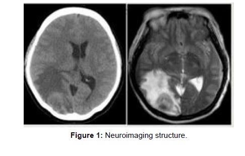
Neuroimaging is the utilization of quantitative (computational) methods to concentrate on the design and capability of the central nervous system, created as an objective approach to logically concentrating on the healthy human brain in a non-invasive manner [1,2]. Progressively it is likewise being utilized for quantitative investigations of brain disease and psychiatric illness. Neuroimaging is a profoundly multidisciplinary research field and is not a medical specialty (Figure 1).
Neuroimaging varies from neuroradiology which is a medical specialty and uses cerebrum imaging in a clinical setting. Neuroradiology is drilled by radiologists who are medical practitioners. Neuroradiology fundamentally focuses on recognizing cerebrum injuries, like vascular disease, strokes, tumors and inflammatory disease. As opposed to neuroimaging, neuroradiology is subjective (based on subjective impressions and extensive clinical training) yet in some cases utilizes essential quantitative techniques. Functional brain imaging techniques, for example, functional magnetic resonance imaging (fMRI), are normal in neuroimaging but rarely used in neuroradiology. Neuroimaging falls into two general classes:
Structural imaging which is utilized to quantify brain structure using example: Voxel based morphometry.
Functional imaging which is utilized to study brain function, frequently utilizing fMRI and different procedures like PET and MEG.
References
- Abrahams S, Goldstein LH, Simmons A, Brammer M, Williams SCR, et al. (2004) Word retrieval in amyotrophic lateral sclerosis: a functional magnetic resonance imaging study. Brain 127: 1507-1517.
- Agosta F, Henry RG, Migliaccio R, Neuhaus J, Miller BL, et al. (2010) Language networks in semantic dementia. Brain 133: 286-299.
Indexed at, Google Scholar, Crossref
Citation: Todt D (2022) Neuroimaging Techniques in Humans and Neurologic Diseases. Arch Sci 6: 131. DOI: 10.4172/science.1000131
Copyright: © 2022 Todt D. This is an open-access article distributed under the terms of the Creative Commons Attribution License, which permits unrestricted use, distribution, and reproduction in any medium, provided the original author and source are credited.
Share This Article
Open Access Journals
Article Tools
Article Usage
- Total views: 734
- [From(publication date): 0-2022 - Jan 12, 2025]
- Breakdown by view type
- HTML page views: 565
- PDF downloads: 169

