Case Report Open Access
Neuroblastoma in the Adult: Which Therapy is Effective?
| Feryal Karaca1, Çi�?�?dem Usul Af�?�?ar2*, Meral Günald�?±2, and Berksoy �?�?ahin2 | |
| 1Department of Radiation Oncology, Cukurova University Medical Faculty, Adana, Turkey | |
| 2Department of Medical Oncology, Cukurova University Medical Faculty, Adana, Turkey | |
| Corresponding Author : | Çi�?�?dem Usul Af�?�?ar, MD Department of Medical Oncology Çukurova University Medical Faculty Balcali Hospital, Adana, Turkey Tel: +903223386060 (3142) E-mail:cigdemusul@yahoo.com |
| Received June 19, 2014; Accepted August 04, 2014; Published August 08, 2014 | |
| Citation: Karaca F, Af�?�?ar CU, Günald�?± M, �?�?ahin B (2014) Neuroblastoma in the Adult: Which Therapy is Effective? Clin Pharmacol Biopharm 3:119. doi:10.4172/2167-065X.1000119 | |
| Copyright: © 2014 Karaca F, et al. This is an open-access article distributed under the terms of the Creative Commons Attribution License, which permits unrestricted use, distribution, and reproduction in any medium, provided the original author and source are credited. | |
Visit for more related articles at Clinical Pharmacology & Biopharmaceutics
Abstract
The examination of a twenty-eight-year-old male patient with lower back pain revealed a mass that extended through the spinal canal at the L1-L2 level and exhibited left paravertebral extension which was indistinguishable from the psoas muscle. Neuroblastoma was diagnosed after performing a biopsy. The disease progressed in that same year and in the following years, even after performing surgery, radiotherapy, chemotherapy, and bone marrow transplantation. The course of the disease was resistant to different high-dose chemotherapies and radiotherapy. The patient died four years after diagnosis. Neuroblastoma, a tumor which more frequently appears in children, is seen as an advanced or metastatic disease in adults. Currently there is no standard treatment for neuroblastoma in adults. The treatment protocols applied in children are insufficient for adults. Generally neuroblastoma is characterized as a localized disease, but in adults it is more aggressive.
| Keywords |
| Neuroblastoma; Extracranial malignant tumor |
| Introduction |
| Neuroblastoma is a common, extracranial malignant tumor in children. It is observed, 90% of the time, among children younger than five years old [1,2]. It is rarely seen after the age of twenty. Neuroblastoma originates from neural crest cells. In adults, it presents in the abdominal and retroperitoneal area, although it most frequently occurs in the adrenal gland among children. It can appear anywhere in the sympathetic nervous system [3,4]. |
| Case Report |
| In 2006, a twenty-eight-year-old male patient had lower back pain which had persisted for two months. He also experienced difficulty walking and loss of feeling in his left leg. A large soft tissue mass, which extended through the spinal canal at the L1-L2 level and exhibited left paravertebral extension while being indistinguishable from the psoas muscle, was found. A metastatic medullary infiltration in the lumbar vertebrae and iliac crest were detected in the patient’s upper abdomen and pelvic tomography. |
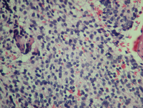
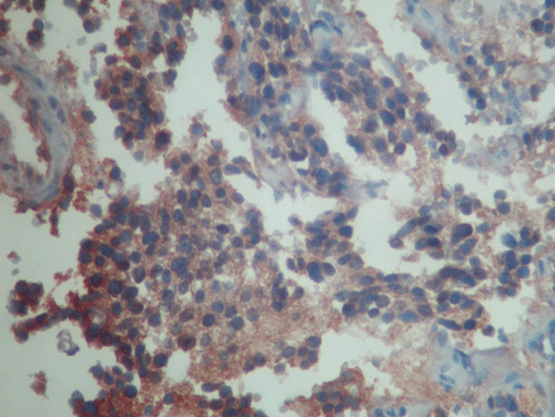
| Routine laboratory tests of the patient (complete blood count, liver and kidney function tests) were normal. No increase in homovanillic acid or vanilmandelic acid level was detected in the urine. A biopsy of the mass putting pressure on the lumbar spine was performed and the patient was diagnosed with neuroblastoma (Figures 1 and 2). Our patient was identified as a high risk patient according to INSS (International Neuroblastoma Staging System) and INRG (International Neuroblastoma Risk Group). The patient underwent an epidural mass excision, followed by a T12-L1-L2 laminectomy. Six courses of adjuvant cisplatin, doxorubicin, etoposide, and cyclophosphamide chemotherapy were administered. External radiotherapy of 3000 cGy was administered to the operation zone after chemotherapy. In the same year, the patient underwent autologous stem cell rescue after high dose chemotherapy and radiotherapy. |
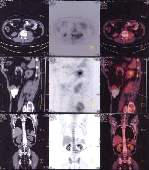
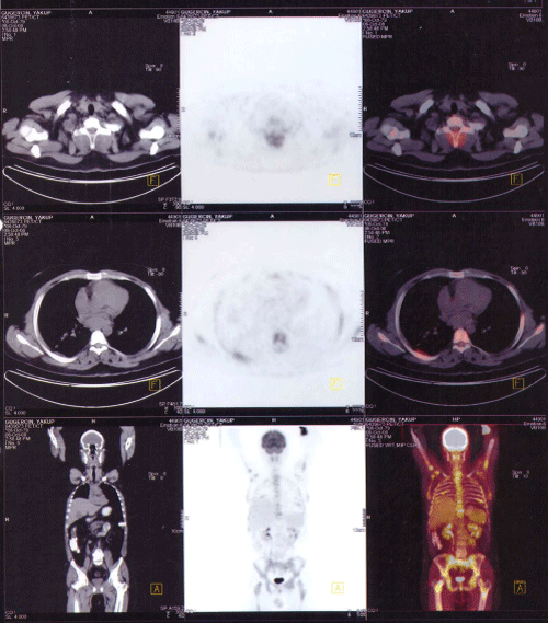
| Three months later, during the patient’s routine checks and an assessment made due to back pain, a 12 × 6 × 6 cm mass that extended through the epidural space at the cervical-6 and the thoracic-1 vertebra level was detected. Laminectomy and mass excision were performed. After the second operation, the patient was staged as having metastatic neuroblastoma and six courses of chemotherapy with cisplatin, doxorubicin, etoposide, and cyclophosphamide were administered. An additional dose of 3000 cGy palliative radiotherapy was given. However, the patient was later admitted to hospital with neutropenic fever, thus piperacilline/tazobactam was provided. The physical examination revealed a mass in the psoas muscle. Lytic lesions of various sizes, the largest in the left iliac bone, were found in all long bones and vertebrae. Increased activity in the bone marrow was observed on PET-CT (Figure 3). F-18 FDG uptake was observed in the 5.5 × 2.5 cm mass that extended through the L3 vertebrae to the left iliopsoas muscle (Figure 3). Six cures of bevacizumab, irinotecan, prednisolone, and docetaxel chemotherapy were administered to the patient with primary prophylaxis of febrile neutropenia (filgrastim). The patient had increased bone pain in 2008 (Figure 4). A 4 × 4 cm mass that extended through the C7 vertebrae and lytic metastases throughout the skeletal system were found by using PET-CT imaging. Neuroblastoma metastasis was identified in the bone marrow biopsy. There were no tumoral cells in the CSF cytology. Pachymeningeal contrast enhancement in an MRI of the brain and a contrast-enhancing metastatic mass in the external occipital protuberans were detected. Three cures of cisplatin, doxorubicin, etoposide and cyclophosphamide therapy were administered. When the patient did not respond to the previous treatment, topotecan, cyclophosphamide, and filgrastim were given. Palliative care was provided for the patient due to a low ECOG performance score and resistance to chemotherapy. The patient died four years after diagnosis, despite all treatments. |
| Discussion |
| Neuroblastoma is common in the first years of life, although it is seen less frequently in adolescents and young adults. In children, it can develop within the adrenal medulla or sympathetic ganglion. In adults, it is most frequently seen in the abdomen, pelvis, thoracic spinal cord, mediastinum, and olfactory region [5,6]. Patients who have a primary tumor in the neck and upper mediastinum can develop Horner’s syndrome. However, tumors along the spinal cord can result in compression, paraplegia, and pain. Metastasis sites include bone, bone marrow, lung, brain, breast, lymph nodes, liver and pleura [5,7]. Urinary catecholamine levels of adolescent patients are quite low, even though they seek medical help at an advanced stage. Generally adolescent neuroblastoma is mortal [8]. The most important positive prognostic factors in cases of neuroblastoma are low-age group and the stage of the disease at the time of diagnosis. Neuroblastoma is a malignancy which tends to spread to vascular and lymphogenic areas. In adults, it is inclined to metastasize in parenchymal tissues such as the brain and lungs. The incidence of metastases in the cranial nerve system is very high. However, new metastases are very difficult to diagnose without symptoms [9]. |
| Currently there is no standard treatment for neuroblastoma in adults. A complete resection is sufficient in low-risk patients. In high-risk patients, however, combined treatment modalities such as operation, chemotherapy and radiotherapy should be administered [10]. Induction chemotherapy and surgery on the primary tumour are therapeutic options. If there is a partial response; radiotherapy on the remaining tumour is suggested. After induction, high dose chemotherapy and autologous stem cell transplantation are the best approaches. All treatment modalities were carried out with our highrisk patient. |
| In a new study done by the M.D. Anderson Cancer Center; adult versus pediatric neuroblastoma patients were compared according to classification and therapy. There were no differences in PFS (progression free survivial) or OS (overall survival) between stage-matched risk categories of pediatric and adult patients. As a result, they emphasized that adult and pediatric patients with neuroblastoma achieve similar survival outcomes. INRG classification should be employed to stratify adult neuroblastoma patients and help select appropriate treatment [11]. |
| Neuroblastoma, which is extremely rare and quite aggressive in adults, can be managed with different modalities of treatment at different stages. We have just presented a case study which outlines a treatment approach for adults. While true that it was ultimately ineffective, it is a treatment modality. Modalities used in children such as chemotherapy, surgery, bone marrow transplantation, and radiotherapy could be used in adults as well [5]. The most relevant chemotherapeutics are cyclophosphamide, ifosfamide, vincristine, adriamycin, cisplatin, carboplatin, and etoposide. Also, bone marrow transplantation is an option for high-risk patients [12]. |
| References |
|
References
- Park JR, Eggert A, Caron H (2008) Neuroblastoma: biology, prognosis, and treatment. Pediatr Clin North Am 55: 97-120.
- Brodeur GM, MarisJ M (2011) Neuroblastoma. In: Pizzo PA, Poplack DG, Principles and Practice of Pediatric Oncology. 6th edition, Lippincott Williams & Wilkins, Philadelphia, PA, 895-937.
- Menegaux F, Olshan AF, Reitnauer PJ, Blatt J, Cohn SL (2005) Positive association between congenital anomalies and risk of neuroblastoma. Pediatr Blood Cancer 45: 649-655.
- McLean TW, Iskandar SS, Shimada H, Hall MC (2004) Neuroblastoma in an adult. Urology 64: 1232.
- Franks LM, Bollen A, Seeger RC, Stram DO, Matthay KK (1997) Neuroblastoma in adults and adolescents: an indolent course with poor survival. Cancer 79: 2028-2035.
- Wang HL, Fan GK, Lin ZH (2009) [Clinical analysis of 10 cases of olfactory neuroblastoma]. Zhejiang Da Xue Xue Bao Yi Xue Ban 38: 103-106.
- Panovska-Stavridis I, Ivanovski M, Hadzi-Pecova L, Ljatifi A, Trajkov D, et al. (2010) A case report of aggressive adult neuroblastoma mimicking acute leukemia with fulminant course and fatal outcome. Prilozi 31: 349-359.
- Conte M, De Bernardi B, Milanaccio C, Michelazzi A, Rizzo A, et al. (2005) Malignant neuroblastic tumors in adolescents. Cancer Lett 228: 271-274.
- Conte M, Parodi S, De Bernardi B, Milanaccio C, Mazzocco K, et al. (2006) Neuroblastoma in adolescents: the Italian experience. Cancer 106: 1409-1417.
- Schalk E, Mohren M, Koenigsmann M, Buhtz P, Franke A, et al. (2005) Metastatic adrenal neuroblastoma in an adult. Onkologie 28: 353-355.
- Conter HJ, Gopalakrishnan V, Ravi V, Ater JL, Patel S, et al. (2014) Adult versus Pediatric Neuroblastoma: The M.D. Anderson Cancer Center Experience. Sarcoma 2014.
- Ishola TA, Chung DH (2007) Neuroblastoma. Surg Oncol 16: 149-156.
Figures at a glance
 |
 |
 |
 |
| Figure 1 | Figure 2 | Figure 3 | Figure 4 |
Relevant Topics
- Applied Biopharmaceutics
- Biomarker Discovery
- Biopharmaceuticals Manufacturing and Industry
- Biopharmaceuticals Process Validation
- Biopharmaceutics and Drug Disposition
- Clinical Drug Trials
- Clinical Pharmacists
- Clinical Pharmacology
- Clinical Research Studies
- Clinical Trials Databases
- DMPK (Drug Metabolism and Pharmacokinetics)
- Medical Trails/ Drug Medical Trails
- Methods in Clinical Pharmacology
- Pharmacoeconomics
- Pharmacogenomics
- Pharmacokinetic-Pharmacodynamic (PK-PD) Modeling
- Precision Medicine
- Preclinical safety evaluation of biopharmaceuticals
- Psychopharmacology
Recommended Journals
Article Tools
Article Usage
- Total views: 16229
- [From(publication date):
November-2014 - Apr 02, 2025] - Breakdown by view type
- HTML page views : 11672
- PDF downloads : 4557
