Nature of Organ and Tissue Function and Medical Imaging
Received: 04-Jan-2023 / Manuscript No. roa-23-87489 / Editor assigned: 06-Jan-2023 / PreQC No. roa-23-87489 (PQ) / Reviewed: 20-Jan-2023 / QC No. roa-23-87489 / Revised: 24-Jan-2023 / Manuscript No. roa-23-87489 (R) / Published Date: 31-Jan-2023 DOI: 10.4172/2167-7964.1000422
Image Article
The process and interaction of imaging the interior of a body for clinical examination and mediation, as well as the visual representation of the capacity of particular organs or tissues, is known as medical imaging (physiology). Medical imaging is used to diagnose and treat infections as well as to reveal hidden internal structures beneath the skin and bones. In addition, medical imaging creates a database of typical life structures and physiology so that abnormalities can be identified. Even though it is possible to perform imaging of removed organs and tissues for clinical purposes, these techniques are typically regarded as a feature of pathology rather than medical imaging [1].
X-ray radiography, magnetic resonance imaging, ultrasound, endoscopy, elastography, tactile imaging, thermography, medical photography, and nuclear medicine functional imaging strategies like positron discharge tomography and single-photon emission computed tomography are just a few of the imaging innovations utilized in this field, which is essential for biological imaging in its broadest sense [2].
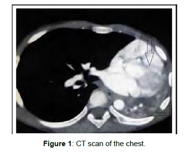
Electroencephalography (EEG), Magnetoencephalography (MEG), electrocardiography (ECG), and other methods of measuring and recording that are not primarily intended to produce images address various innovations that produce information that cannot be presented in the form of a parameter graph versus time or maps that contain information about the measurement locations. These innovations may be considered subsets of medical imaging in a different field with a limited correlation (Figure 1).
Five billion medical imaging studies had been conducted worldwide by 2010. In 2006, approximately half of all ionizing radiation exposure in the United States came from medical imaging.
References
- Roobottom CA, Mitchell G, Morgan-Hughes G (2010) "Radiation-reduction strategies in cardiac computed tomographic angiography". Clin Radiol 65: 859-867.
- Jr Mettler FA, Thomadsen BR, Bhargavan M, Gilley DB, Gray GE, et al. (2008) Medical radiation exposure in the U.S. in 2006: preliminary results. Health Phys 95: 502-507.
Indexed at, Crossref, Google Scholar
Citation: Akirha T (2023) Nature of Organ and Tissue Function and MedicalImaging. OMICS J Radiol 12: 422. DOI: 10.4172/2167-7964.1000422
Copyright: © 2023 Akirha T. This is an open-access article distributed under theterms of the Creative Commons Attribution License, which permits unrestricteduse, distribution, and reproduction in any medium, provided the original author andsource are credited.

