Case Report Open Access
Multiseptate Gallbladder in an Asymptomatic Pediatric Patient
Ortolá P*, Carazo ME, Cortés J and Rodríguez L
Department of Pediatric Surgery, Hospital Universitario y Politécnico La Fe, Valencia, Spain
- *Corresponding Author:
- Paula Ortolá Fortes
Hospital Universitario y Politécnico La Fe
Department of Pediatric Surgery
2F, Avenida de Fernando Abril Martorell
106, 46026, Valencia, Spain
Tel: +34 650034818
E-mail: paula.ortola.fortes@gmail.com
Received date: August 22, 2015 Accepted date: November 12, 2015 Published date: November 18, 2015
Citation: Ortola P, Carazo ME, Cortes J, Rodríguez L (2015) Multiseptate Gallbladder in an Asymptomatic Pediatric Patient. J Gastrointest Dig Syst 5:353. doi:10.4172/2161-069X.1000353
Copyright: © 2015 Ortola P, et al. This is an open-access article distributed under the terms of the Creative Commons Attribution License, which permits unrestricted use, distribution, and reproduction in any medium, provided the original author and source are credited.
Visit for more related articles at Journal of Gastrointestinal & Digestive System
Abstract
Multiseptate gallbladder is a rare biliary anomaly that physicians should be aware of in the differential diagnosis of recurrent right upper quadrant pain. Investigation with ultrasound is recommended and magnetic resonance cholangiopancreatography is used to rule out associated anomalies. Although symptomatic patients have significant relief after surgical treatment, regular follow-up should be considered in selected patients. We report the case of an asymptomatic child that had an incidental diagnosis of multiseptate gallbladder and is being followed-up.
Keywords
Multiseptate gallbladder; Ultrasonography; Pediatrics; Asymptomatic diseases
Introduction
Multiseptate gallbladder (MSG) is a really rare congenital anomaly of the biliary tree, first described in 1963 by Simon and Tandon [1]. Since this reference in the literature, less than 50 cases have been published, only 13 of them in paediatric patients [2]. In children, this disease is usually diagnosed before adolescence (mean age nine years) and the male to female ratio have been 1:2 until now [2].
The majority of children affected present with abdominal pain or discomfort, usually located in right upper quadrant or epigastrium. Nausea and vomiting are also frequent. However, some cases are asymptomatic and difficult to diagnose.
Abdominal ultrasound is the most useful and safety implement for confirming the diagnosis. Nevertheless, other radiologic examinations can be employed to better study this rare disease and its associated anomalies. Generally, blood tests are also performed initially in order to assist outcomes.
Several treatments are currently available. Surgery is the most often chosen therapy. However, conservative attitude with regular follow-up can be considered in some cases.
We report here a case of a paediatric patient with an asymptomatic septate gallbladder, diagnosed incidentally at ultrasound, who is being followed-up without complications.
Case History
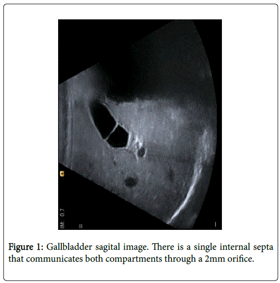
A five-month-old girl underwent ultrasound examination because of kidney anomalies, already detected during pregnancy controls. The study revealed a normal volume gallbladder with a single internal septa that communicated both compartments through a two millimetres orifice. Wall thickness of the gallbladder was also normal. No biliary sludge, stones, surrounding fluid or tenderness during the examination were found. Pancreas, liver and biliary tree had no abnormalities. Bilateral kidney duplicity was identified (Figure 1).
Another ultrasound test was performed three months later, as a follow-up, and the same anomalies were detected within the gallbladder.
Based on the two imaging tests performed until now, MSG has been diagnosed. Our patient has never presented symptoms and laboratory tests have been normal. Magnetic resonance cholangiopancreatography (MRCP) has not been done yet, although it is a possibility to definitively rule out other abnormalities. Currently, the patient is ten months old and she is being followed-up annually. No surgical treatment is considered at the moment.
Discussion
MSG is a developmental abnormality of the gallbladder characterized by multiple columnar epithelized thin septa that divide the gallbladder in several compartments [3], providing it a honeycomb-like appearance [2]. These compartments, usually of different sizes, are not completely independent, as there are small orifices in septa allowing communication among them, as in our case. Septa can be complete, involving the entire lumen of gallbladder, or only partial [3]. Three theories have been propounded to explain this anomaly [2,3,4]. Incomplete cavitation or vacuolization of the embryonic gallbladder bud [1]. Wrinkling theory: during its development, the solid gallbladder bud creates invaginations that give it an irregular wrinkled appearance. These invaginations can lately merge with the solid intraepithelial structure [5]. Phrygian cap theory: the solid gallbladder bud may raise faster than its surrounding bed and peritoneum, developing aberrant wrinkling of the gallbladder due to the lack of space [5].
The first theory does not explain the presence of muscular tissue within the septa, but the latter theories could explain it. Since the presence of this tissue is not a constant finding, there could be more than one mechanism involved.
Anomalies of the biliary ducts have been reported in patients with MSG (gallbladder ectopy or hypoplasia4, choledocal cysts, anomalous junction of biliary and pancreatic ducts [3]). It has also been associated with an increment in the possibility of developing gallbladder cancer and cholangiocarcinoma, as some of the anomalies associated are risk factors for malignancy [2,3].
As in our patient, some cases can be asymptomatic and MSG can be diagnosed as a casual finding during imaging studies performed for other reasons. However, the majority of patients usually present with recurrent right upper quadrant or epigastric pain (often irradiated to the back), nausea and vomiting or, in some cases, with symptoms of associated complications (biliary sludge, cholecystitis, cholelithiasis or even pancreatitis) [3,4]. It is still not clear why some patients are completely asymptomatic and other cases present with recurrent pain episodes. There are two explanations of the mechanism that might cause the symptoms [2,3,4,6]. Transient episodes of flow obstruction of the bile through the holes that exist within the septa and communicate the different compartments [5]. Uncoordinated contractions of the gallbladder which increase intraluminal pressure of some of the compartments [7].
Ultrasound is the most useful image source for diagnosis. However, MRCP is also helpful to confirm it and search for associated anomalies of the biliary tree2. Despite the image source, the most frequent radiological findings are: lobed shape normal size gallbladder and multiple compartments separated by several complete or partial thin septa4, which have fine echogenic bands without acoustic shadowing6.
Differential diagnosis is with acute cholecystitis, as necrosis of the wall might cause pseudomembranes that can mimic MSG in ultrasound. Other diagnoses include polypoid cholesterolosis, desquamated gallbladder mucosa, hydatid cyst, adenomyomatosis and acute hepatitis [2].
In symptomatic patients, surgical treatment (laparoscopic cholecystectomy preferably) has made disappear the symptoms in the reported cases4. On the other hand, as we are performing with our case, patients without severe symptoms or complications and normal laboratory values probably do not need this treatment, at least immediately, and can be managed as outpatients with regular followup [3].
Conclusion
Multiseptate gallbladder, although rare, is a biliary anomaly that physicians should be aware of in the differential diagnosis of recurrent right upper quadrant pain attacks in children [8]. Associated anomalies of the biliary tree must be ruled out. For that reason, investigation with ultrasound and MRCP is recommended [2]. However, definitive diagnosis is anatomopathological [4]. Although symptomatic patients have significant relief after surgical treatment4, regular follow-up should be considered in patients without severe symptoms or complications and normal laboratory values3, as we report with our case.
References
- simon M, Tandon BN (1963) Multiseptate gallbladder. A case report.Radiology 80: 84-86.
- Wanaguru D, Jiwane A, Day AS, Adams S (2011) Multiseptate gallbladder in an asymptomatic child. Case Rep Gastrointest Med 2011: 470658.
- Geremia P, Tomà P, Martinoli C, Camerini G, Derchi LE (2013) Multiseptate gallbladder: clinical and ultrasonographic follow-up for 12 years.J PediatrSurg 48: e25-28.
- Menocal N, Garrote A, García D, Alonso JM, Santos JA (2011) Multiseptate gallbladder. An unusual congenital anomaly. Rev. Chil. Radiol. Santiago 176-178.
- Bhagavan BS, Amin PB, Land AS, Weinberg T (1970) Multiseptate gallbladder. Embryogenetic hypotheses.Arch Pathol 89: 382-385.
- Rivera-Troche EY, Hartwig MG, Vaslef SN (2009) Multiseptate gallbladder.J GastrointestSurg 13: 1741-1743.
- Toombs BD, Foucar E, Rowlands BJ, Strax R (1982) Multiseptate gallbladder.South Med J 75: 610-612.
- Demirpolat G, Duygulu G, Tamsel S (2010) Multiseptate gallbladder in a child with recurrent abdominal pain.DiagnIntervRadiol 16: 306-307.
Relevant Topics
- Constipation
- Digestive Enzymes
- Endoscopy
- Epigastric Pain
- Gall Bladder
- Gastric Cancer
- Gastrointestinal Bleeding
- Gastrointestinal Hormones
- Gastrointestinal Infections
- Gastrointestinal Inflammation
- Gastrointestinal Pathology
- Gastrointestinal Pharmacology
- Gastrointestinal Radiology
- Gastrointestinal Surgery
- Gastrointestinal Tuberculosis
- GIST Sarcoma
- Intestinal Blockage
- Pancreas
- Salivary Glands
- Stomach Bloating
- Stomach Cramps
- Stomach Disorders
- Stomach Ulcer
Recommended Journals
Article Tools
Article Usage
- Total views: 11780
- [From(publication date):
December-2015 - Jul 04, 2025] - Breakdown by view type
- HTML page views : 10915
- PDF downloads : 865

