Multiple Nabothians Cysts in a 52 Years Old Woman with Multiple Myofibromas
Received: 23-Jan-2022 / Manuscript No. roa-22-55844 / Editor assigned: 03-Mar-2022 / PreQC No. roa-22-55844(PQ) / Reviewed: 17-Mar-2022 / QC No. roa-22- 55844 / Revised: 22-Mar-2022 / Manuscript No. roa-22-55844(R) / Published Date: 29-Mar-2022 DOI: 10.4172/2167-7964.1000368
Abstract
Nabothian cysts are mucinous benign cysts that are formed due to the accumulation of cervical mucus inside blocked cervical crypts. While the presence of small-sized Nabothian cysts is usually clinically asymptomatic and requires no treatment or intervention, the diagnosis of larger or multiple Nabothian cysts can be mistaken with malignant tumors, including mucin producing carcinomas such as Adenoma malignum. We report the case of a large multiple Nabothian cyst that was correctly diagnosed preoperatively using ultrasonography and magnetic resonance imaging (MRI)in a woman with 2 large myofibromas.
Keywords
Naboth; Multiple cysts; Magnetic Resonance Imaging.
Image Article
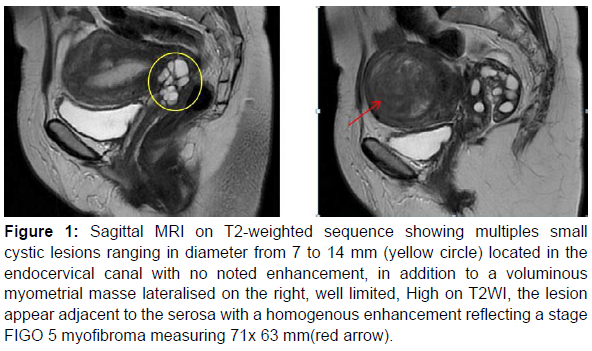
Nabothian cysts are small, asymptomatic, non-neoplastic cervical cysts that are frequently seen in women of reproductive age. No medical intervention is needed since the resolve themselves spontaneously [1]. In our rare case in which a woman of 52 years old with a recent diagnosis of 2 myofibroma via pelvic ultrasound has underwent an MRI to further characterize those lesions. The MRI showed in addition to 2 voluminous fibromas multiple cysts that caused compression of surrounding viscera and further aggravate her symptoms related to mass effect such as constipation and urinary incontinence (Figure 1).
Figure 1: Sagittal MRI on T2-weighted sequence showing multiples small cystic lesions ranging in diameter from 7 to 14 mm (yellow circle) located in the endocervical canal with no noted enhancement, in addition to a voluminous myometrial masse lateralised on the right, well limited, High on T2WI, the lesion appear adjacent to the serosa with a homogenous enhancement reflecting a stage FIGO 5 myofibroma measuring 71x 63 mm(red arrow).
Large and/or multiple nabothian cysts, especially deep nabothian cysts, are difficult to distinguish from minimal-deviation adenocarcinoma (MDA) [2]. Furthermore, neither cytology nor histology helps substantially discern the two entities. Very few case reports featuring those too entities has been trying to characterize them using MRI. Overall nabothians cysts are usually low on T1WI and high on T2WI, the cyst walls are smooth and not enhanced with intravenous Gadolinium In contrast to MDA. The absence of a watery discharge and an MR image displaying a round or oval cyst without enhancement after intravenous gadolinium are helpful in the diagnosis of nabothian cysts.
References
- Shroff N, Bhargava P (2021) Giant nabothian cysts: A rare incidental diagnosis on MRI. Radiol Case Rep 16: 1473-1476.
- Oguri H, Nagamasa M, Chiaki I, Tomoaki K, Yorito Y, et al. (2004) MRI of endocervical glandular disorders: three cases of a deep nabothian cyst and three cases of a minimal-deviation adenocarcinoma. Magn Reson Imaging 22: 1333-1337.
Indexed at, Google Scholar, Crossref
Citation: Najwa A, Zaynab I, Ismail HM, Nabil MB (2022) Multiple Nabothians Cysts in a 52 Years Old Woman with Multiple Myofibromas. OMICS J Radiol 11: 367. DOI: 10.4172/2167-7964.1000368
Copyright: © 2022 Najwa A, et al. This is an open-access article distributed under the terms of the Creative Commons Attribution License, which permits unrestricted use, distribution, and reproduction in any medium, provided the original author and source are credited.
Share This Article
Open Access Journals
Article Tools
Article Usage
- Total views: 3772
- [From(publication date): 0-2022 - Feb 01, 2025]
- Breakdown by view type
- HTML page views: 3359
- PDF downloads: 413

