Make the best use of Scientific Research and information from our 700+ peer reviewed, Open Access Journals that operates with the help of 50,000+ Editorial Board Members and esteemed reviewers and 1000+ Scientific associations in Medical, Clinical, Pharmaceutical, Engineering, Technology and Management Fields.
Meet Inspiring Speakers and Experts at our 3000+ Global Conferenceseries Events with over 600+ Conferences, 1200+ Symposiums and 1200+ Workshops on Medical, Pharma, Engineering, Science, Technology and Business
Case Report Open Access
Multimodality Molecular Imaging Correlation of a GLUT1-Positive Myopericytic Lesion with a Typical Features-A Case Study
| Faiq Shaikh1*, Benjamin Huang1, Craig Buchman2 and Arif Sheikh1 | |
| 1Departments of Radiology, University of North Carolina Hospitals, Chapel Hill, NC, USA | |
| 2Head and Neck Surgery, University of North Carolina Hospitals, Chapel Hill, NC, USA | |
| Corresponding Author : | Faiq Shaikh UNC Department of Radiology CB # 7510, 101 Manning Drive 104 Physician Office Building Chapel Hill, NC 27599-7510, USA Tel: (919) 966-5105 Fax: (919) 843-8740 E-mail: fshaikh@unch.unc.edu |
| Received March 20, 2013; Accepted March 26, 2013; Published March 30, 2013 | |
| Citation: Shaikh F, Huang B, Buchman C, Sheikh A (2013) Multimodality Molecular Imaging Correlation of a GLUT1-Positive Myopericytic Lesion with a Typical Features-A Case Study. OMICS J Radiology. 2:111. doi: 10.4172/2167-7964.1000111 | |
| Copyright: © 2013 Shaikh F, et al. This is an open-access article distributed under the terms of the Creative Commons Attribution License, which permits unrestricted use, distribution, and reproduction in any medium, provided the original author and source are credited. | |
Visit for more related articles at Journal of Radiology
| Keywords |
| Myopericytoma; AIDS; FDG-PET; MRI; GLUT1 |
| Introduction |
| Myopericytoma is a distinct perivascular, myoid neoplasm of skin and soft tissues with characteristic immunohistochemical features, and is histologically characterized by a concentric perivascular proliferation of plump spindled to round cells [1-4]. These are tumors show apparent differentiation towards myoid cells and exist along a spectrum of tumors that show myopericytic differentiation [1]. The term was first adopted in 1998 and has since been felt to be the appropriate term for a number of entities including myofibroma and infantile hemangiopericytoma [1]. Prior to this description, they were included in the broad descriptive category of hemangiopericytomas as defined by Stout and Murray in 1942 [5]. |
| Myopericytomas represent about 1% of all vascular tumors. They are seen more commonly in middle-aged adults and may arise from a wide range of anatomic sites, typically in the subcutaneous tissue of the extremities, [2,3,5]. About 25% arise from the head and neck [6]. Though these are usually benign tumors, and the local recurrence rate in these tumors is considered to be about 17% [7]. In addition, rare cases of myopericytomas that demonstrate atypical features of high cellularity, frequent mitotic figures, pleomorphism and necrosis have been described, and have been termed as malignant myopericytomas [3]. The malignant variant of myopericytoma shares the same basic histological features with their benign counterpart, but is distinguished by features such as high cellularity, pleomorphism, atypical mitotic figures and necrosis [3]. Malignant myopericytoma invariably demonstrates myoid differentiation and therefore stains positively for smooth muscle actin in the nodular as well as in the areas of perivascular infiltration [3]. Very few reports of malignant myopericytomas have been reported since McMenamin and Fletcher’s original article in 2002 [3]. |
| We describe a case of a patient with Acquired Immunodeficiency Syndrome (AIDS) who developed a biopsy-proven myopericytic lesion with atypical features, in his left ear. During the clinical workup, the mass was imaged with several modalities, including Computed Tomography (CT), Magnetic Resonance Imaging (MRI) and 18-fluorodeoxyglucose Positron Emission Tomography (FDGPET). The histopathologic picture comprised of atypical features suggestive of unclear malignant potential. While there have been a number of reports on the multimodality imaging features of the benign variant of myopericytoma [8], to the best of our knowledge, this is the first report describing the Molecular Imaging features along with pathological correlation using GLUT-1 receptor status of a myopericytic lesion. |
| Case Report |
| A 55 year old male with poorly controlled AIDS (CD4 count of 10), presented with progressive otalgia, hearing loss and worsening left temporomandibular joint pain. On physical exam, a large mass filling the left external auditory canal was identified. During his workup, the patient underwent a diagnostic biopsy and numerous imaging studies including, contrast enhanced CT, MRI, and FDG-PET/CT. |
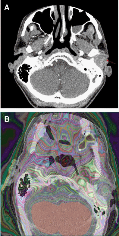
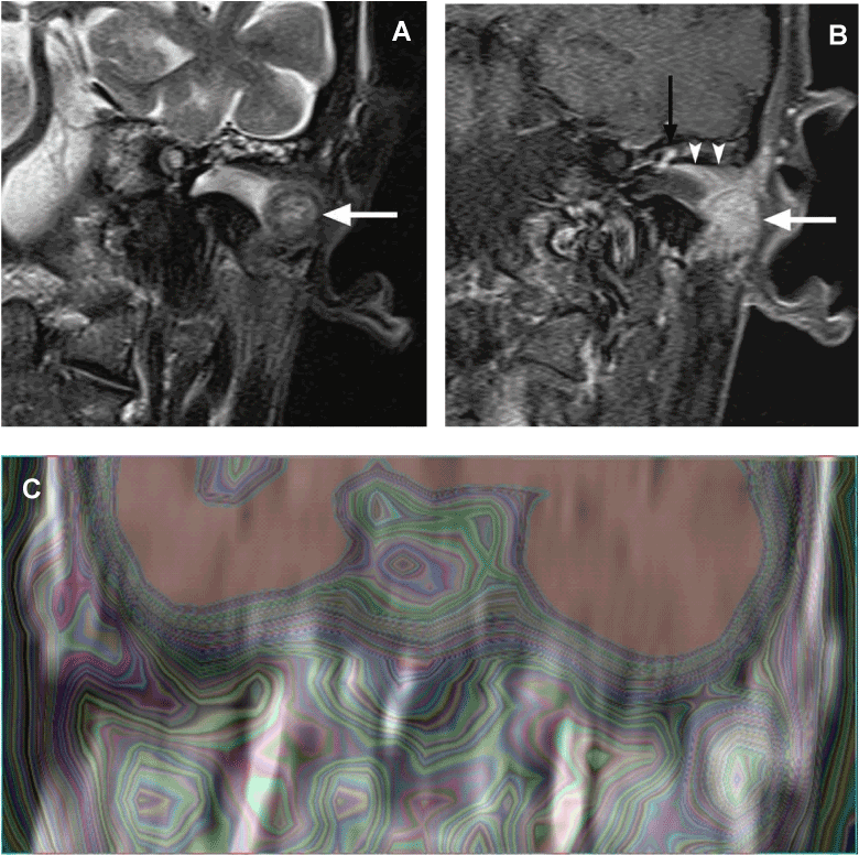
| On the initial CT, a large mass was demonstrated in the left auditory canal extending into the middle ear through the tympanic membrane through the external auditory canal (Figure 1A). On MRI, the tumor was nearly isointense to muscle on T1-weighted images and mildly hyperintense and heterogeneous on T2-weighted images. Following administration of intravenous gadolinium, the mass demonstrated strong mostly homogeneous enhancement, and there was evidence of a tail of enhancing tumor extending along the roof of the external auditory canal into the epitympanum (Figures 2A and 2B). |
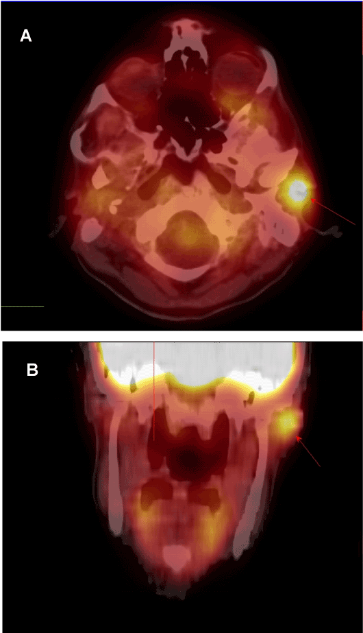
| An FDG-PET/CT was also performed on this patient to exclude any evidence of metastatic disease. The tumor was identified as being hypermetabolic with intensely increased FDG uptake (SUVavg 3.4), with no evidence of metastatic involvement of regional lymph nodes or distant organs (Figures 3A and 3B). Additionally, FDG-PET images were fused with the contrast enhanced. |
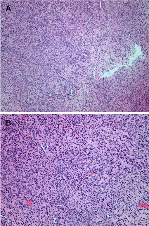
| CT (Figure 1B) and with MR images (Figure 2C), and a moderate extent of heterogeneity in FDG uptake and density on CT, and signal intensity on MRI was appreciated. On light microscopy the lesion displayed irregular pleomorphic and hyperchromatic atypical cells demonstrating marked heterogeneity. These cells ranged in shape from epithelioid to spindled and were arranged around stellate vessels. They were found to be immunoreactive for smooth muscle actin and focally positive for CD31 and caldesmon. The tumor was also Found to be moderately positive for GLUT-1 (Figures 4A, 4B and 4C). Given the extreme rarity of malignant myopericytoma, the diagnosis is made with some hesitation, in favour of “atypical myopericytic neoplasm with uncertain malignant potential.” In view of the uncontrolled immunocompromised state of the patient, surgery was deferred. |
| Discussion |
| Myopericytic lesions are lesions felt to be derived from or showing differentiation toward perivascularmyoid cells. Since myopericytoma was first distinguished as a separate histopathological entity, the clinical, microscopic and radiological features of this benign form have been described [1-9]. However, the radiological correlation (including PET/CT) of malignant myopericytoma is essentially undescribed. From a histological stand point, our case has atypical features, and clinical and Molecular Imaging features suggestive of malignancy given its locally aggressive nature and intense tracer uptake. We offer important radiological findings by detailing the PET/CT, contrasted CT and MR imaging correlation of this interesting myopericytic lesion. |
| The CT and MRI imaging features of the tumor in the present case have been shown to be nonspecific, and similar to those which have been described previously in the literature for benign myopericytomas [9]. On CT, myopericytomas are usually iso- to hypodense relative to muscle and can show mild to strong enhancement with intravenous contrast. Enhancement may be homogeneous or heterogeneous. The shape and margins of myopericytomas can also be variable and can range from ovoid and well-defined to irregular and poorly marginated. Occasionally, myopericytomas can also present as lytic bone lesions [10]. |
| On MRI, benign myopericytomas show low to intermediate signal intensity on T1-weighted images and increased heterogeneous signal intensity on T2-weighted images. At least one report describes the presence of T1 hyperintense intratumoral foci suggestive of hemorrhage within a myopericytoma of the lower extremity, with heterogeneous-to-strong homogeneous enhancement following intravenous gadolinium administration [9]. |
| In the original case series by Menamin and Fletcher one patient out of 5 with malignant myopericytoma had a biopsy-proven liver metastasis [3]. In our patient, PET/CT demonstrated that the primary tumor was FDG-avid, but no metastatic foci were seen. |
| This tumor was found to be GLUT-1 positive, which correlates with its FDG avidity. Glucose is the main energy substrate for many tumors, and due to its hydrophilic nature, it depends on transmembrane receptors for its transport into cells, such as GLUT-1. Overexpression of GLUT-1 receptors is seen in many FDG-avid tumors, which can be assessed through histopathologic staining [11]. |
| Molecular Imaging is based on an understanding of the physiologic activity associated with molecular pathways, histopathologic features, anatomic radiographic behavior, as well as the clinical presentation and course of a disease. Understanding disease will require an intimate knowledge of all these areas to appropriately classify and treat it. Newer entities could also be better understood, especially if their malignancy potential and their clinical course is indeterminate, particularly when the disease entity as we have described is rare. Thus, we hope that understanding myopericytic lesions and through their Molecular Imaging behavior will lead to better clinical classification of these entities when correlated with their histopathologic findings. |
| Acknowledgements |
| We would like to thank Dr Christopher Fletcher, Chief of Surgical Pathology at Dana Farber Cancer Institute, Brigham and Women’s Hospital, Boston, MA for his contribution. |
References |
|
Figures at a glance
 |
 |
 |
 |
| Figure 1 | Figure 2 | Figure 3 | Figure 4 |
Post your comment
Relevant Topics
- Abdominal Radiology
- AI in Radiology
- Breast Imaging
- Cardiovascular Radiology
- Chest Radiology
- Clinical Radiology
- CT Imaging
- Diagnostic Radiology
- Emergency Radiology
- Fluoroscopy Radiology
- General Radiology
- Genitourinary Radiology
- Interventional Radiology Techniques
- Mammography
- Minimal Invasive surgery
- Musculoskeletal Radiology
- Neuroradiology
- Neuroradiology Advances
- Oral and Maxillofacial Radiology
- Radiography
- Radiology Imaging
- Surgical Radiology
- Tele Radiology
- Therapeutic Radiology
Recommended Journals
Article Tools
Article Usage
- Total views: 13818
- [From(publication date):
March-2013 - Jul 03, 2025] - Breakdown by view type
- HTML page views : 9254
- PDF downloads : 4564
